Review
. 2013 Jul;126(1):49-61.
doi: 10.1016/j.bandl.2012.10.001. Epub 2012 Dec 1.

Frontal-thalamic circuits associated with language

Affiliations
Review

Frontal-thalamic circuits associated with language

Helen Barbas et al. Brain Lang. 2013 Jul.

Abstract

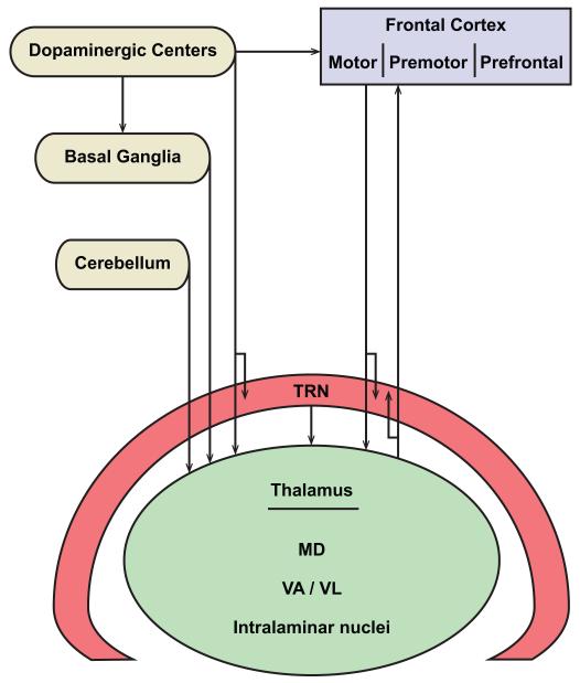
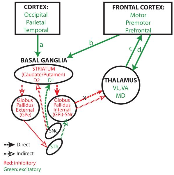
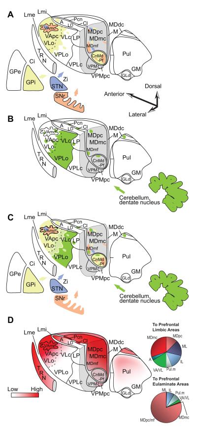
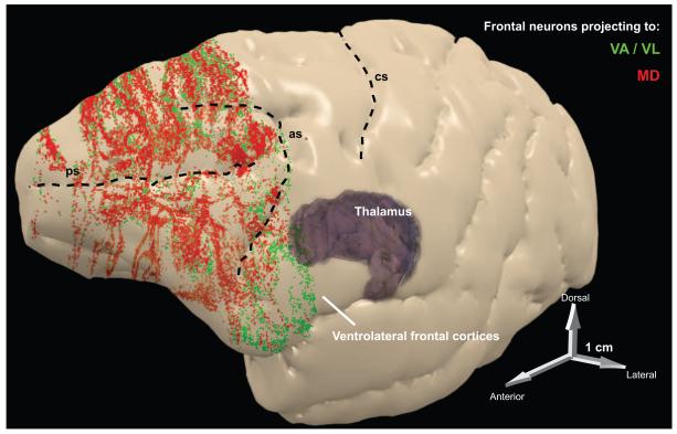
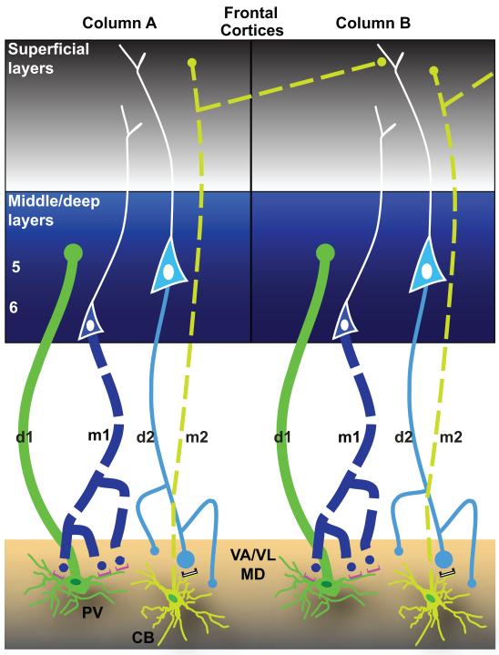
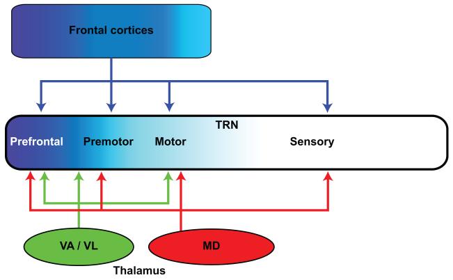
Thalamic nuclei associated with language including the ventral lateral, ventral anterior, intralaminar and mediodorsal form a hub that uniquely receives the output of the basal ganglia and cerebellum, and is connected with frontal (premotor and prefrontal) cortices through two parallel circuits: a thalamic pathway targets the middle frontal cortical layers focally, and the other innervates widely cortical layer 1, poised to recruit other cortices and thalamic nuclei for complex cognitive operations. Return frontal pathways to the thalamus originate from cortical layers 6 and 5. Information through this integrated thalamo-cortical system is gated by the inhibitory thalamic reticular nucleus and modulated by dopamine, representing a specialization in primates. The intricate dialogue of distinct thalamic nuclei with the basal ganglia, cerebellum, and specific dorsolateral prefrontal and premotor cortices associated with language, suggests synergistic roles in the complex but seemingly effortless sequential transformation of cognitive operations for speech production in humans.


Figures

Figure 1
Figure 1
Thalamic nuclei that serve as a hub for distributed neural circuits associated with language. The nodes of networks that interact with the thalamus include: the frontal cortex, which consists of motor, premotor, and prefrontal areas; basal ganglia; cerebellum; dopaminergic groups from the mesencephalic substantia nigra pars compacta and the ventral tegmental area; and the inhibitory thalamic reticular nucleus (TRN).
Figure 2
Figure 2
The frontal cortex in the primate brain. A, B, Medial (top) and lateral (bottom) views of the rhesus monkey (A) and human (B) brains show cingulate, orbital, and lateral prefrontal, premotor cortices and the primary motor cortex. Abbreviations: ACC, anterior cingulate cortices; as, arcuate sulcus; cc, corpus callosum; cgs, cingulate sulcus; cs, central sulcus; ifs, inferior frontal sulcus; LPFC, lateral prefrontal cortices; M1, primary motor cortex; OFC, orbitofrontal cortices; Pre-SMA, anterior supplementary motor area; ps, principal sulcus; SMA, supplementary motor area; sfs, superior frontal sulcus; Thal, thalamus.
Figure 3
Figure 3
Direct and indirect basal ganglia loops. The entire cortex projects to the basal ganglia. The diagram shows the direct and indirect pathways through the basal ganglia. Excitatory pathways are shown in green and inhibitory in red. Pathways that are common to the direct and indirect pathways are shown by solid green arrows. The cortex projects to the striatum (arrows a, b). Cortical axons terminate on inhibitory neurons of either the caudate or putamen, depending on their origin in the cortex. The direct pathway (heavy dotted line, solid arrowheads) projects from the striatum to GPi/SNr, which projects to the thalamus. Neurons in the GPi/SNr are inhibited by the direct pathway and therefore they can’t inhibit the thalamus (X, inhibited pathway). The thalamus remains free to project to the frontal cortex and back for movement. The indirect pathway (fine dotted line, silhouette arrowheads) takes a different route: from the striatum it projects and inhibits GPe. The GPe projects to the excitatory subthalamic nucleus (STN). The inhibited GPe leaves the STN free to activate GPi/SNr, which inhibits the thalamus, preventing activation and movement. Dopaminergic pathways from the substantia nigra pars compacta (SNc) have opposite effects on the two parallel pathways because they interact with distinct types of dopamine receptors found on neurons in the striatum. In the direct pathway, dopaminergic axons interact with D1 receptors, which are excitatory. In the indirect pathway, dopaminergic axons interact with D2 receptors, which are inhibitory. Abbreviations: GPe, external segment of the globus pallidus; GPi, internal segment of the globus pallidus. SNc, substantia nigra pars compacta; STN, subthalamic nucleus; MD, mediodorsal thalamic nucleus; VA, ventral anterior thalamic nucleus; VL, ventral lateral thalamic nucleus.
Figure 4
Figure 4
Innervation of the primate thalamus from the basal ganglia, cerebellum, and connections with prefrontal cortices, based on pathway studies in non-human primates [e.g. (Francois, Tande, Yelnik, & Hirsch, 2002; Sidibe, Pare, & Smith, 2002; Rouiller, Liang, Babalian, Moret, & Wiesendanger, 1994; Sakai, Inase, & Tanji, 1996; Sakai, Stepniewska, Qi, & Kaas, 2000; Erickson, Melchitzky, & Lewis, 2004)]. A, Matched coloring shows projections from the output nuclei of the basal ganglia. B, Shows projections from the output of the cerebellum through the dentate nucleus. C, Overlap of A and B. D (left), Prefrontal connections with the thalamus. D (right), Relative distribution of thalamic projection neurons directed to cingulate and orbital limbic areas (top, limbic) and to the eulaminate areas 46, 8 and 9 (bottom). Quantitative data are from: (Barbas, Henion, & Dermon, 1991; Dermon & Barbas, 1994). Abbreviations: A, anterior nuclei; Ci, internal capsule; Cl, centrolateral nucleus; CnMd, centromedian nucleus; GLd, lateral dorsal nucleus geniculate; GM, medial nucleus geniculate; GPe, globus pallidus external; GPi, globus pallidus internal; LD, lateral dorsal nucleus; Lme, external lamina; Lmi, internal lamina; LP, lateral posterior nucleus; M, midline nuclei; MDdc/mc/mf/pc, the densocellular (dc), magnocellular (mc), multiform (mf), and parvicellular (pc) subdivisions of the mediodorsal (MD) nucleus; Pcn, paracentral nucleus; Pul, pulvinar nucleus; TRN, thalamic reticular nucleus; SNr, substantia nigra reticulata; STN, subthalamic nucleus; VAmc/pc, magnocellular (mc) and parvocellular (pc) subdivision of the ventral anterior (VA) nucleus; VL, ventrolateral nucleus; VLc, caudal ventrolateral nucleus; VLo, oral ventrolateral nucleus; VPLc, caudal subdivision of the ventral posterior-lateral nucleus; VPLo, oral subdivision of the ventral posterior-lateral nucleus;VPM, medial ventroposterior nucleus; VPMpc, parvocellular subdivision of the medial ventroposterior nucleus; Zi, zona incerta.
Figure 5
Figure 5
Summary of frontal projections to thalamic nuclei associated with language. Three-dimensional reconstruction of the left hemisphere of the rhesus monkey cortex, which was rendered semi-transparent to show frontal neurons in layers 5 and 6 that project to MD (red) or to VA/VL (green). The projection zones include prefrontal areas (in front of the arcuate sulcus), as well as premotor areas situated behind the upper and lower limbs of the arcuate sulcus (as). Abbreviations: as, arcuate sulcus; cs, central sulcus; MD, mediodorsal nucleus; ps, principal sulcus; VA, ventral anterior nucleus; VL, ventrolateral nucleus.
Figure 6
Figure 6
Two parallel systems link the thalamus with the cortex. Schematic diagram summarizes the features of reciprocal driving and modulatory corticothalamic pathways linking the frontal cortex with VA/VL thalamic nucleus. The thickness of the lines indicates the strength of the projection. The size of the dot denotes the size of the terminals. Solid lines (d1 and d2) represent driving pathways, and dotted lines (m1 and m2) represent modulatory pathways. Dark green represents PV, and light green CB thalamic projection neurons. There are two parallel circuits between prefrontal cortex and thalamus. One originates mostly from PV thalamic projection neurons (dark green, bottom panels), and terminates focally with large terminals in the middle layers (3b – 5a, center blue panels) of PFC (d1). In turn, layer 6 neurons project (m1) and terminate as numerous small terminals that form synapses mainly with PV thalamic projection neurons (dark green, bottom panels), through metabotropic receptors (mGluR1a, magenta). The other circuit originates in layer 5 (d2) and forms synapses through a mix of large and small terminals mainly on CB thalamic projection neurons (light green) with ionotropic receptors, white). The CB thalamic neurons, in turn, send widespread projections (m2) mainly to the superficial layers (1 – 3a) of frontal cortex (grey), and terminate mostly as small terminals that cross borders of neighboring regions and are intermingled with the apical dendrites of neighboring layer 5 neurons. All depicted pathways are excitatory and glutamatergic. Adapted from: Zikopoulos and Barbas, 2007b.
Figure 7
Figure 7
Frontal cortical and associated thalamic nuclei project to TRN. Frontal cortices and the VA/VL thalamic nuclei predominantly target the frontal sectors of TRN (prefrontal, premotor, and motor; shades of blue). Some dorsolateral prefrontal cortices (areas 9 and 46), and their principal thalamic nucleus, MD, target all TRN sectors, including the posterior sensory-related regions.
Figure 8
Figure 8
Dopaminergic fiber distribution in the primate thalamus. Dark shades represent high, and light shades represent sparser innervation. For list of abbreviations see legend to Figure 4.

References

    1. Alexander GE, Delong MR, Strick PL. Parallel organization of functionally segregated circuits linking basal ganglia and cortex. Annual Review of Neuroscience. 1986;9:357–381. - PubMed
    1. Anderson ME. Pallidal and cortical detriments of thalamic activity. In: Kultas-Ilinsky K, Ilinsky IA, editors. Basal ganglia and thalamus in health and movement disorders. Kluwer Academic/ Plenum Publishers; New York: 2001. pp. 93–104.
    1. Arikuni T, Kubota K. The organization of prefrontocaudate projections and their laminar origin in the Macaque monkey: A retrograde study using HRP-Gel. Journal of Comparative Neurology. 1986;244:492–510. - PubMed
    1. Arnsten AF, Li BM. Neurobiology of executive functions: catecholamine influences on prefrontal cortical functions. Biological Psychiatry. 2005;57:1377–1384. - PubMed
    1. Bachevalier J, Meunier M, Lu MX, Ungerleider LG. Thalamic and temporal cortex input to medial prefrontal cortex in rhesus monkeys. Experimental Brain Research. 1997;115:430–444. - PubMed

Publication types

MeSH terms