. 2013 Feb;27(2):224-37.
doi: 10.1210/me.2012-1310. Epub 2012 Dec 4.

β-catenin regulates GnRH-induced FSHβ gene expression

Affiliations

β-catenin regulates GnRH-induced FSHβ gene expression

Qian Wang et al. Mol Endocrinol. 2013 Feb.

Abstract

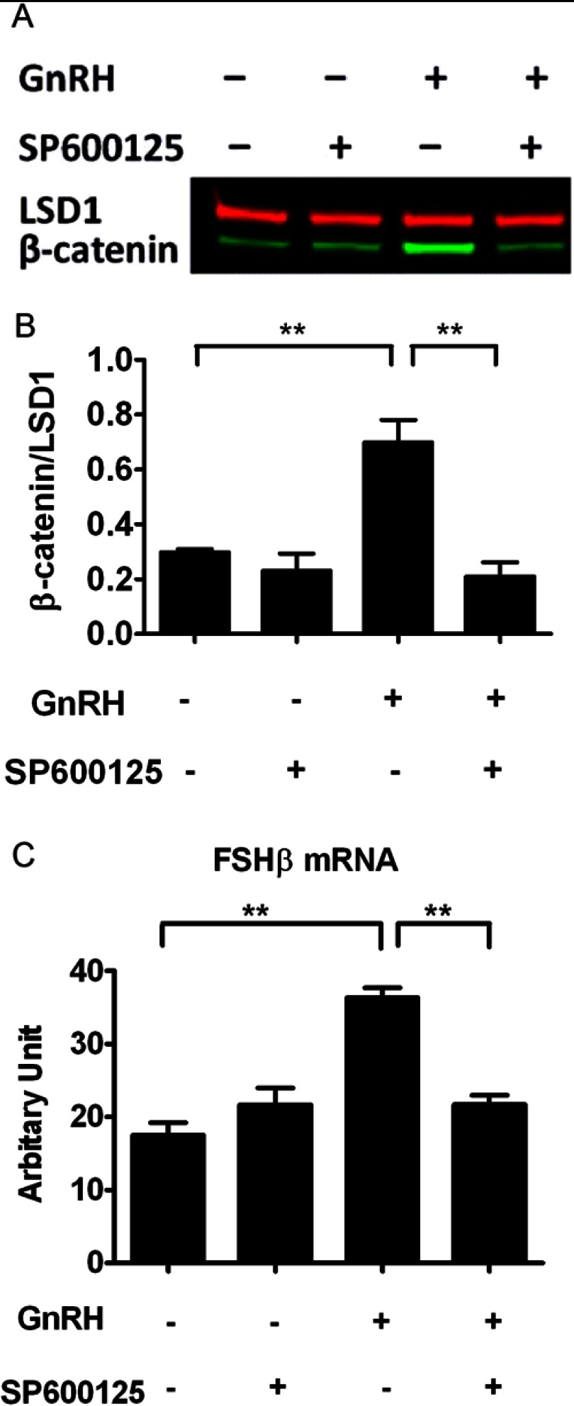
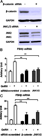
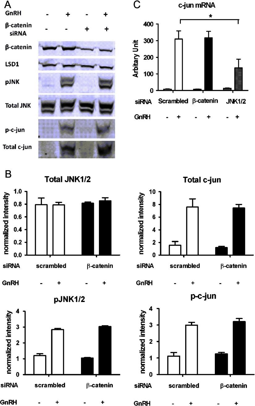
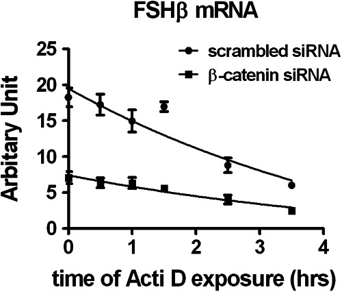
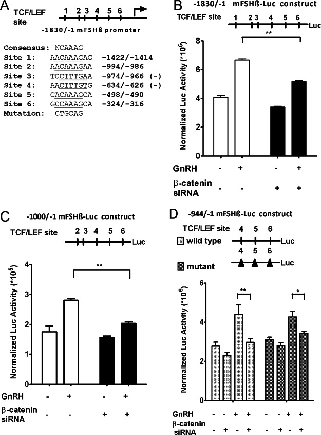
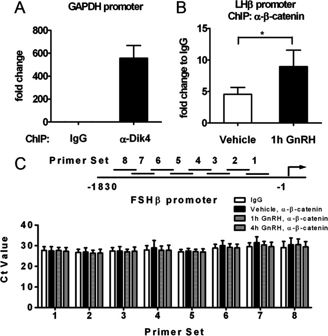
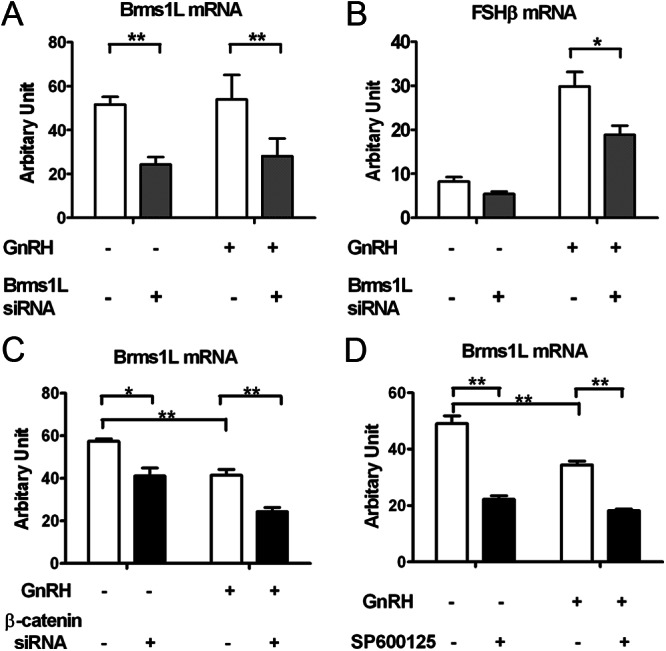
The regulation of gonadotropin synthesis by GnRH plays an essential role in the neuroendocrine control of reproduction. The known signaling mechanisms involved in gonadotropin synthesis have been expanding. For example, involvement of β-catenin in LHβ induction by GnRH has been discovered. We examined the role of β-catenin in FSHβ gene expression in LβT2 gonadotrope cells. GnRH caused a sustained increase in nuclear β-catenin levels, which was significantly reduced by c-Jun N-terminal kinase (JNK) inhibition. Small interfering RNA-mediated knockdown of β-catenin mRNA demonstrated that induction of FSHβ mRNA by GnRH depended on β-catenin and that regulation of FSHβ by β-catenin occurred independently of the JNK-c-jun pathway. β-Catenin depletion had no impact on FSHβ mRNA stability. In LβT2 cells transfected with FSHβ promoter luciferase fusion constructs, GnRH responsiveness was conferred by the proximal promoter (-944/-1) and was markedly decreased by β-catenin knockdown. However, none of the T-cell factor/lymphoid enhancer factor binding sites in that region were required for promoter activation by GnRH. Chromatin immunoprecipitation further corroborated the absence of direct interaction between β-catenin and the 1.8-kb FSHβ promoter. To elucidate the mechanism for the β-catenin effect, we analyzed approximately 1 billion reads of next-generation RNA sequencing β-catenin knockdown assays and selected the nuclear cofactor breast cancer metastasis-suppressor 1-like (Brms1L) as one candidate for further study. Subsequent experiments confirmed that Brms1L mRNA expression was decreased by β-catenin knockdown as well as by JNK inhibition. Furthermore, knockdown of Brms1L significantly attenuated GnRH-induced FSHβ expression. Thus, our findings indicate that the expression of Brms1L depends on β-catenin activity and contributes to FSHβ induction by GnRH.


Figures

Fig. 1.
Fig. 1.
Attenuation of GnRH-induced β-catenin nuclear accumulation and FSHβ mRNA expression by JNK inhibition. A, Effect of SP600125 on GnRH-induced nuclear accumulation of β-catenin in LβT2 cells. Cells were serum starved overnight, pretreated with 40 μm SP600125 or vehicle for 30 min, and stimulated with 10 nm GnRH or vehicle for 15 min. Nuclear extracts were subjected to a quantitative Western blot analysis using a β-catenin-specific antibody. LSD1 (in red) was used as a loading control. B, Quantification of Western blot densitometry from three independent experiments, plotted as mean ± sem. One-way ANOVA (n = 3; *, P < 0.05). C, Effect of SP600125 on GnRH-induced FSHβ mRNA levels. Cells were serum starved overnight, pretreated with 40 μm SP600125 or vehicle for 30 min, and stimulated with 5 nm GnRH or vehicle for 6 h. Relative mRNA copy numbers of FSHβ were determined by quantitative real time-PCR. Two-way ANOVA (n = 4; **, P < 0.01).
Fig. 2.
Fig. 2.
Effect of siRNA-mediated silencing of β-catenin and JNK on GnRH-induced FSHβ mRNA levels. A, LβT2 cells were transfected with either scrambled, β-catenin, JNK1, or JNK2 siRNA for 72 h using the nucleofection method (as described in Materials and Methods). Whole-cell lysates were subjected to a Western blot analysis using JNK- and β-catenin-specific antibodies. GAPDH was used as a loading control. B, Effect of β-catenin or JNK1/2 knockdown on FSHβ gene expression. LβT2 cells were transfected with either scrambled, β-catenin, or JNK1/2 siRNA for 48 h, serum starved overnight, and stimulated with 5 nm GnRH or vehicle for 8 h. RNA copy numbers of FSHβ were determined by quantitative real time-PCR. Two-way ANOVA (n = 4; **, P < 0.01). C, Effect of β-catenin or JNK1/2 knockdown on FSHβ gene expression under low-frequency GnRH pulses. LβT2 cells were transfected with either scrambled, β-catenin, or JNK1/2 siRNA for 48 h, serum-starved overnight, and stimulated with slow pulses of 5 nm GnRH or vehicle for 6 h. Each pulse lasted 5 min. The time intervals between GnRH pulses (media replacement) were 2 h. Samples were collected 30 min after the last pulse. Relative mRNA copy numbers of FSHβ were determined by quantitative real time-PCR. Two-way ANOVA (n = 4; **, P < 0.01).
Fig. 3.
Fig. 3.
Effect of β-catenin knockdown on GnRH-induced JNK-c-jun activation. A, LβT2 cells were transfected with either scrambled or β-catenin siRNA for 48 h, serum-starved overnight, and stimulated with 10 nm GnRH or vehicle for 30 min (for JNK) and 1 h (for c-jun). Whole-cell lysates were subjected to a Western blot analysis using β-catenin-, JNK-, phospho-JNK-, c-jun-, and phospho-c-jun-specific antibodies 72 h after transfection. LSD1 was used as a loading control. B, Quantification of Western blot densitometry from three independent experiments, plotted as mean ± sem. Two-way ANOVA. C, Effect of β-catenin or JNK1/2 knockdown on c-jun gene expression. LβT2 cells were transfected with either scrambled, β-catenin, or JNK1/2 siRNA for 48 h, serum starved overnight, and stimulated with 10 nm GnRH or vehicle for 1 h. RNA copy numbers of c-jun were determined by quantitative real time-PCR. Two-way ANOVA with post Bonferroni corrections (n = 3; *, P < 0.05).
Fig. 4.
Fig. 4.
Effect of β-catenin knockdown on FSHβ mRNA stability. LβT2 cells were transfected with either scrambled or β-catenin for 48 h, serum starved overnight, and stimulated with 5 nm GnRH for 2 h. Medium was replaced with fresh culture medium for another 4 h. Cells were then exposed to 2 μg/ml actinomycin D (Acti D, 1.6 μm) and harvested at the indicated time points. RNA copy numbers of FSHβ were determined by quantitative real time-PCR. Decay curves were fitted to the data using GraphPad Prism (n = 4); Student's t test.
Fig. 5.
Fig. 5.
Effect of β-catenin gene silencing on GnRH-stimulated FSHβ gene promoter activity. A, The 1.8-kb (−1830/−1) FSHβ promoter region was cloned from mouse genomic DNA upstream of a luciferase reporter gene. Using the TRANSFAC database, analysis against the T-cell factor/lymphoid enhancer factor (TCF/LEF) family core binding signature revealed six putative TCF/LEF binding sites. Sequences of the consensus and the six putative TCF/LEF sites are shown. “Mutation” designates the PstI restriction site that was used in block replacement mutagenesis in D. B (top panel), Structure of the 1.8-kb FSHβ promoter luciferase (Luc) construct. The TCF/LEF sites are numbered 1–6, as in panel A. B (bottom panel), LβT2 cells were cotransfected with the 1.8-kb FSHβ promoter luciferase construct, a control β-galactosidase expression vector (internal standard for transfection efficiency), and either scrambled or β-catenin siRNA. Forty eight hours after transfection, cells were serum starved for 4 h and stimulated with 10 nm GnRH or vehicle for 16 h. Luciferase activity was measured and normalized to β-galactosidase. Two-way ANOVA (n = 3; **, P < 0.01). C (top panel), Structure of the −1000/−1 FSHβ promoter luciferase construct. C (bottom panel), Cells were cotransfected with a −1000/−1 FSHβ promoter luciferase construct, a control β-galactosidase expression vector, and either scrambled or β-catenin siRNA. Cells were then treated as described in panel B. Two-way ANOVA (n =3; **, P < 0.01). D (top panel), Structure of the −944/−1 FSHβ promoter luciferase construct and its mutant counterpart. Block replacement mutations symbolized by black triangles were introduced at the three most proximal TCF/LEF sites in the context of the −944/−1 construct. D (bottom panel), Either the wild-type or the mutated −944/−1 promoter construct was transfected into LβT2 cells, as described in panel B. Two-way ANOVA (n = 3; *, P < 0.05; **, P < 0.01). The data shown are mean ± sem of triplicate samples from one experiment and are representative of three independent experiments.
Fig. 6.
Fig. 6.
Chromatin immunoprecipitation of the β-catenin protein to the FSHβ promoter. Quantification of the ChIP assays performed on LβT2 cells is shown. A, The presence of dimethyl-histone H3-Lys4 binding to the GAPDH promoter was assessed with an antidimethylhistone H3-Lys4 antibody (α-Dik4, as indicated) and served as a positive control for the ChIP assay. A nonspecific antibody, normal mouse IgG was used as a negative control. Data were normalized to the IgG control and represent the mean ± sem of four experiments. B, The binding of β-catenin to the LHβ promoter in the presence of GnRH was assessed with an anti-β-catenin antibody (α-β-catenin, as indicated) and served as a positive control for β-catenin immunoprecipitation. Cells were serum starved overnight and stimulated with GnRH (10 nm) or vehicle for 1 h. Chromatin immunoprecipitation was performed on genomic DNA fragments from those cell nuclei. Data were normalized to the IgG control and represent the mean ± sem of four experiments. Paired t test (n = 4; *, P < 0.05). C, Cells were serum-starved overnight and stimulated with either vehicle or GnRH (10 nm) for 1 h or 4 h. The presence of β-catenin binding to the FSHβ promoter under GnRH stimulation was assessed with the same antibody as in panel B, through the use of eight primer sets (1–8, as indicated) encompassing the six putative TCF/LEF sites within the 1.8-kb promoter region. Results are expressed as Ct values and represent the mean ± sem of three independent experiments.
Fig. 7.
Fig. 7.
Role of Brms1L in β-catenin regulation of GnRH-induced FSHβ expression. A, Knockdown efficiency of Brms1L siRNA in LβT2 cells. LβT2 cells were transfected with either scrambled or Brms1L siRNA for 48 h, serum starved overnight, and stimulated with 5 nm GnRH or vehicle for 2 h. Medium was replaced with fresh culture medium for another 4 h. B, Effect of siRNA-mediated silencing of Brms1L on GnRH-induced FSHβ mRNA levels. Cells were treated as described in panel B. C, Effect of siRNA-mediated silencing of β-catenin on Brms1L mRNA levels. Cells were transfected with either scrambled or β-catenin siRNA for 48 h, serum starved overnight, and stimulated with 5 nm GnRH or vehicle for 2 h. D, Effect of JNK inhibition on Brms1L mRNA levels. Cells were serum starved overnight, pretreated with 40 μm SP600125 or vehicle for 30 min, and stimulated with 5 nm GnRH or vehicle for 6 h. Relative mRNA copy numbers of Brms1L and FSHβ were determined by quantitative real time-PCR. Two-way ANOVA (n = 4; **, P < 0.05). Results represent the mean ± sem of three independent experiments.

Similar articles

Cited by

References

    1. Pierce JG, Parsons TF. 1981. Glycoprotein hormones: structure and function. Annu Rev Biochem 50:465–495 - PubMed
    1. Burger LL, Haisenleder DJ, Dalkin AC, Marshall JC. 2004. Regulation of gonadotropin subunit gene transcription. J Mol Endocrinol 33:559–584 - PubMed
    1. Alarid ET, Windle JJ, Whyte DB, Mellon PL. 1996. Immortalization of pituitary cells at discrete stages of development by directed oncogenesis in transgenic mice. Development 122:3319–3329 - PubMed
    1. Pernasetti F, Vasilyev VV, Rosenberg SB, Bailey JS, Huang HJ, Miller WL, Mellon PL. 2001. Cell-specific transcriptional regulation of follicle-stimulating hormone-β by activin and gonadotropin-releasing hormone in the LβT2 pituitary gonadotrope cell model. Endocrinology 142:2284–2295 - PubMed
    1. Thomas P, Mellon PL, Turgeon J, Waring DW. 1996. The L β T2 clonal gonadotrope: a model for single cell studies of endocrine cell secretion. Endocrinology 137:2979–2989 - PubMed

Publication types

MeSH terms