. 2013 Jan 23;32(2):290-302.
doi: 10.1038/emboj.2012.322. Epub 2012 Dec 4.

Initiation of DNA damage responses through XPG-related nucleases

Affiliations

Initiation of DNA damage responses through XPG-related nucleases

Karen Kuntz et al. EMBO J. .

Abstract

Lesion-specific enzymes repair different forms of DNA damage, yet all lesions elicit the same checkpoint response. The common intermediate required to mount a checkpoint response is thought to be single-stranded DNA (ssDNA), coated by replication protein A (RPA) and containing a primer-template junction. To identify factors important for initiating the checkpoint response, we screened for genes that, when overexpressed, could amplify a checkpoint signal to a weak allele of chk1 in fission yeast. We identified Ast1, a novel member of the XPG-related family of endo/exonucleases. Ast1 promotes checkpoint activation caused by the absence of the other XPG-related nucleases, Exo1 and Rad2, the homologue of Fen1. Each nuclease is recruited to DSBs, and promotes the formation of ssDNA for checkpoint activation and recombinational repair. For Rad2 and Exo1, this is independent of their S-phase role in Okazaki fragment processing. This XPG-related pathway is distinct from MRN-dependent responses, and each enzyme is critical for damage resistance in MRN mutants. Thus, multiple nucleases collaborate to initiate DNA damage responses, highlighting the importance of these responses to cellular fitness.


Figures

Figure 1
Figure 1
ast1, an XPG-related nuclease, is a high copy suppressor of chk1-E472D. (A) Ten-fold serial dilutions of chk1-E472D cdc27-P11 transformed with an empty vector or the indicated plasmids and were grown at 25 and 32.5°C. A plasmid overexpressing ast1 is able to rescue the temperature sensitivity of chk1-E472D cdc27-P11 at 32.5°C. (B) A schematic of the domain structures of the XPG-related family members in S. pombe. The XPG-related nucleases all contain conserved N-terminal (XPG-N) and Internal (XPG-I) domains required for their nuclease activity. (C) Ten-fold serial dilutions of chk1-E472D transformed with an empty vector or the indicated plasmids (Ast1ND is the nuclease-dead derivative) were grown at 25 and 36°C in the presence and absence of 0.01% MMS. A plasmid overexpressing wild-type ast1 is able to rescue the DNA damage sensitivity of chk1-E472D at 36°C. (D) Ten-fold serial dilutions of chk1-D155A transformed with an empty vector or the indicated plasmids were grown at 30°C in the presence and absence of 0.0075% MMS. A plasmid overexpressing ast1 is unable to rescue the DNA damage sensitivity of chk1-D155A. (E) Western blot with anti-HA antibody and extracts from a wild-type strain containing HA-tagged chk1 and from an HA-tagged-chk1-E472D strain transformed with an empty vector or a plasmid overexpressing ast1. Strains were grown in the presence and absence of 0.01% MMS. Overexpression of ast1 restored a mobility shift to chk1-E472D following DNA damage. (F) Ten-fold serial dilutions of the indicated strains were grown in the presence or absence of 0.005% MMS at 30°C. The chk1Δ ast1Δ double mutant is not more sensitive to MMS than the chk1Δ single mutant. (G) A UV-C survival curve was performed with the indicated strains. Graph shows the mean±s.d. for each data point (n=3). The chk1Δ ast1Δ double mutant is not more sensitive to UV than the chk1Δ single mutant.
Figure 2
Figure 2
Lethal cell-cycle arrest of exo1Δ rad2Δ is dependent on chk1 and ast1. (A) Single colonies from tetrad analysis are shown at the indicated magnifications, and are photographed 2 days following dissection on YES plates containing the vital dye phloxin B, which stains dead cells red. Strains are indicated above each photo. A wild-type colony is shown for comparison. The rad2Δ exo1Δ double mutant shows a cell-cycle arrest (elongated cells) that are viable (phloxin negative) at this time point, though inviable after further incubation. The rad2Δ exo1Δ chk1Δ triple mutant dies (phloxin positive, arrowed) with a ‘cut’ phenotype following 1–3 cell cycles indicating a checkpoint defect. The rad2Δ exo1Δ ast1Δ triple mutant dies as a micro-colony with many inviable (phloxin positive, examples arrowed) cells showing a ‘cut’ phenotype, also indicating a checkpoint defect. For this analysis, the ‘cut’ phenotype is defined as septated, phloxin-positive (dead) cells, or septated cells in which one daughter is dead (phloxin positive) and one alive (phloxin negative), which is derived from unequal chromosome segregation. (B) Ten-fold serial dilutions of the indicated strains were grown at 30°C in the presence and absence of 0.008% MMS. The exo1Δ ast1Δ and rad2Δ ast1Δ double mutants are not more sensitive to MMS than the exo1Δ and rad2Δ single mutants, respectively. (C) A UV-C survival curve was performed with the indicated strains. Graph shows the mean±s.d. for each data point (n=3). The exo1Δ ast1Δ and rad2Δ ast1Δ double mutants are not more sensitive to UV than the exo1Δ and rad2Δ single mutants, respectively. (D) Western blot with anti-HA antibody and extracts from strains of the indicated genotype containing HA-tagged chk1. The exo1Δ ast1Δ and rad2Δ ast1Δ double mutants do not have increased Chk1 activation above the levels seen in the exo1Δ and rad2Δ single mutants, respectively.
Figure 3
Figure 3
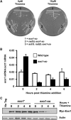
Creation of a thiamine repressible exo1 shut-off allele. (A) The indicated strains were streaked on plates in the presence or absence of 10 μM thiamine. The exo1-so strain is shown as a control. In agreement with the tetrad analysis, both the double mutant rad2Δ exo1-so and the triple mutant ast1Δ rad2Δ exo1-so are dead in the presence of thiamine when exo1 expression is abolished. (B) cDNA synthesis followed by qPCR with primers in the exo1 open reading frame is shown for a wild-type strain and a strain containing the exo1-so allele. All qPCR data are normalized to data obtained with a primer in the actin open reading frame. Graph shows the mean±standard error (n=3–5) for each data point. By 2 h following the addition of thiamine, exo1 expression is reduced to about 25% of wild-type levels. (C) Western blots using an anti-Myc antibody and an anti-Actin control with extracts from a wild-type control and cells containing a Myc-tagged exo1-so allele. By 6 h following the addition of thiamine, exo1 expression is almost undetectable at the protein level.
Figure 4
Figure 4
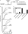
Checkpoint defects in strains lacking ast1, exo1 and rad2. (A) Strains containing the cdc25-22 mutation were grown at 25°C, thiamine was added to inactivate exo1 and cells were incubated for 2 h. Cells were shifted to 36°C for 4 h and then were treated with 30 J/m2 UV-C or left untreated. Cell septation was then monitored by microscopy following a shift back to 25°C. (B) The percentage of cells entering mitosis for each indicated strain following the time course described in (A). Wild-type cells show a delay in mitotic entry following UV-C treatment while chk1Δ cells show no delay. The cell-cycle delay and checkpoint arrest of the rad2Δ exo1-so double mutant both before and after UV-C treatment are dependent on ast1. Graphs show the mean±standard error (n=3) for each data point. (C) Cultures of the indicated strains were grown at 30°C. The percentage of aberrant mitoses or ‘cut’ cells was monitored by microscopy both before and after the addition of thiamine. Graph shows the mean±s.d. (n=3) for each data point. The cell-cycle arrest of the rad2Δ exo1-so strain is dependent on chk1 and partially dependent on ast1. The triple mutant rad2Δ exo1-so chk1-S345A shows an intermediate percentage of ‘cut’ cells following thiamine addition revealing a partial dependence on the phosphorylation of Chk1 on Serine 345 following DNA damage.
Figure 5
Figure 5
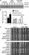
Multiple pathways are necessary to activate the DNA damage checkpoint. (A) Western blot with anti-HA antibody and extracts from the indicated strains containing HA-tagged chk1. Samples were taken at the indicated times following the time course in Figure 4. The wild-type strain shows Chk1 activation following UV treatment while the rad2Δ exo1-so strain shows no Chk1 activation. The ast1Δ rad2Δ exo1-so strain has an intermediate level of Chk1 activation. (B) Chk1 kinase assay performed on the indicated strains containing HA-tagged chk1. Graph shows the mean±s.d. for each data point (n=3). The wild-type strain shows an increase in Chk1 activation following UV treatment while the rad2Δ exo1-so strain shows no increase in Chk1 activation above basal levels. The triple mutant ast1Δ rad2Δ exo1-so has an intermediate amount of Chk1 activation following UV treatment. (C) Ten-fold serial dilutions of the indicated strains were plated on media containing 0.001% MMS or a control plate. Note that pku70Δ suppresses the increased MMS sensitivity of rad32Δ ast1Δ cells, but not rad32Δ exo1Δ cells, however, pku70Δ does suppress the slow growth phenotype of rad32Δ exo1Δ cells.
Figure 6
Figure 6
Ast1, Exo1 and Rad2 play a role in DSB repair. (A) ChIP and qPCR of HA-Ast1, Myc-Exo1 and Myc-Rad2 strains before and after the induction of a site-specific DSB using an HO endonuclease. Graph shows the mean±s.e.m. (n=3–6) for each data point. RPA-GFP and Rad52-YFP are used as positive controls. Following induction of the break, all nucleases and positive controls are recruited to the DSB. A control primer, act1, was used for qPCR. Cutting efficiency for the inducible HO endonuclease in this system is ∼20% of chromatids. (B) Quantification of RPA-GFP and Rad51 focus formation before and after treatment with 50 J/m2 UV or 0.5 mU Bleomycin for the indicated strains by microscopy following the time course described in Figure 4. Graph shows the mean±s.d. for each data point (n=3). Fewer foci are seen in the rad2Δ exo1-so and ast1Δ rad2Δ exo1-so strains following UV and Bleomycin treatment than in the wild-type strain. (C) Quantification of the recombinants produced in a single-stranded annealing assay for the indicated strains. Both ast1Δ and exo1Δ single mutants had a reduced capacity for single-strand annealing as measured by this assay.
Figure 7
Figure 7
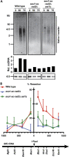
Cells lacking ast1, exo1 and rad2 have a defect in ssDNA production. (A) Non-denaturing Southern blot of the indicated strains treated with the indicated doses of UV following the time course described in Figure 4. The blot performed under native conditions (20 × SSC) is shown at the top of the figure while the denaturing Southern blot loading control is shown underneath. As seen in the native Southern blot, the wild-type strain had an increase in ssDNA production following UV treatment. The rad2Δ exo1-so strain did not have an increase in ssDNA production while the ast1Δ rad2Δ exo1-so strain had a small increase. The denaturing Southern blot shows a 4.5-kb band of the same intensity for all strains under all conditions as a loading control. The probe used for both Southern blots is an XhoI and KpnI fragment that recognizes the non-transcribed spacer (NTS) region of the S. pombe rDNA locus. The quantification of signal is in arbitrary phosphorimager units, corrected to the loading control (the corresponding track in the denatured transfer), and normalized to unirradiated wild-type cells. (B) qPCR analysis of in vivo resection of an I-PpoI-induced DSB within the 28S gene of the rDNA repeats. Cutting efficiency in this system is ⩾90% of rDNA repeats. The restriction map shows the position of sites on each side of the I-PpoI site used in the analysis where numbers indicate nucleotides, and the arrow depicts the direction of transcription. Data are mean±s.d. for three independent experiments.

Similar articles

Cited by

References

    1. Al-Khodairy F, Carr AM (1992) DNA repair mutants defining G2 checkpoint pathways in Schizosaccharomyces pombe. EMBO J 11: 1343–1350 - PMC - PubMed
    1. Al-Khodairy F, Fotou E, Sheldrick KS, Griffiths DJF, Lehman AR, Carr AM (1994) Identification and characterisation of new elements involved in checkpoint and feedback controls in fission yeast. Mol Biol Cell 5: 147–160 - PMC - PubMed
    1. Alseth I, Osman F, Korvald H, Tsaneva I, Whitby MC, Seeberg E, Bjoras M (2005) Biochemical characterization and DNA repair pathway interactions of Mag1-mediated base excision repair in Schizosaccharomyces pombe. Nucleic Acids Res 33: 1123–1131 - PMC - PubMed
    1. Ampatzidou E, Irmisch A, O’Connell MJ, Murray JM (2006) Smc5/6 is required for repair at collapsed replication forks. Mol Cell Biol 26: 9387–9401 - PMC - PubMed
    1. Araki H, Leem SH, Phongdara A, Sugino A (1995) Dpb11, which interacts with DNA polymerase II(epsilon) in Saccharomyces cerevisiae, has a dual role in S-phase progression and at a cell cycle checkpoint. Proc Natl Acad Sci USA 92: 11791–11795 - PMC - PubMed

Publication types

MeSH terms