Comparative Study
. 2013 Jan 7;54(1):85-95.
doi: 10.1167/iovs.12-10050.

A1120, a nonretinoid RBP4 antagonist, inhibits formation of cytotoxic bisretinoids in the animal model of enhanced retinal lipofuscinogenesis

Affiliations
Comparative Study

A1120, a nonretinoid RBP4 antagonist, inhibits formation of cytotoxic bisretinoids in the animal model of enhanced retinal lipofuscinogenesis

Nicoleta Dobri et al. Invest Ophthalmol Vis Sci. .

Abstract

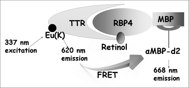
Purpose: Excessive accumulation of lipofuscin is associated with pathogenesis of atrophic age-related macular degeneration (AMD) and Stargardt disease. Pharmacologic inhibition of the retinol-induced interaction of retinol-binding protein 4 (RBP4) with transthyretin (TTR) in the serum may decrease the uptake of serum retinol to the retina and reduce formation of lipofuscin bisretinoids. We evaluated in vitro and in vivo properties of the new nonretinoid RBP4 antagonist, A1120.

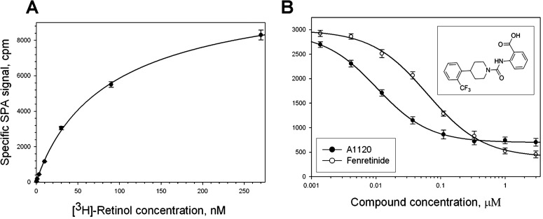
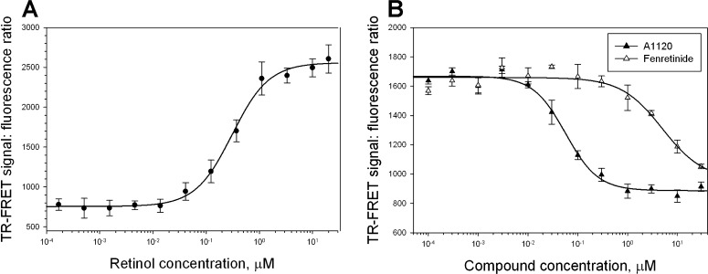
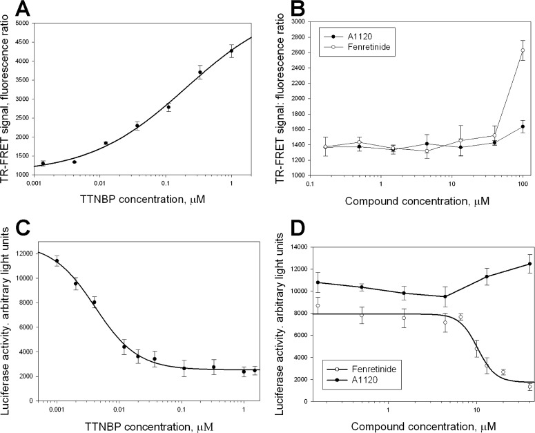
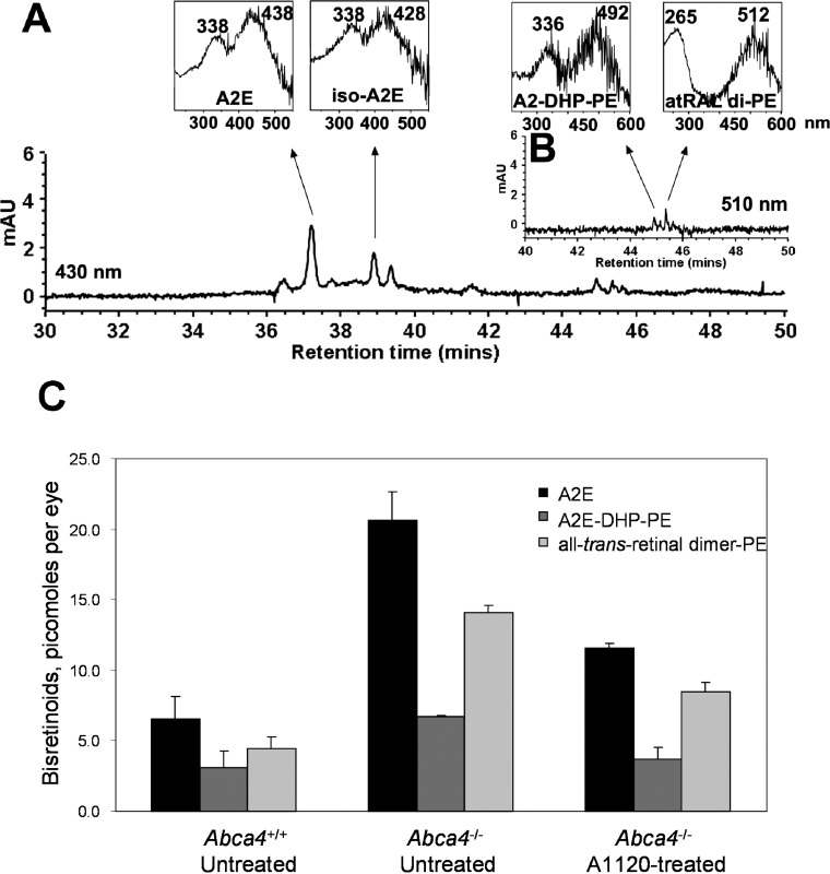
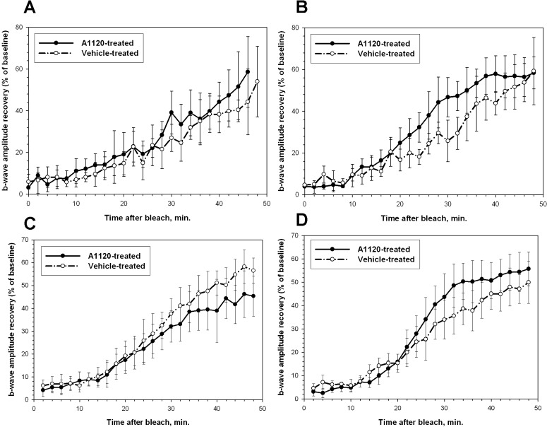
Methods: RBP4 binding potency, ability to antagonize RBP4-TTR interaction, and compound specificity were analyzed for A1120 and for the prototypic RBP4 antagonist fenretinide. A1120 ability to inhibit RPE65-mediated isomerohydrolase activity was assessed in the RPE microsomes. The in vivo effect of A1120 administration on serum RBP4, visual cycle retinoids, lipofuscin bisretinoids, and retinal visual function was evaluated using a combination of biochemical and electrophysiologic techniques.

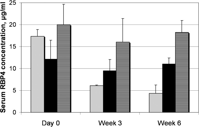
Results: In comparison to fenretinide, A1120 did not act as a RARα agonist, while exhibiting superior in vitro potency in RBP4 binding and RBP4-TTR interaction assays. A1120 did not inhibit isomerohydrolase activity in the RPE microsomes. A1120 dosing in mice induced 75% reduction in serum RBP4, which correlated with reduction in visual cycle retinoids and ocular levels of lipofuscin fluorophores. A1120 dosing did not induce changes in kinetics of dark adaptation.

Conclusions: A1120 significantly reduces accumulation of lipofuscin bisretinoids in the Abca4(-/-) animal model. This activity correlates with reduction in serum RBP4 and visual cycle retinoids confirming the mechanism of action for A1120. In contrast to fenretinide, A1120 does not act as a RARα agonist indicating a more favorable safety profile for this nonretinoid compound.


Conflict of interest statement

Disclosure: N. Dobri, None; Q. Qin, None; J. Kong, None; K. Yamamoto, None; Z. Liu, None; G. Moiseyev, None; J. Ma, None; R. Allikmets, P; J.R. Sparrow, P; K. Petrukhin, P

Figures

Figure 1.
Figure 1.
Characterization of test compounds in SPA-based RBP4 binding experiments. (A) Saturation isotherm for the binding of 3H-retinol to human RBP4. Biotinylated RBP4 (50 nM) was incubated with increasing concentrations of 3H-retinol (0.37–270 nM) in a total volume of 100 μL. Nonspecific binding was assessed in the presence of 20 μM nonradioactive retinol. Presented results are from the representative experiment performed in triplicate. Similar titration data were obtained in two additional experiments. (B) Representative isotherms of A1120 and fenretinide binding to human RBP4. 3H-retinol at 10 nM was used as a radioligand. Upper inset shows the A1120 structure.
Figure 2.
Figure 2.
Schematic depiction of the TR-FRET based assay format for characterization of compounds antagonizing retinol-induced RBP4-TTR interaction. MBP-tagged RBP4 bound to the d2-conjugated anti-MBP antibody interacts with europium-labeled TTR in the presence of retinol. Retinol-induced formation of the RBP4-TTR complex brings europium to the close proximity of d2 initiating energy transfer registered as a FRET signal. Compounds antagonizing retinol-dependent RBP4-TTR interaction induce the reduction of FRET signal.
Figure 3.
Figure 3.
Characterization of A1120 and fenretinide in the TR-FRET RBP4-TTR interaction assay. (A) Dose-dependent induction of RBP4-TTR interaction by retinol in the TR-FRET assay. (B) Antagonist inhibition of retinol-dependent RBP4-TTR interaction by A1120 and fenretinide. In addition to test compounds, retinol at 1 μM was present in the reaction mix. TR-FRET results are shown as fluorescence ratio (Fl668/Fl620 ×10,000).
Figure 4.
Figure 4.
Assessment of specificity for A1120 and fenretinide in two RARα assays. Titration of the control RARα agonist, TTNPB, (A, C) and two test compounds (B, D) in the TR-FRET RARα-SRC2-2 interaction assay (A, B), and in the mammalian two-hybrid RARα-NCoR interaction assay (C, D). TTNBP, as well as fenretinide at higher concentrations, induce conformational changes in the ligand binding domain of RARα that stimulate the release of transcriptional corepressors, such as NCoR, while favoring the interaction with transcriptional co-activators, such as SRC1. In the TR-FRET assay (A, B) agonist-induced interaction of GST-tagged RARα fragment with biotinylated co-activator peptide, SRC2-2, is registered as a FRET signal generated by energy transfer from europium-labeled anti-GST antibody to Streptavidin-XL665. In the mammalian two-hybrid assay (C, D) co-expression of GAL4-RARα and VP16-NCoR fragments resulted in induction of luciferase expression from the reporter plasmid containing Gal4 UAS elements due to constitutative RARα-NcoR interaction that brings VP16 activation function to the vicinity of the luciferase promoter. TTNPB (C) as well as fenretinide (D) induce dose-dependent release of NCoR from RARα, which is registered as a decrease in luciferase expression.
Figure 5.
Figure 5.
Effect of long-term oral A1120 administration on serum RBP4 in Abca4−/− mice. Serum RBP4 levels were measured with ELISA test in vehicle-treated wild-type mice (hatched columns), vehicle-treated Abca4−/− mice (black columns), and A1120-treated Abca4−/− mice (gray columns) at indicated time points. A1120 formulated in a chow was dosed at 30 mg/kg. Compared to day 0, statistically significant 64% RBP4 reduction at week 3 and 75% RBP4 reduction at week 6 is seen in the A1120 treatment group (P < 0.05). Changes in RBP4 levels at different time points within the vehicle-treated wild-type and vehicle-treated Abca4−/− groups were not statistically significant.
Figure 6.
Figure 6.
Effect of A1120 treatment on the levels of lipofuscin fluorophores in eyes of the Abca4−/− mice. Bisretinoids were extracted from the eyecups of vehicle-treated wild-type mice, vehicle-treated Abca4−/− mice, and A1120-treated Abca4−/− mice after 6 weeks of dosing and analyzed by HPLC. (A) The representative reverse phase HPLC chromatogram (monitoring at 430 nm) of an extract from eyecups of A1120-treated Abca4−/− mice. Insets on the top show UV-visible absorbance spectra of A2E and iso-A2E. (B) Chromatographic monitoring at 510 nm, retention time 40 to 50 minutes, for A2-DHP-PE and atRALdi-PE detection, with insets on the top showing absorbance UV-visible spectra of A2-DHP-PE and atRALdi-PE. (C) Levels of A2E, A2-DHP-PE, and atRALdi-PE in vehicle-treated wild-type mice, vehicle-treated Abca4−/− mice, and A1120-treated Abca4−/− mice after 6 weeks of dosing showing 45% to 50% reduction in bisretinoid levels in response to A1120 treatment.
Figure 7.
Figure 7.
Effect of A1120 on isomerohydrolase activity in bovine RPE microsomes. Bovine RPE microsomes (30 μg of protein) were incubated with DMSO (A) or 200 μM A1120 (B) in the presence of all-trans 3H-retinol (0.2 μM) for 2 hours. The generated retinoids were analyzed by HPLC with peak 1 representing retinyl ester, peak 2 representing all-trans retinol, peak 3 representing 11-cis retinol, and peak 4 representing all-trans retinol.
Figure 8.
Figure 8.
Effect of chronic A1120 administration on kinetics of rod b-wave amplitude recovery after photobleaching. Analysis of the recovery of b-wave amplitude after photobleaching was conducted in Abca4−/− (A, B) and wild-type (C, D) mice as described in Materials and Methods. (A, C) Kinetics of b-wave recovery in the A1120-treated and control groups before the start of the compound administration. (B, D) Kinetics of b-wave recovery in A1120- and vehicle-treated groups after 6 weeks (B) and 3 weeks (D) of compound administration at the 30 mg/kg per day oral dose.

References

    1. Petrukhin K. New therapeutic targets in atrophic age-related macular degeneration. Expert Opin Ther Targets. 2007; 11: 625–639 - PubMed
    1. Spaide RF. Fundus autofluorescence and age-related macular degeneration. Ophthalmology. 2003; 110: 392–399 - PubMed
    1. von Ruckmann A, Fitzke FW, Bird AC. Fundus autofluorescence in age-related macular disease imaged with a laser scanning ophthalmoscope. Invest Ophthalmol Vis Sci. 1997; 38: 478–486 - PubMed
    1. Holz FG, Bellman C, Staudt S, Schutt F, Volcker HE. Fundus autofluorescence and development of geographic atrophy in age-related macular degeneration. Invest Ophthalmol Vis Sci. 2001; 42: 1051–1056 - PubMed
    1. Feeney-Burns L, Hilderbrand ES, Eldridge S. Aging human RPE: morphometric analysis of macular, equatorial, and peripheral cells. Invest Ophthalmol Vis Sci. 1984; 25: 195–200 - PubMed

Publication types

MeSH terms