. 2013 Jan;93(1):81-95.
doi: 10.1038/labinvest.2012.162. Epub 2012 Nov 19.

A physiological role for connective tissue growth factor in early wound healing

Affiliations

A physiological role for connective tissue growth factor in early wound healing

Maria P Alfaro et al. Lab Invest. 2013 Jan.

Abstract

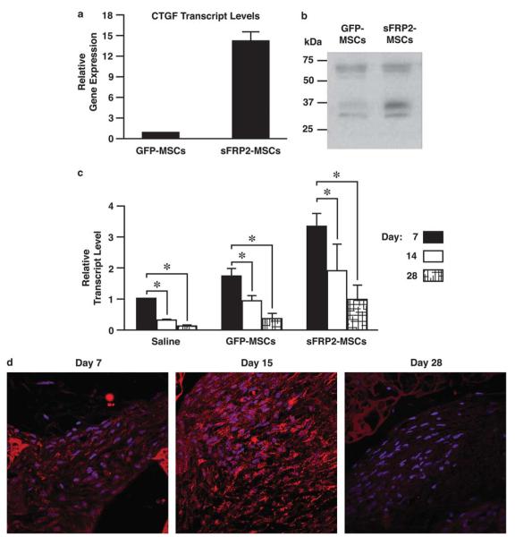
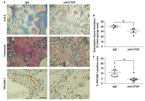
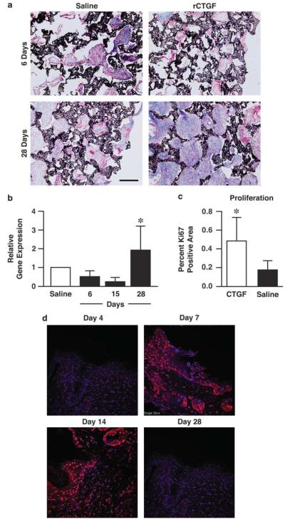
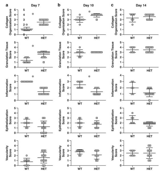
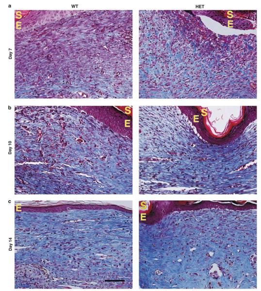
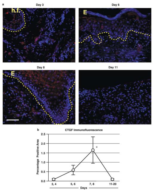
Mesenchymal stem cells (MSCs) that overexpress secreted frizzled-related protein 2 (sFRP2) exhibit an enhanced reparative phenotype. The secretomes of sFRP2-overexpressing MSCs and vector control-MSCs were compared through liquid chromatography tandem mass spectrometry. Proteomic profiling revealed that connective tissue growth factor (CTGF; CCN2) was overrepresented in the conditioned media of sFRP2-overexpressing MSCs and MSC-derived CTGF could thus be an important paracrine effector. Subcutaneously implanted, MSC-loaded polyvinyl alcohol (PVA) sponges and stented excisional wounds were used as wound models to study the dynamics of CTGF expression. Granulation tissue generated within the sponges and full-thickness skin wounds showed transient upregulation of CTGF expression by MSCs and fibroblasts, implying a role for this molecule in early tissue repair. Although collagen and COL1A2 mRNA were not increased when recombinant CTGF was administered to sponges during the early phase (day 1-6) of tissue repair, prolonged administration (>15 days) of exogenous CTGF into PVA sponges resulted in fibroblast proliferation and increased deposition of collagen within the experimental granulation tissue. In support of its physiological role, CTGF immunoinhibition during early repair (days 0-7) reduced the quantity, organizational quality and vascularity of experimental granulation tissue in the sponge model. However, CTGF haploinsufficiency was not enough to reduce collagen deposition in excisional wounds. Similar to acute murine wound models, CTGF was transiently present in the early phase of human acute burn wound healing. Together, these results further support a physiological role for CTGF in wound repair and demonstrate that when CTGF expression is confined to early tissue repair, it serves a pro-reparative role. These data also further illustrate the potential of MSC-derived paracrine modulators to enhance tissue repair.


Figures

Figure 1
Figure 1
Proteomic analysis reveals connective tissue growth factor (CTGF) upregulation in sFRP2-MSCs and its levels decrease with time within granulation tissue. (a) Quantitative real-time PCR analysis revealed a 14.3 ± 1.3-fold increase in CTGF transcript levels in sFRP2-MSCs compared with GFP-MSCs, normalized to 18S content, n = 3. *P<0.05 by Student’s t-test. (b) Representative blot demonstrating increased CTGF protein in the TCA-precipitated conditioned media (CM) of sFRP2-MSCs. About 20 μg of protein; conditioned medium of 1 × 10⁶ cells from each cell type were analyzed. The average intensity increase of the ~37 kDa band is 2.57±0.8 as determined by ImageJ analysis of band density, n = 3. (c) The transcript levels of CTGF within GFP-MSC, sFRP2-MSC- and saline-loaded sponges decrease with time as assessed by qRT-PCR; normalized to 18S content, n = 6 sponges per time point. *P≤0.05 by one-way ANOVA with Bonferroni post test. (d) Representative immunofluorescent staining with anti-CTGF of granulation tissue generated within sFRP2-MSC-loaded sponges after designated days following implantation.
Figure 2
Figure 2
Connective tissue growth factor (CTGF)-blocking antibody inhibited advanced granulation tissue formation. (a) Representative low power (× 10) trichrome images showed decreased granulation tissue in anti-CTGF-treated vs IgG-treated- MSC-loaded sponges (top panel). High power (× 20) H&E images showed that mesenchymal stem cell (MSC)-loaded sponges treated with anti-CTGF were less organized and exhibited decreased cellularity and red cell containing blood vessels as compared with IgG control (middle panels). Representative immunostained sections from anti-CTGF vs IgG-treat-sponges using anti-PECAM-1 to designate vascular density (× 40, bottom panels). (b) Graph of granulation tissue area in histological sponge sections quantified from each experimental animal presented as a percentage of total sponge area. (c) Average of the vascular density graphed as percentage of immunopositive PECAM-1 area/total tissue area in n = 10 histological sections from each animal. Unpaired Student’s t-test was used to compare data between groups, n = 4 in each group. *P<0.05 designates statistical significance.
Figure 3
Figure 3
Addition of recombinant connective tissue growth factor (CTGF) to polyvinyl alcohol (PVA) sponges enhances a proliferative, collagenous granulation tissue. (a) Representative ×10 images of trichrome-stained granulation tissue after 6 or 28 days of recombinant CTGF (rCTGF) (1 μg/day/sponge) or saline injections. Black=sponge, blue=collagen, red=granulation tissue. (b) Quantitative real-time PCR analysis of mouse collagen1a2 demonstrates that CTGF exposure for the initial stages (days 0–15) of wound repair yields decreased collagen deposition normalized to 18S content, whereas prolonged exposure (28 days) increased collagen transcript levels. n=6 sponges per group. *P<0.001 one-way analysis of variance (ANOVA) with Bonferroni post test. (c) rCTGF addition for 28 days (1 μg/sponge) increased the proliferative index of the granulation tissue as quantified by the threshold values of Ki67-positive areas in ×20 representative images of PVA sponges. ANOVA *P=0.0392 vs saline. (d) CTGF was temporally regulated in murine excisional wounds (n=4 animals for each time point). Murine stented excisional wounds were immunolabeled for CTGF. Representative images stained for CTGF in red and DAPI in blue demonstrate a spike in CTGF protein levels after day 4 (days 7–14). The staining was faint to absent at day 28. Both epidermal and dermal staining can be appreciated at days 7 and 14.
Figure 4
Figure 4
CTGFLacZ/+ mice have statistically similar wound healing parameters as WT 10 days after wounding. Histopathological scores assessing collagen content, amount of granulation tissue and vascularity within sections of excisional wounds from CTGF+/+ mice (WT) vs CTGFLacZ/+ (HET) mice 7 days (a), 10 days (b) and 14 days (c) after injury. Improved collagen organization in 7- and 10-day-old wounds of HET mice was observed. n≥6 wounds per group; paired t-test *P≤0.05.
Figure 5
Figure 5
Connective tissue growth factor (CTGF) HET mice have no changes in wound healing. Representative ×40 images of trichrome-stained excisional wounds within WT and HET mice 7 days (a) 10 days (b), and 14 days (c) post injury. Collagen is visualized by blue staining. E, epithelial layer, S, stratum corneum buildup; dark blue signify more mature collagen, light blue signify nascent collagen fibrils.
Figure 6
Figure 6
Connective tissue growth factor (CTGF) is temporally regulated in human burn wounds. Human burn wounds at different time points after injury were immunolabeled for CTGF. (a) Representative images of human burn wounds stained for CTGF in red and DAPI in blue demonstrate a spike in CTGF protein levels around days 7 and 8. The dotted line indicates the dermal-epithelial interface. (b) Average percentage positive areas positive for CTGF (±s.d.) as determined by thresholded images. n≥4 for each time point, *P<0.001 ANOVA with Bonferroni post test. E, epithelial layer; h.f., hair follicle.

References

    1. Gnecchi M, Zhang Z, Ni A, et al. Paracrine mechanisms in adult stem cell signaling and therapy. Circ Res. 2008;103:1204–1219. - PMC - PubMed
    1. Caplan AI, Dennis JE. Mesenchymal stem cells as trophic mediators. J Cell Biochem. 2006;98:1076–1084. - PubMed
    1. Alfaro MP, Pagni M, Vincent A, et al. The Wnt modulator sFRP2 enhances mesenchymal stem cell engraftment, granulation tissue formation and myocardial repair. Proc Natl Acad Sci USA. 2008;105:18366–18371. - PMC - PubMed
    1. Mirotsou M, Zhang Z, Deb A, et al. Secreted frizzled related protein 2 (Sfrp2) is the key Akt-mesenchymal stem cell-released paracrine factor mediating myocardial survival and repair. Proc Natl Acad Sci USA. 2007;104:1643–1648. - PMC - PubMed
    1. Estrada R, Li N, Sarojini H, et al. Secretome from mesenchymal stem cells induces angiogenesis via Cyr61. J Cell Physiol. 2009;219:563–571. - PMC - PubMed

Publication types

MeSH terms