

Subarachnoid hemorrhage-induced hydrocephalus in rats
- PMID: 23212164
- PMCID: PMC3552015
- DOI: 10.1161/STROKEAHA.112.662312
Subarachnoid hemorrhage-induced hydrocephalus in rats
Abstract
Background and purpose: Hydrocephalus is an important complication of subarachnoid hemorrhage (SAH). We investigated the occurrence of acute hydrocephalus in a rat SAH model.
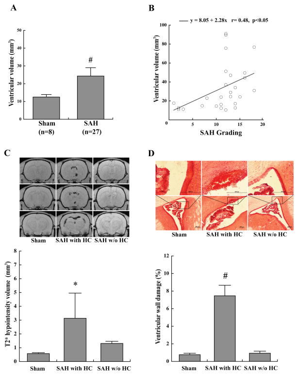
Methods: SAH was induced by endovascular perforation in adult male Sprague-Dawley rats (n=36). Sham rats (n=8) underwent the same procedure without perforation. MRI was performed 24 hours after SAH and the volume of the ventricular system and extent of T2* hypointensity lesions were measured. We defined hydrocephalus as ventricular volume > +3 SDs above the mean in sham animals. SAH grade was determined and brains were used for histology, immunohistochemistry, Perls staining, and Western blot analysis. Ventricular wall damage was defined as percentage of ependymal surface disruption.
Results: All surviving rats (n=27) after SAH had ventricular enlargement (33.6 ± 4.7 versus 13.5 ± 1.4 mm(3) in sham animals, P<0.01). Ventricular volume correlated with SAH severity (r=0.48; P<0.05). Out of 27 SAH rats, 12 demonstrated hydrocephalus and all had intraventricular blood accumulation. Rats with hydrocephalus had more severe ventricular wall damage (7.4 ± 1.2%) than the sham animals (0.6 ± 0.2%; P<0.01) and rats without hydrocephalus (1.1 ± 0.2%; P<0.01). Periventricular iron deposition was observed and heme oxygenase-1 and Iba-1 expression were markedly increased in hydrocephalus rats.
Conclusions: SAH causes ventricular enlargement in a rat endovascular perforation model, with hydrocephalus occurring in 44% of animals at 24 hours. Rats with hydrocephalus had more severe SAH, intraventricular hemorrhage, and greater ventricular wall damage.
Figures


References
-
- Del Bigio MR. Neuropathological changes caused by hydrocephalus. Acta Neuropathol. 1993;85:573–585. - PubMed
-
- Hasan D, Vermeulen M, Wijdicks EF, Hijdra A, van Gijn J. Management problems in acute hydrocephalus after subarachnoid hemorrhage. Stroke. 1989;20:747–753. - PubMed
-
- Bederson JB, Germano IM, Guarino L. Cortical blood flow and cerebral perfusion pressure in a new noncraniotomy model of subarachnoid hemorrhage in the rat. Stroke. 1995;26:1086–1091. - PubMed
Publication types
MeSH terms
Grants and funding
LinkOut - more resources
Full Text Sources
Other Literature Sources
Medical
