. 2012:3:1248.
doi: 10.1038/ncomms2262.

Identification and characterization of a human mitochondrial NAD kinase

Affiliations
Free PMC article

Identification and characterization of a human mitochondrial NAD kinase

Kazuto Ohashi et al. Nat Commun. 2012.
Free PMC article

Abstract

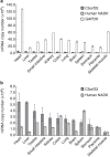
NAD kinase is the sole NADP(+) biosynthetic enzyme. Despite the great significance of NADP(+), to date no mitochondrial NAD kinase has been identified in human, and the source of human mitochondrial NADP(+) remains elusive. Here we present evidence demonstrating that a human protein of unknown function, C5orf33, is a human mitochondrial NAD kinase; this protein likely represents the missing source of human mitochondrial NADP(+). The C5orf33 protein exhibits NAD kinase activity, utilizing ATP or inorganic polyphosphate, and is localized in the mitochondria of human HEK293A cells. C5orf33 mRNA is more abundant than human cytosolic NAD kinase mRNA in almost all tissues examined. We further show by database searches that some animals and protists carry C5orf33 homologues as their sole NADP(+) biosynthetic enzyme, whereas plants and fungi possess no C5orf33 homologue. These observations provide insights into eukaryotic NADP(+) biosynthesis, which has pivotal roles in cells and organelles.


Figures

Figure 1
Figure 1. Structure of C5orf33 protein.
(a) Primary structures of the C5orf33 protein and its truncations. Predicted mitochondrial-targeting sequence and NADK motif are in black and grey, respectively. The amino-acid residue numbers are indicated on the structures. Asterisk in parentheses (*) indicates the start codon found in pMK3270 and pMK3243, but not in the sequence of C5orf33 variant 1. (b) mRNA structures of C5orf33 transcript variants 1 and 2. Grey arrows indicate open reading frames encoding the indicated proteins. The nucleotide (nt) base and amino-acid (aa) residue numbers are shown. The nucleotide sequences indicated by double-headed arrows are identical between variants 1 and 2. The putative start codon (ATG) is denoted by an asterisk (a,b). The nucleotide sequence of C5orf33 variant 2 (62CTTGCATTGAAAGGCTCTAGTTAC85) corresponds to the N-terminal region of the Δ100C5orf33 protein (101LALKGSSY108) (a,b).
Figure 2
Figure 2. NADK activity of C5orf33 protein.
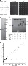
(a) In vivo assay of NADK activity of C5orf33 protein. S. cerevisiae MK1598 cells (an NADK triple mutant having YCplac33-UTR1) carrying the indicated plasmids were spotted onto the solid media and were incubated at 30 °C for 4 days. C5orf33 protein was less effective than Δ62C5orf33 protein in reverting the lethality. We speculate that this is because in the yeast, human C5orf33 is less able to supply cytosolic NADP+ than the yeast mitochondrial Pos5 itself. (b) SDS–PAGE of purified C5orf33 protein (arrowhead; 43 kDa). (c) NADK activity of purified C5orf33 protein analysed by TLC in the presence (+) or absence (−) of indicated compounds as described in the Methods. (d) Saturation curves for NAD+ of purified human NADK (dashed line) and C5orf33 protein (line) determined under 8 and 2 mM ATP, respectively. The specific activity (U μmol−1; 1 μmol NADP+ produced per 1 min and 1 μmol subunit of the enzyme) was calculated using the subunit molecular mass of C5orf33 protein (45 kDa) or human NADK (43 kDa) (Table 1). Each point represents the average of three determinations; error bars represent s.d.
Figure 3
Figure 3. Localization of C5orf33 protein in mitochondria of human cells.
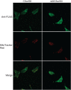
HEK293A cells were transiently transfected with plasmid expressing C terminally FLAG-tagged C5orf33 or Δ62C5orf33 protein. The cells were fixed and immunostained with a rabbit anti-FLAG primary antibody and AlexaFluor 488-conjugated anti-rabbit secondary antibody. Mitochondria were stained with MitoTracker Red.
Figure 4
Figure 4. Tissue-specific mRNA levels determined by absolute qPCR.
mRNAs of C5orf33 protein (dark grey), human NADK (light grey) and GAPDH (white bars) (a); only those of the C5orf33 protein and human NADK are shown in panel b. Means and s.d. of three determinations are shown.
Figure 5
Figure 5. siRNA transfection.
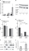
HEK293A cells were transfected with siRNA#2 against C5orf33 protein (C5orf33 siRNA) or control siRNA, and incubated for 1, 2 and 3 days. (a) Levels of C5orf33 mRNA in the transfected cells are shown as relative values normalized against the level in control siRNA-transfected cells incubated for 1 day after transfection. Knockdowns of 69, 63 and 79% relative to the control, as indicated, were obtained. (b) Expression of C5orf33 protein in the transfected cells. Proteins were analysed by western blotting using anti-C5orf33 and anti-GAPDH antibodies. (c) Viability. Transfected cells incubated for 3 days were treated with 12.5 μM menadione (black bar) or vehicle (0.62% (v/v) dimethyl sulfoxide (DMSO), grey bar) for 20 h, and viability was measured using calcein AM as in Supplementary Methods. Viability was also measured using MTT as in Supplementary Fig. S5. (d) Intracellular ROS levels. Transfected cells incubated for 3 days were incubated with 10 μM 5-(and-6)-chloromethyl-2′,7′-dichlorodihydrofluorescein diacetate, acetyl ester (CM-H2DCFDA) for 30 min, and then treated without (grey bar) or with (black bar) 100 μM menadione for 30 min; intracellular ROS was measured as described in Supplementary Methods. t-test; *P=0.0085, **P=0.0026, ***P=0.0022, ****P=0.0044 × 10⁻⁴. Viability and intracellular ROS levels are presented as relative values normalized against their respective levels in control siRNA-transfected cells not treated with menadione; each independent experiment was conducted in duplicate (c,d). (e) Mitochondrial and cytosolic fractions of HEK293A cells that had been incubated for 3 days after transfections. Mitochondrial (M; 1.9 μg) and cytosolic (C; 1.9 μg) fractions and whole cells (W; from 3.3 × 10⁴ transfected HEK293A cells) were analysed by western blotting using anti-CoxII, anti-GAPDH and anti-C5orf33 antibodies. CoxII and GAPDH are mitochondrial and cytosolic markers, respectively. (f) NADK activity of mitochondrial fraction obtained in panel e. Mitochondrial NADK activity is expressed as pmol of NADP+ formed in 1 h. NADK activity was decreased to 47% upon knockdown of C5orf33. t-test; *P=0.024. Means and s.d. of three independent determinations are shown (a,c,d,f).

References

    1. Pollak N., Dolle C. & Ziegler M. The power to reduce: pyridine nucleotides—small molecules with a multitude of functions. Biochem. J. 402, 205–218 (2007). - PMC - PubMed
    1. Ziegler M. New functions of a long-known molecule. Emerging roles of NAD in cellular signaling. Eur. J. Biochem. 267, 1550–1564 (2000). - PubMed
    1. Outten C. E. & Culotta V. C. A novel NADH kinase is the mitochondrial source of NADPH in Saccharomyces cerevisiae. EMBO J. 22, 2015–2024 (2003). - PMC - PubMed
    1. Smith J. S. et al.. A phylogenetically conserved NAD+-dependent protein deacetylase activity in the Sir2 protein family. Proc. Natl Acad. Sci. USA 97, 6658–6663 (2000). - PMC - PubMed
    1. Yamasaki M. et al.. Organelle selection determines agonist-specific Ca2+ signals in pancreatic acinar and beta cells. J. Biol. Chem. 279, 7234–7240 (2004). - PubMed

Publication types

MeSH terms