Review
. 2012 Dec;192(4):1165-202.
doi: 10.1534/genetics.112.145516.

Mitotic exit and separation of mother and daughter cells

Affiliations
Review

Mitotic exit and separation of mother and daughter cells

Eric L Weiss. Genetics. 2012 Dec.

Abstract

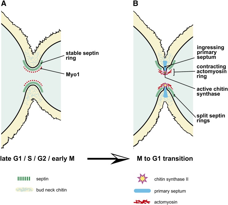
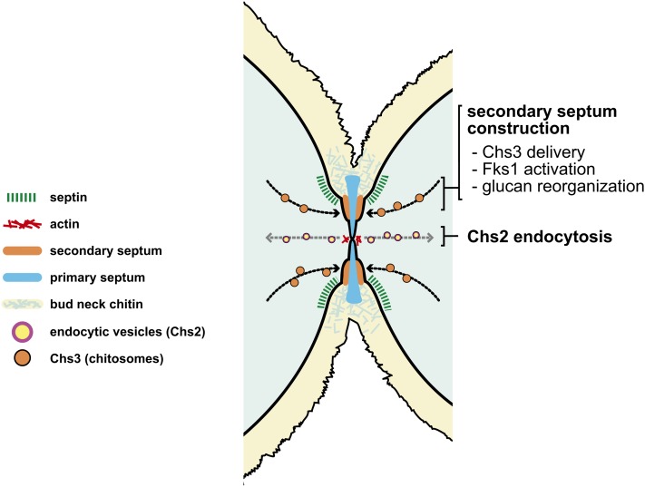
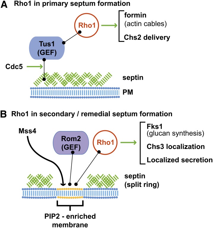
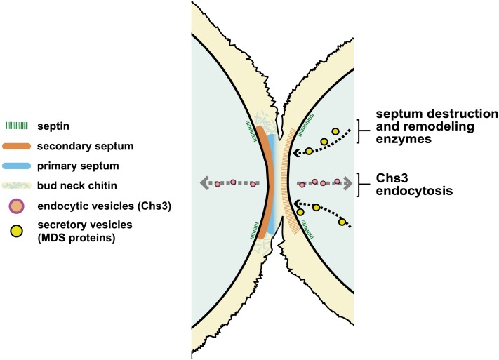
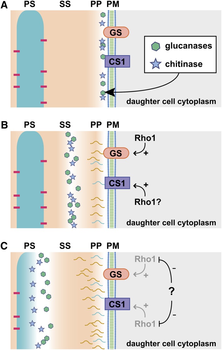
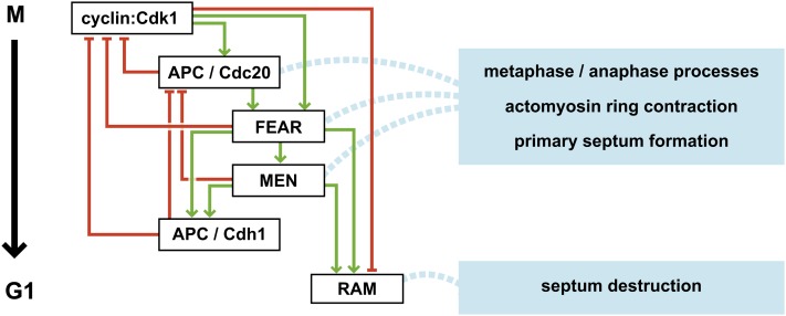
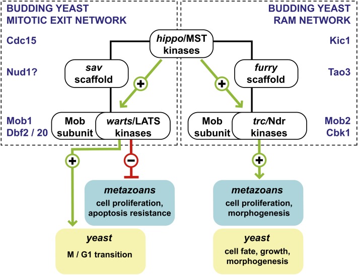
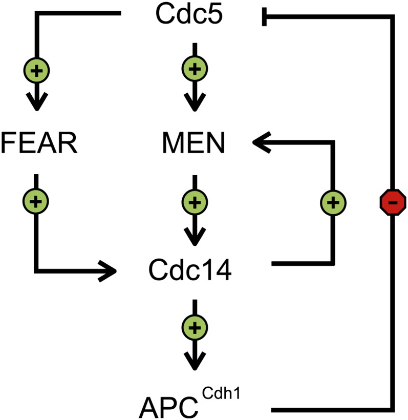
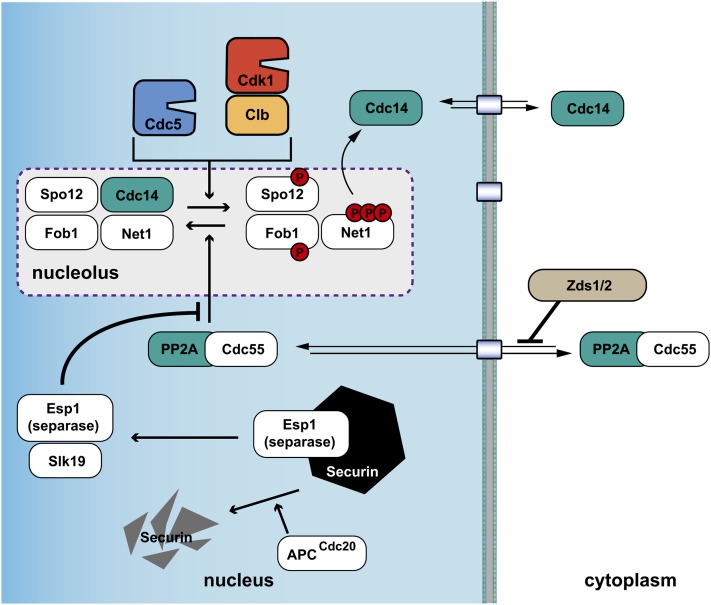
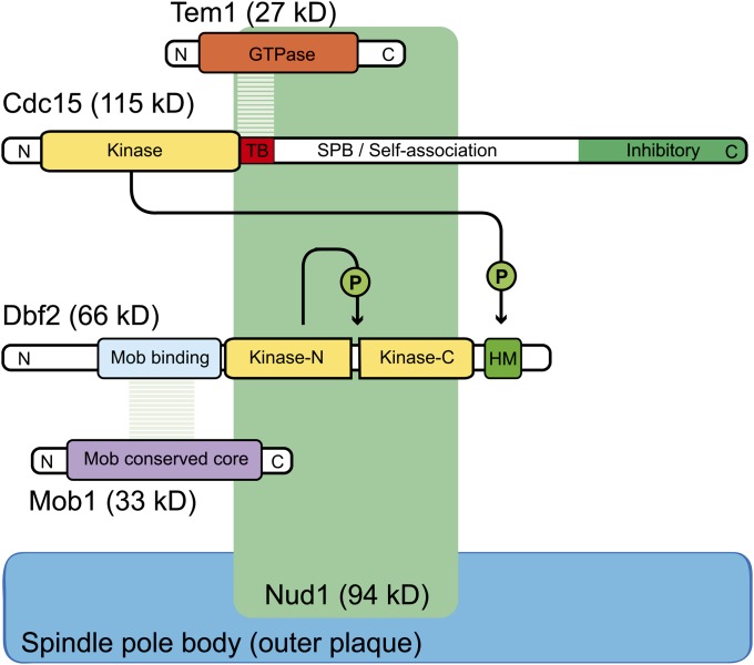
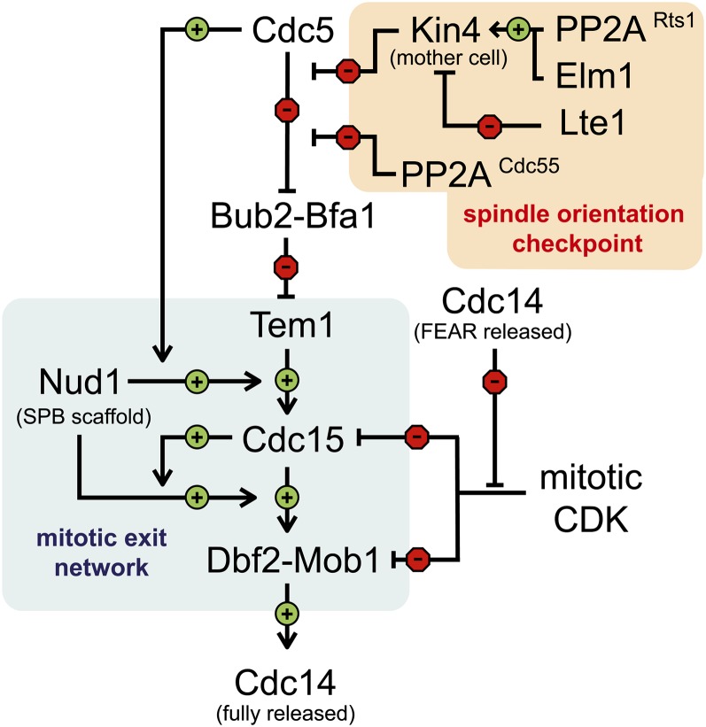
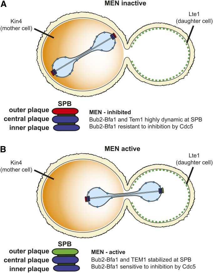
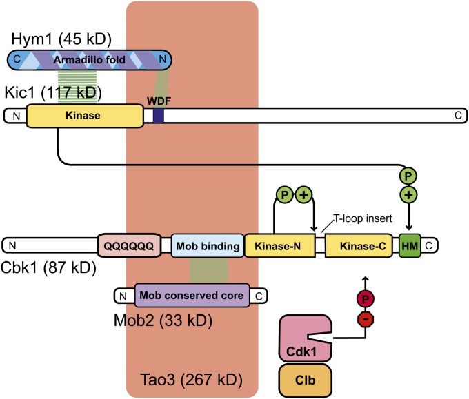
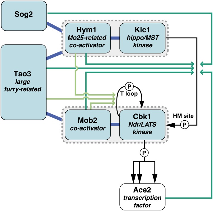
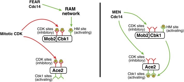
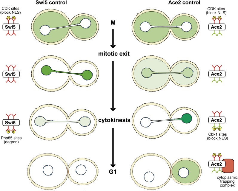
Productive cell proliferation involves efficient and accurate splitting of the dividing cell into two separate entities. This orderly process reflects coordination of diverse cytological events by regulatory systems that drive the cell from mitosis into G1. In the budding yeast Saccharomyces cerevisiae, separation of mother and daughter cells involves coordinated actomyosin ring contraction and septum synthesis, followed by septum destruction. These events occur in precise and rapid sequence once chromosomes are segregated and are linked with spindle organization and mitotic progress by intricate cell cycle control machinery. Additionally, critical paarts of the mother/daughter separation process are asymmetric, reflecting a form of fate specification that occurs in every cell division. This chapter describes central events of budding yeast cell separation, as well as the control pathways that integrate them and link them with the cell cycle.


Figures

Figure 1
Figure 1
Early organization of the cytokinesis site and initiation of cytokinesis. (A) When the bud is formed in G1/S, septin filaments (green dashes) help organize the bud neck and promote recruitment of Myo1 (red dashes); filamentous actin is not markedly present. Chitin (blue lines) is present in the bud neck from early stages in its formation. The septin lattice also serves as a barrier that prevents diffusion of material associated with the daughter cell cortex into the mother cell. (B) During early cytokinesis, a contractile actomyosin ring (red lines) is assembled that helps guide deposition of a primary septum (blue) through the localized action of Chs2 (yellow star).
Figure 2
Figure 2
Completion of the primary septum and secondary septum synthesis. Coincident with completion of the primary septum (blue), the actomyosin ring (red lines) is disassembled and Chs2 is internalized through endocytosis (yellow circles). Deposition of the secondary septum (orange) occurs on both mother and daughter sides of the primary septum. This involves synthesis of glucan and chitin by Fks1 (which is resident in the plasma membrane) and Chs3, which is delivered in by exocytic vesicles (orange circles) along with other material that helps build up the structure.
Figure 3
Figure 3
Model for Rho1 action in coordination of primary and secondary septum synthesis. (A) Early in cytokinesis, Cdc5-mediated GEF recruitment localizes Rho1–GTP, which promotes assembly of actin cables. It is not clear if direct association with septins (green) mediates this. (B) Splitting of the septin ring permits formation of a spatially constrained PIP2-rich membrane domain generated by Mss4. This concentrates Rho1 to the site via binding to phospholipids and the Rom2 GEF. Rho1 promotes multiple events, indicated at right.
Figure 4
Figure 4
Septum destruction. After the secondary septum is complete, Chs3 is endocytosed (orange circles)and presumably Rho1–Fks1 is partially inactivated (not shown). Coincident with this, mother/daughter separation enzymes (MDS proteins) that reorganize and degrade the septum are produced in the daughter cell and delivered to the division site. The chitin synthase Chs1 is also delivered to the site (not shown), where it repairs damage caused by degradative enzymes.
Figure 5
Figure 5
A model for enzymatic destruction of the septum. (A) Upon completion of septation, in response to an unknown signal, chitinase and a suite of glucanases are exocytosed into the small periplasmic space (PP) between the secondary septum (SS) and the outer leaflet of the plasma membrane (PM). Coincident with this, glucan and chitin are synthesized by transmembrane enzymes Fks1 (GS) and Chs1 (CS1), respectively. These oligosaccharide chains are transferred enzymatically to other wall components. (B) Exocytosis of septum destruction enzymes ceases. Digestion and chain rearrangement by glucanases and glucanosyltransferases creates a zone into which chitinase can diffuse. This is pushed forward by new wall synthesis. (C) New wall synthesis is attenuated, and chitinase reaches the primary septum (PS). Digestion breaks the link between mother and daughter.
Figure 6
Figure 6
Overall organization of mitotic exit control. Mitotic exit is controlled by a series of systems that both activate (green pointed arrows) and inhibit (red block arrows) one another as cells pass from M into G1 phase. The temporal sequence in which the systems function during mitotic exit is ordered from top to bottom. A recurring theme is that a regulator will activate a downstream process that ultimately antagonizes the activator. For example, mitotic cyclin–Cdk1 promotes APCCdc20 function and the FEAR pathway, which both then oppose M-phase CDK function. The large-scale processes occurring sequentially during mitotic exit are shown in blue boxes on the right, connected by dashed lines to the regulatory systems that promote them.
Figure 7
Figure 7
Conserved structure of Mst/hippo signaling pathways. Pathways similar to the MEN and the RAM network are broadly conserved in eukaryotes. Activating phosphorylations are shown as green arrows, while inhibitory phosphorylation is shown as a red block arrow. The MEN is most similar to the pathway in the left box, although no sav ortholog is present. The RAM is most similar to the pathway in the right box.
Figure 8
Figure 8
Cdc5 turns on its own destruction. The overall signaling interactions downstream of Cdc5 activation converge to activate Cdc14. This activates the APCCdh1 form of the APC, which promotes degradation of Cdc5 and thereby closes a negative feedback loop.
Figure 9
Figure 9
The FEAR pathway. The balance of phosphorylation and dephosphorylation of RENT components, most notably Net1, determines whether Cdc14 is sequestered or released from the nucleolus. Nucleocytoplasmic trafficking determines how much Cdc14 gets out of the nucleus and how much PP2ACdc55 gets in. Esp1 inhibits PP2ACdc55 dephosphorylation of RENT components.
Figure 10
Figure 10
Molecular organization of the core MEN pathway. Activating phosphorylations are noted, and the domain organization of the Cdc15–Tem1 module and the Mob1–Dbf2 module are shown. Cdc15 is an Mst/hippo-related kinase that binds the Ras-family GTPase Tem1 through a region C-terminal to its kinase domain. Other parts of Cdc15 may act to promote self-association and MEN inhibition. Dbf2 is an Ndr/LATS family protein kinase that binds to Mob1 via a region N-terminal to its kinase domain. Nud1 acts as a scaffold that brings elements of the pathway together at the outer plaque of the SPB.
Figure 11
Figure 11
The logic of the MEN. Positive interactions in the broader MEN are indicated as pointed arrows, and negative ones as block arrows. The core MEN (Tem1–Cdc15–Dbf2 complexed with Nud1) is negatively regulated by both mitotic CDK and the Tem1 GTPase activating protein (GAP) Bub2–Bfa1. Cdc14 reverses the effects of CDK phosphorylation. Cdc5 turns Bub2–Bfa1 off, and Kin4 and PP2ACdc55 negatively regulate this. Cdc5 also activates the MEN more directly.
Figure 12
Figure 12
MEN activation is closely tied to the mitotic spindle. Activation of the MEN is closely tied to the location of the mitotic spindle. Kin4, which antagonizes Bub2–Bfa1 inactivation, is localized to the mother cell; Lte1, which acts positively on the pathway, is located in the daughter cell. (A) When SPBs are present in the mother cell cytoplasm, the MEN is inhibited at the outer plaque (red). (B) Movement of one of the SPBs into the daughter is permissive for MEN activation (green SPB outer plaque), as long as Cdc5 is active.
Figure 13
Figure 13
The RAM network. This figure represents the overall organization of the core components of the RAM network. Activating phosphorylations are noted, and the domain organization of the Hym1–Kic1 module and the Mob2–Cbk1 module are shown. Kic1 is an Mst/hippo-related kinase that likely binds the MO25 ortholog Hym1 through its kinase domain and a C-terminal peptide motif. Cbk1 is an Ndr/LATS family protein kinase that binds to Mob2 via a region N-terminal to its kinase domain. Cbk1’s N-terminal region contains a polyglutamine tract, but is not highly conserved. Mitotic CDK phosphorylation likely inhibits Cbk1. Tao3 may act as a scaffold that brings elements of the pathway together, but there is little evidence for or against this notion.
Figure 14
Figure 14
Signaling structure of the RAM network. Protein–protein interactions, (shown as thick blue lines) and importance for in vivo phosphorylation levels of individual sites (shown as green lines) suggest how modules in the RAM network relate to Cbk1 phosphoregulation. Kic1 probably directly phosphorylates Cbk1’s C-terminal HM site, and Cbk1 autophosphorylates its own activation loop in cis. All RAM network components are required for normal HM site phosphorylation, as is Ace2. Tao3, Mob2, and Hym1 are required for normal activation loop autophosphorylation.
Figure 15
Figure 15
MEN and FEAR control of the RAM network. The FEAR pathway activates phosphorylation of Cbk1’s HM site in early anaphase, while direct phosphorylation by mitotic CDK inhibits Ace2 nuclear import as well as Cbk1/RAM network function. Activation of the MEN then releases large amounts of Cdc14, resulting in removal of inhibitory CDK phosphorylations on Cbk1 (and possibly other RAM network components) and Ace2. MEN activation also likely sustains RAM-network mediated phosphorylation of Cbk1’s HM site.
Figure 16
Figure 16
Comparison of Ace2 and Swi5 localization. Swi5 and Ace2 are represented separately as green color in the left and right columns of cell diagrams, and cartoons of the proteins with relevant modifications or proposed protein–protein interactions are shown next to the corresponding cells. In anaphase and very early telophase neither protein enters the nucleus; both proteins’ NLS’s are inactive due to phosphorylation by mitotic CDK. Later in telophase, both proteins are dephosphorylated by Cdc14 and can enter both mother and daughter nuclei. This is sufficient for accumulation of Swi5, which lacks an NES, to high concentrations. Ace2, in contrast, does not accumulate significantly in nuclei. Prior to actomyosin ring contraction, Ace2’s NES is phosphorylated, inhibiting its nuclear export. This probably occurs specifically in the daughter cell. Ace2 then accumulates specifically in the daughter cell nucleus and is depleted in the still-contiguous cytoplasm of mother and daughter cells. In mid-G1, Ace2’s NES is dephosphorylated and the protein is exported from the nucleus. Due to cytoplasmic trapping, Ace2 cannot reenter the nucleus of the newly born daughter cell once it exits.
Figure 17
Figure 17
Domain organization of Ace2. The Ace2 protein contains a C-terminal Zn-finger domain that, in addition to immediately surrounding sequence, is related to Swi5. A C-terminal NLS is regulated by CDK phosphorylation. Ace2’s NES is medially located and is negatively regulated by Cbk1 phosphorylation. An additional site of Cbk1 phosphorylation activates Ace2. A region in the middle of Ace2 mediates association with Cbk1.
Figure 18
Figure 18
Inactivation of Swi5 and Ace2 in G1. In this model, red arrows indicate direct phosphorylation that has an inhibitory effect, and green arrows indicate a positive regulatory interaction. Swi5 is degraded following direct phosphorylation by the G1 CDK Pho85 in complex with an unknown “Pcl” cyclin. For Ace2, following dephosphorylation of its NES it is exported to cytoplasm and sequestered there. This sequestration appears to occur through two mechanisms, one involving direct phosphorylation of CDK sites in Ace2 and another involving a kinase-independent function of Pho85. See text for details.
Figure 19
Figure 19
Domain organization of Ssd1. Ssd1 is an RNaseII-related protein that probably lacks catalytic activity. Canonical Cbk1 consensus motifs (black arrows) are concentrated in the protein’s N-terminal region, including a portion that mediates interaction with phosphorylated RNA polymerase II C-terminal domain (PCTD). A sequence that can act as an NLS is present in the middle part of the protein.
Figure 20
Figure 20
Model for septum destruction control. A simplified structure of Ace2 and RAM network mitotic control shows that Cdc14 activates both systems, in a coherent feed-forward loop (FFL1). Cbk1 control of both Ace2 and Ssd1 constitutes a second feed-forward loop (FFL2). In contrast, Cdc14 regulation of Swi5 appears to involve simple activation, consistent with relative kinetics of the two transcription factors’ activities.

Similar articles

Cited by

References

    1. Adames N. R., Cooper J. A., 2000. Microtubule interactions with the cell cortex causing nuclear movements in Saccharomyces cerevisiae. J. Cell Biol. 149: 863–874 - PMC - PubMed
    1. Adames N. R., Oberle J. R., Cooper J. A., 2001. The surveillance mechanism of the spindle position checkpoint in yeast. J. Cell Biol. 153: 159–168 - PMC - PubMed
    1. Adams D. J., 2004. Fungal cell wall chitinases and glucanases. Microbiology 150: 2029–2035 - PubMed
    1. Alon U., 2007. Network motifs: theory and experimental approaches. Nat. Rev. Genet. 8: 450–461 - PubMed
    1. Amon A., 2008. A decade of Cdc14–a personal perspective. Delivered on July 9, 2007 at the 32nd FEBS Congress in Vienna, Austria. FEBS J. 275: 5774–5784 - PMC - PubMed

Publication types

MeSH terms