. 2012 Dec;192(4):1203-34.
doi: 10.1534/genetics.112.141267.

Mitochondrial protein synthesis, import, and assembly

Affiliations

Mitochondrial protein synthesis, import, and assembly

Thomas D Fox. Genetics. 2012 Dec.

Abstract

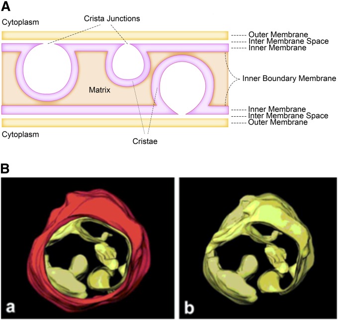
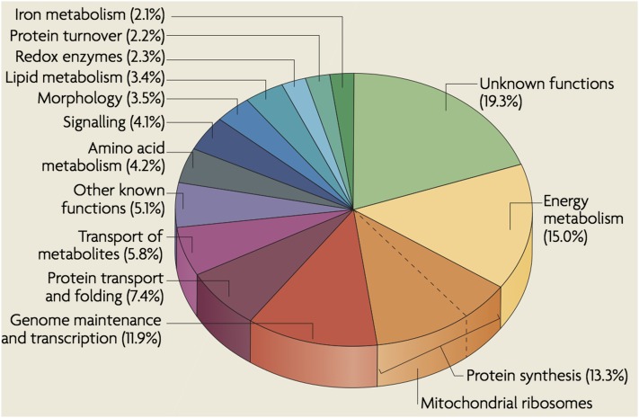
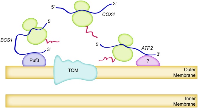
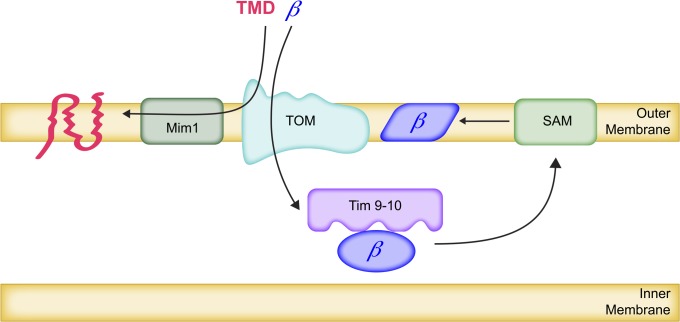
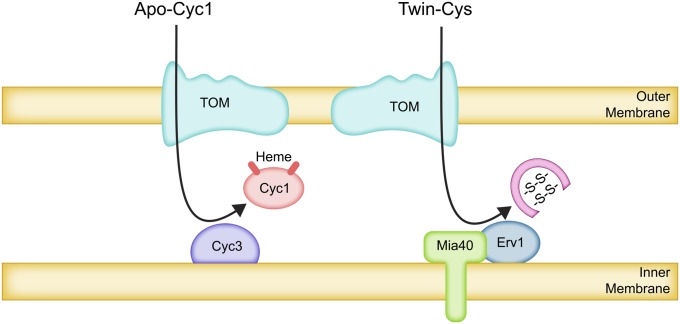
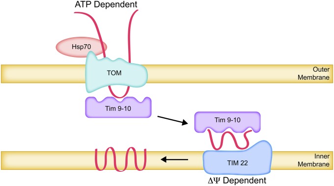
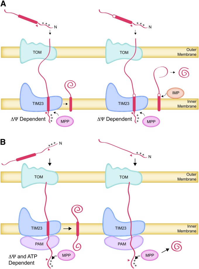
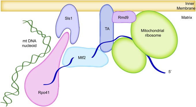
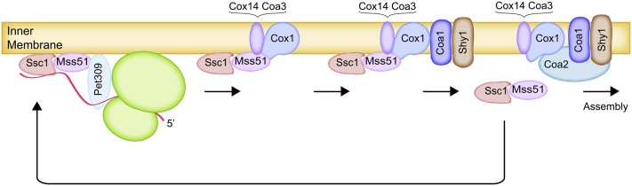
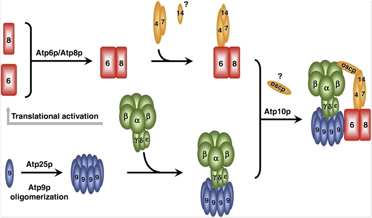
The mitochondrion is arguably the most complex organelle in the budding yeast cell cytoplasm. It is essential for viability as well as respiratory growth. Its innermost aqueous compartment, the matrix, is bounded by the highly structured inner membrane, which in turn is bounded by the intermembrane space and the outer membrane. Approximately 1000 proteins are present in these organelles, of which eight major constituents are coded and synthesized in the matrix. The import of mitochondrial proteins synthesized in the cytoplasm, and their direction to the correct soluble compartments, correct membranes, and correct membrane surfaces/topologies, involves multiple pathways and macromolecular machines. The targeting of some, but not all, cytoplasmically synthesized mitochondrial proteins begins with translation of messenger RNAs localized to the organelle. Most proteins then pass through the translocase of the outer membrane to the intermembrane space, where divergent pathways sort them to the outer membrane, inner membrane, and matrix or trap them in the intermembrane space. Roughly 25% of mitochondrial proteins participate in maintenance or expression of the organellar genome at the inner surface of the inner membrane, providing 7 membrane proteins whose synthesis nucleates the assembly of three respiratory complexes.


Figures

Figure 1
Figure 1
Overview of mitochondrial structure in yeast. (A) Schematic of compartments comprising mitochondrial tubules. The outer membrane surrounds the organelle. The inner membrane surrounds the matrix and consists of two domains, the inner boundary membrane and the cristae membranes, which are joined at cristae junctions. The intermembrane space lies between the outer membrane and inner membrane. (B) Electron tomograph image of a highly contracted yeast mitochondrion observed en face (a) with the outer membrane (red) and (b) without the outer membrane. Reprinted by permission from John Wiley & Sons from Mannella et al. (2001).
Figure 2
Figure 2
Classification of identified mitochondrial proteins according to function. Reprinted by permission from Nature Publishing Group from Schmidt et al. (2010).
Figure 3
Figure 3
Cytoplasmic synthesis of some mitochondrial proteins is localized to the organelles, while the synthesis of others is not. The figure depicts three examples: (1) The ATP2 mRNA is highly localized to mitochondria-bound polysomes (Garcia et al. 2007b), although factors required for this localization are unknown. (2) The BCS1 mRNA is also selectively found in mitochondria-bound polysomes, and its localization is partially dependent upon the mitochondrially localized RNA-binding protein Puf3 and the Puf3-binding sites in its 3′-UTR (Saint-Georges et al. 2008). (3) The COX4 mRNA is exclusively found on free polysomes, unassociated with mitochondria (Garcia et al. 2007b). The Atp2, Bcs1, and Cox4 proteins all traverse the outer membrane via the TOM complex pore.
Figure 4
Figure 4
Insertion of proteins into the outer membrane. β-Barrel proteins are imported through the pores of the TOM complex in the outer membrane and then bound by IMS chaperone complexes comprising Tim9 and Tim10. The β-barrel-Tim9-Tim10 complexes bind to the inner surfaces of SAM complexes in the outer membrane, leading to insertion of β-barrel proteins into the outer membrane lipid bilayer. Some integral outer membrane proteins with multiple transmembrane domains (TMD) contact the Tom70 receptor and are then inserted into the bilayer from the outside through their interaction with multimeric complexes of Mim1.
Figure 5
Figure 5
Trapping of proteins in the IMS by covalent modification. Apo-cytochrome c (Cyc1) traverses the outer membrane via the TOM complex by an unusual and poorly understood mechanism (see text). Covalent attachment of heme by the lyase (Cyc3), bound to the outer surface of the inner membrane, generates holo-cytochrome c. Holo-cytochrome c cannot translocate through the TOM complex and remains in the IMS. In an analogous mechanism, IMS proteins with twin-Cys residue pairs in reduced form are imported through the TOM complex and then oxidized by the Mia40-Erv1 disulfide relay system bound to the inner membrane. The internal disulfide bonds formed in the twin-Cys proteins prevent reverse translocation.
Figure 6
Figure 6
Insertion of multi-spanning carrier proteins into the inner membrane. Newly synthesized multi-spanning carrier proteins, complexed with cytoplasmic Hsp70, are recognized by the Tom70 receptor subunit of the TOM complex. ATP-dependent release from cytoplasmic Hsp70 leads to translocation through the TOM complex in a looped configuration and binding to the Tim9-Tim10 IMS chaperone complex. The multi-spanning proteins are delivered to the TIM22 insertase complex in the inner membrane, released from Tim9-Tim10, and inserted into the inner membrane by reactions that depend upon the Δψ potential across the inner membrane.
Figure 7
Figure 7
Import of proteins with amphipathic positively charged cleavable presequences. (A) ΔΨ-Dependent transport of proteins containing hydrophobic sorting signals (thick bars) to the inner membrane and IMS. The presequences are recognized by the Tom22 receptor of the TOM complex, pass through the TOM complex, and are recognized by the Tim50 subunit of the TIM23 complex. The presequence translocates through the TIM23 complex, driven electrophoretically by ΔΨ. This brings the hydrophobic sorting signal, located immediately downstream of the presequence, into the TIM23 complex. The presequence is removed by proteolysis and the sorting signal is inserted laterally into the inner membrane where it can function as a membrane anchor (left). Proteolytic cleavage at the outer surface of the inner membrane can release a soluble protein into the inner membrane space (right). (B) ΔΨ- and ATP-dependent transport to the inner membrane and the matrix. Presequences traverse the TOM and TIM23 complexes and are removed by proteolysis, as in A. If there is no hydrophobic sorting signal immediately downstream of the presequence, the incoming polypeptide is engaged by the PAM complexed with the inner surface of the TIM23 translocon. ATP hydrolysis by the Hsp70 (Ssc1) subunit of PAM translocates the polypeptide into the matrix. If a downstream hydrophobic sorting signal enters the TIM23 complex, it is released laterally into the inner membrane and translocation ceases (left). If there is no sorting signal, the entire polypeptide is translocated into the matrix (right).
Figure 8
Figure 8
Channeling of mitochondrially coded mRNAs from RNA polymerase (Rpo41) to membrane-bound ribosomes by Sls1, Mtf2, Rmd9, and mRNA-specific translational activators (TA). (The figure is not intended to suggest that mRNAs are translated while still emerging from RNA polymerase.)
Figure 9
Figure 9
Assembly feedback control of Cox1 synthesis by Mss51 activities as a translational activator and assembly factor. Mitochondria translation of the COX1 mRNA is activated by the mRNA-specific activators Pet309 and Mss51 [Mss51 is associated with Hsp70 (Ssc1) throughout]. Synthesis of a new Cox1 polypeptide nucleates an early assembly intermediate containing Mss51 and the assembly factors Cox14 and Coa3. As additional assembly factors associate with newly synthesized Cox1, Mss51 is released from the assembly intermediates and is then available to initiate additional Cox1 synthesis.
Figure 10
Figure 10
Assembly of ATP synthase from modular subassemblies. The imported subunits of the F1 complex (green) are assembled in the matrix with the help of specific assembly chaperones (see text for details). Assembled F1 activates mitochondrial translation of the dicistronic mRNA encoding Atp8 and Atp6, which nucleate the assembly of imported subunits into the stator module (red and tan). The Atp9 ring (blue) is assembled from monomers and then joined with F1. Finally, association of the stator with the F1-Atp9 ring subassembly generates the ATP synthase proton pore at the same time that ATPase activity is coupled to the inner membrane proton gradient. Reprinted by permission from Macmillan Publishers Ltd. from Rak et al. (2011).

References

    1. Abe Y., Shodai T., Muto T., Mihara K., Torii H., et al. , 2000. Structural basis of presequence recognition by the mitochondrial protein import receptor Tom20. Cell 100: 551–560 - PubMed
    1. Ackerman S. H., Tzagoloff A., 1990. Identification of two nuclear genes (ATP11, ATP12) required for assembly of the yeast F1-ATPase. Proc. Natl. Acad. Sci. USA 87: 4986–4990 - PMC - PubMed
    1. Ackerman S. H., Tzagoloff A., 2005. Function, structure, and biogenesis of mitochondrial ATP synthase. Prog. Nucleic Acid Res. Mol. Biol. 80: 95–133 - PubMed
    1. Ackerman S. H., Gatti D. L., Gellefors P., Douglas M. G., Tzagoloff A., 1991. ATP13, a nuclear gene of Saccharomyces cerevisiae essential for the expression of subunit 9 of the mitochondrial ATPase. FEBS Lett. 278: 234–238 - PubMed
    1. Ades I. Z., Butow R. A., 1980. The products of mitochondria-bound cytoplasmic polysomes in yeast. J. Biol. Chem. 255: 9918–9924 - PubMed

Publication types

MeSH terms

Substances