. 2013 Jan 25;288(4):2223-37.
doi: 10.1074/jbc.M112.391680. Epub 2012 Dec 4.

Phosphatase and tensin homolog (PTEN)-induced putative kinase 1 (PINK1)-dependent ubiquitination of endogenous Parkin attenuates mitophagy: study in human primary fibroblasts and induced pluripotent stem cell-derived neurons

Affiliations

Phosphatase and tensin homolog (PTEN)-induced putative kinase 1 (PINK1)-dependent ubiquitination of endogenous Parkin attenuates mitophagy: study in human primary fibroblasts and induced pluripotent stem cell-derived neurons

Aleksandar Rakovic et al. J Biol Chem. .

Abstract

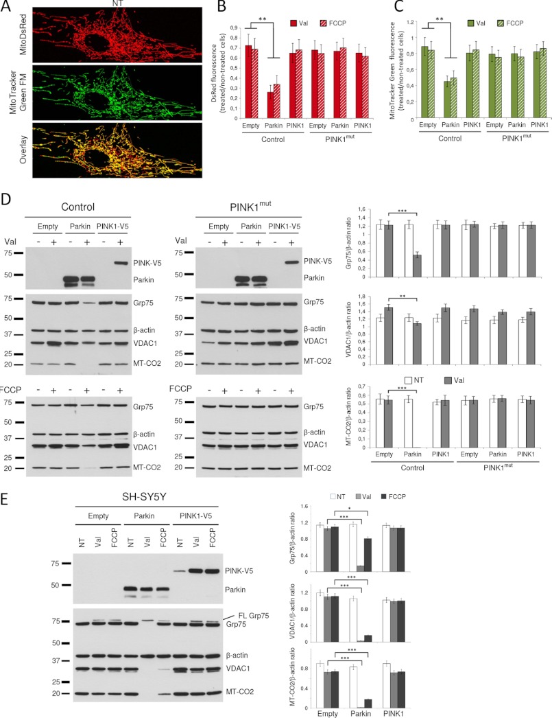
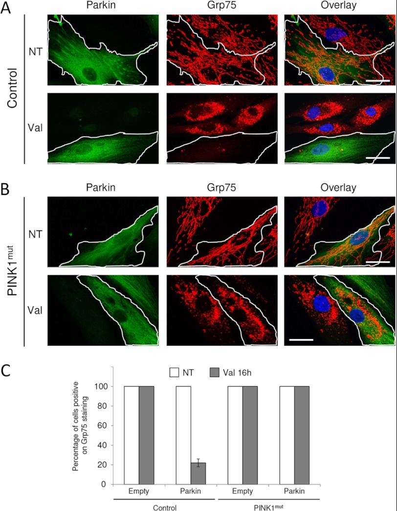
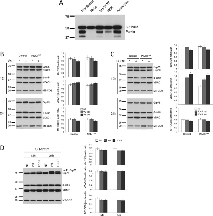
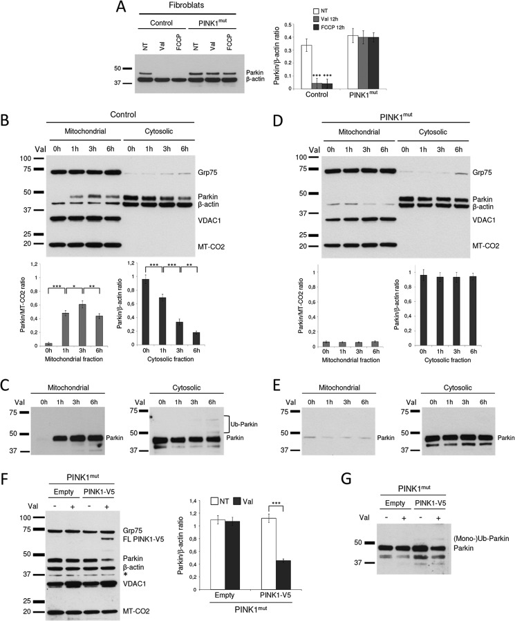
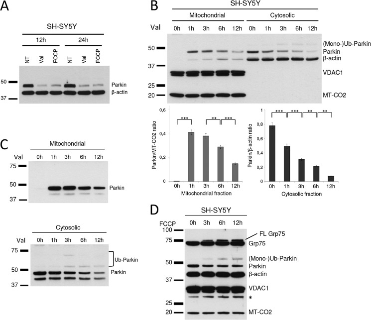
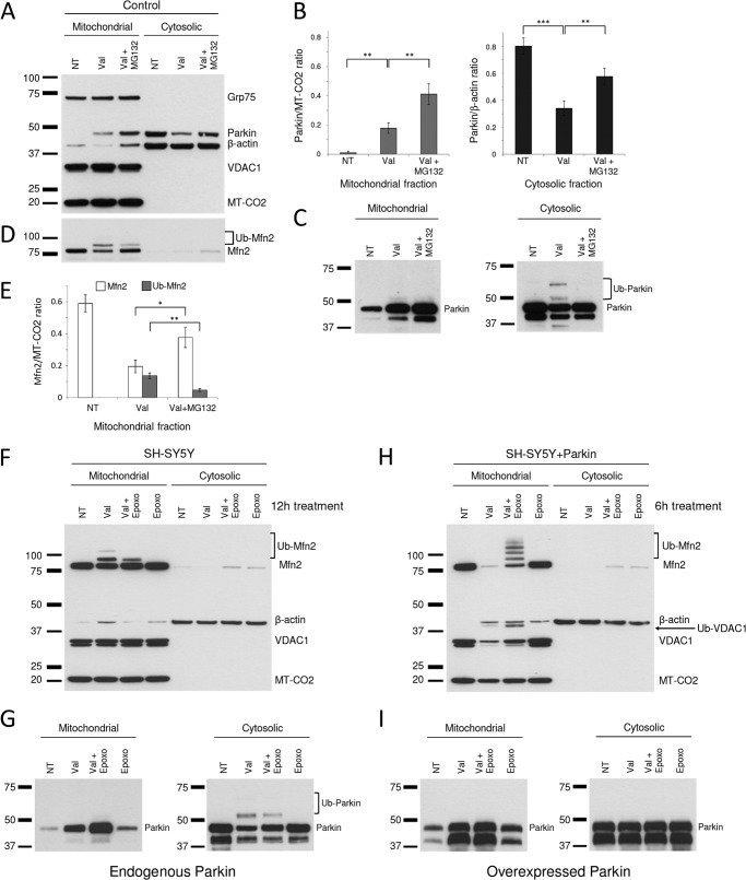
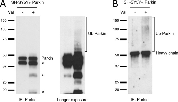
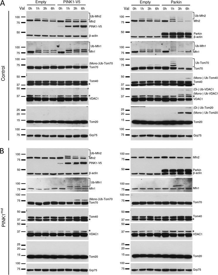
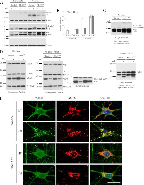
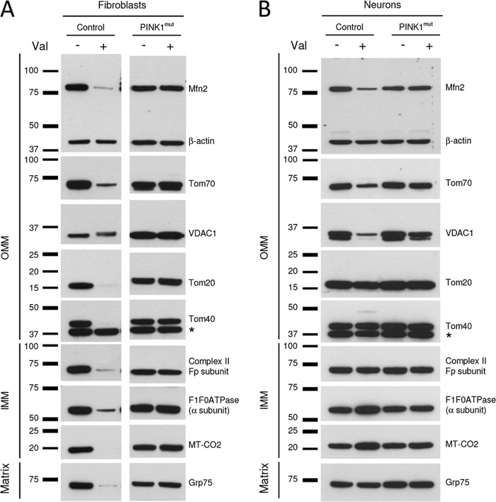
Mutations in the E3 ubiquitin ligase Parkin and the mitochondrial PTEN-induced putative kinase 1 (PINK1) have been identified to cause autosomal recessive forms of familial Parkinson disease, with PINK1 functioning upstream of Parkin in a pathway important for the maintenance of mitochondrial function and morphology. Upon the loss of the mitochondrial membrane potential, Parkin translocates to mitochondria in a PINK1-dependent manner to ubiquitinate mitochondrial proteins. Parkin-mediated polyubiquitination of outer mitochondrial membrane (OMM) proteins recruits the ubiquitin- and LC3-binding adaptor protein p62 to mitochondria and induces mitophagy. Although previous studies examined mitophagy in established cell lines through overexpression approaches, there is an imperative to study the role of endogenous Parkin and PINK1 in human-derived and biologically relevant cell models. Here, we demonstrate in human primary fibroblasts and induced pluripotent stem-derived neurons from controls and PINK1 mutation carriers that endogenous levels of Parkin are not sufficient to initiate mitophagy upon loss of the mitochondrial membrane potential, caused by its (self-)ubiquitination, followed by degradation via the ubiquitin proteasome system. Next, we showed differential PINK1-dependent, Parkin-mediated ubiquitination of OMM proteins, which is Parkin dose-dependent and affects primarily OMM proteins of higher molecular mass. In contrast to the situation fibroblasts, we did not detect mitophagy in induced pluripotent stem-derived neurons even upon overexpression of Parkin. Taken together, our data demonstrate that mitophagy differs between human non-neuronal and neuronal cells and between "endogenous" and "Parkin-overexpressing" cellular models.


Figures

FIGURE 1.
FIGURE 1.
Overexpression of Parkin is required for a significant reduction in levels of mitochondrial proteins and mitochondrial mass. A, nontreated fibroblasts expressing MitoDsRed were stained using MTG. B and C, fibroblasts from a control and a PINK1 mutant stably expressing Parkin or PINK-V5 were transduced with lentiviral particles expressing MitoDsRed or stained using MTG followed by treatment with 1 μm valinomycin or with 10 μm FCCP for 12 h. DsRed (B) or MTG fluorescence (C) was measured using a plate reader. The results were normalized for cell number and expressed as a ratio between valinomycin- or FCCP-treated and nontreated cells. D, fibroblasts from a control and a PINK1 mutant were transduced with lentiviral particles expressing Parkin or C-terminally V5-tagged PINK1 (PINK1-V5) to generate stable cell lines. Next, cells were treated for 12 h with 1 μm valinomycin or with 10 μm FCCP. The proteins were extracted, separated on SDS gels, and immunoblotted using antibodies against Parkin and V5 or against mitochondrial proteins Grp75, VDAC1, and MT-CO2 with β-actin as a loading control. These proteins are reliable mitochondrial markers and mitochondrial loading controls because of their constitutive and ubiquitous expression. Note that endogenous Parkin is not detected in this experiment because of a high dilution of the anti-Parkin antibody (2000× in comparison with the dilution necessary to detect endogenous Parkin). Immunoblots were quantified densitometrically, and the intensity of each band was normalized to β-actin. E, SH-SY5Y cells stably expressing Parkin or PINK-V5 were treated with 1 μm valinomycin or with 10 μm FCCP for 12 h. Proteins were extracted, separated on SDS gels, and immunoblotted using antibodies against Parkin and V5 or against mitochondrial proteins Grp75, VDAC1, and MT-CO2 with β-actin as a loading control. Immunoblots were quantified densitometrically, and the intensity of each band was normalized to β-actin. The error bars represent the means ± standard deviations from three independent experiments. NT, nontreated; Val, valinomycin.
FIGURE 2.
FIGURE 2.
Overexpression of Parkin is required for mitophagy. A and B, fibroblasts from a control (A) and a PINK1 mutant (B) were transiently transfected with Parkin. Upon transfection, the cells were treated with 1 μm valinomycin for 16 h followed by immunostaining using an antibody against Parkin (green, circled cells) and mitochondrial protein Grp75 (red). C, the percentage of cells with detectable mitochondria was determined for 100 cells/condition in three independent experiments. Scale bar, 25 μm. NT, nontreated; Val, valinomycin.
FIGURE 3.
FIGURE 3.
Endogenous Parkin is not sufficient to cause a detectable reduction in levels of mitochondrial proteins. A, whole cell lysate of fibroblasts and of HeLa, SH-SY5Y, HEK cells, and astrocytes was analyzed by Western blotting using antibodies against Parkin and β-tubulin as a loading control. Specificity of the anti-Parkin antibody used in this study was demonstrated previously (11). B and C, fibroblasts from control and PINK1 mutant were treated for 12 or 24 h with 1 μm valinomycin (B) or with 10 μm FCCP (C). Proteins were extracted, separated on SDS gels, and immunoblotted against mitochondrial proteins: Grp75, Hsp60, VDAC1, and MT-CO2. β-Actin served as a loading control. Note an additional band of Grp75 (FL Grp75) upon 24 h of treatment with either valinomycin or FCCP in both controls and PINK1mut. This is due to the fact that loss of the mitochondrial membrane potential prevents mitochondrial import of Grp75 and its mitochondrial processing. Therefore, this protein remains in the cytosol as a nonprocessed pre-protein as shown before (26, 34). Next, we detected a PINK1-independent increase in levels of VDAC1 in both valinomycin- and FCCP-treated cells. This increase was more pronounced in cells treated with FCCP and is in accordance with previous findings that mitochondrial toxins increase the level of the VDAC1 protein but not its mRNA expression (35). Finally, protein levels of the two remaining mitochondrial markers Hsp60 and MT-CO2 were unaffected by treatment and also comparable between controls and PINK1mut. The resulting immunoblots were quantified densitometrically. D, SH-SY5Y cells were treated for 12 or 24 h with 1 μm valinomycin or with 10 μm FCCP. The proteins were extracted, separated on SDS gels, and immunoblotted against mitochondrial proteins: Grp75, VDAC1, and MT-CO2. β-Actin served as a loading control. The intensity of each band was normalized to the intensity of β-actin. The error bars represent the means ± standard deviations from three independent experiments. NT, nontreated; Val, valinomycin; FL, full length.
FIGURE 4.
FIGURE 4.
PINK1-dependent ubiquitination of endogenous Parkin prevents its mitochondrial translocation. A, fibroblasts from a control and a PINK1 mutant were treated for 12 h with 1 μm valinomycin or 10 μm FCCP. B, fibroblasts from a healthy control were treated with 1 μm valinomycin. Cells were harvested at different time points to prepare mitochondrial and cytosolic fractions and analyzed by Western blotting using antibodies against Parkin and mitochondrial proteins Grp75, VDAC1, and MT-CO2 and β-actin as a loading control. After densitometric quantification, the intensity of each band was normalized to the intensity of either MT-CO2 or β-actin. The error bars represent the means ± standard deviations from three independent experiments. C, proteins from each fraction were analyzed by Western blotting using an antibody against Parkin only. D, fibroblasts from a PINK1 mutant were treated with 1 μm valinomycin. Cells were harvested at different time points to prepare mitochondrial and cytosolic fractions and analyzed by Western blotting using antibodies against Parkin and mitochondrial proteins Grp75, VDAC1, and MT-CO2. β-Actin served as a loading control. Densitometric quantification of the immunoblots from three independent experiments normalized to β-actin. E, proteins from each fraction were analyzed by Western blotting using an antibody against Parkin only. F and G, fibroblasts from PINK1 mutant stably expressing PINK-V5 were treated with 1 μm valinomycin for 12 h. The proteins were extracted, separated on SDS gels, and immunoblotted against Grp75, V5 tag, Parkin, VDAC1, and MT-CO2 (F) or against an anti-Parkin antibody only (G). NT, nontreated; Val, valinomycin.
FIGURE 5.
FIGURE 5.
Ubiquitination of endogenous Parkin prevents its mitochondrial translocation in neuroblastoma cells. A, SH-SY5Y cells were treated for 12 and 24 h with 1 μm valinomycin or 10 μm FCCP. B, SH-SY5Y cells were treated with 1 μm valinomycin. The cells were harvested at different time points to prepare mitochondrial and cytosolic fractions and analyzed by Western blotting using antibodies against Parkin and mitochondrial proteins Grp75, VDAC1, and MT-CO2 with β-actin as a loading control. After densitometric quantification, the intensity of each band was normalized to the intensity of either MT-CO2 or β-actin. The error bars represent the means ± standard deviations from three independent experiments. C, proteins from each fraction were analyzed by Western blotting using an antibody against Parkin only. D, SH-SY5Y cells were treated with 1 μm valinomycin. Proteins were extracted at different time points, separated on SDS gels, and immunoblotted against Grp75, Parkin, VDAC1, and MT-CO2. β-Actin served as a loading control. NT, nontreated; Val, valinomycin.
FIGURE 6.
FIGURE 6.
Inhibition of the UPS prevents ubiquitination of endogenous Parkin and increases its levels in mitochondria. A, fibroblasts from controls were treated with 1 μm valinomycin alone or with 1 μm valinomycin plus 10 μm MG132 for 12 h. Cells were harvested to prepare mitochondrial and cytosolic fractions and analyzed by Western blotting using antibodies against Parkin, Grp75, VDAC1, MT-CO2, and β-actin. B, Densitometric analysis of immunoblots normalized to the intensity of either MT-CO2 or β-actin. Error bars represent means ± standard deviations from three independent experiments. C, Proteins from each fraction were analyzed by Western blotting using an antibody against Parkin only. D and E, immunoblots from A were reprobed using an anti-Mfn2 antibody (D) and quantified densitometrically (E). F–I, SH-SY5Y cells expressing endogenous Parkin (F and G) and SH-SY5Y cells overexpressing Parkin (H and I) were treated with 1 μm valinomycin alone or with 1 μm valinomycin plus 4 μm epoxomycin for 12 and 6 h. Cells were harvested to prepare mitochondrial and cytosolic fractions and analyzed by Western blotting using antibodies against Grp75, VDAC1, MT-CO2, and β-actin. G and I, proteins from each fraction were analyzed by Western blotting using an antibody against Parkin only. Val, valinomycin; Mfn2, mitofusin 2; Ub-Mfn2, ubiquitinated mitofusin 2.
FIGURE 7.
FIGURE 7.
Immunoprecipitation confirmed ubiquitination of Parkin. Whole cell lysate from SH-SY5Y cells overexpressing WT Parkin was immunoprecipitated using an anti-Parkin antibody. The resulting immunoprecipitates (IP) were separated by SDS-PAGE and immunoblotted using antibodies against Parkin (A) or ubiquitin (B). Val, valinomycin.
FIGURE 8.
FIGURE 8.
Overexpression of Parkin but not of PINK1 increases ubiquitination rates of outer mitochondrial membrane proteins. Fibroblasts from a control (A) or from a PINK1 mutant (B) stably expressing Parkin or PINK-V5 were treated with 1 μm valinomycin. Cells were harvested at different time points, and total proteins were analyzed by Western blotting using antibodies against the following outer mitochondrial membrane proteins: Mfn1, Mfn2, Tom70, Tom40, VDAC1, and Tom20. Grp75 and β-actin served as a loading controls. Exogenous expression of Parkin and PINK1-V5 was confirmed using antibodies against Parkin and V5 tag. Val, valinomycin; *, nonspecific band; Ub, ubiquitin.
FIGURE 9.
FIGURE 9.
Valinomycin-induced mitophagy is not observed in iPS-generated neurons. A, fibroblasts and iPS-derived neurons from a control and a PINK1 mutant were treated with 1 μm valinomycin for 12 h. Proteins were extracted, separated on SDS gels, and immunoblotted using various antibodies. B, densitometric quantification of immunoblots probed with an antibody against Parkin. C, longer exposure of the immunoblot probed with an antibody against Parkin. D, iPS-derived neurons expressing endogenous levels of Parkin (left panel) or overexpressing Parkin (middle panel) were treated with 1 μm valinomycin for 16 h. Cells were analyzed by Western blotting using antibodies against Grp75, VDAC1, and MT-CO2. β-Actin served as a loading control. Overexpressed Parkin was detected using high dilution of the anti-Parkin antibody (2500× in comparison with the dilution necessary to detect endogenous Parkin) (right panel). E, iPS-derived neurons from a control and a PINK1 mutant stably expressing Parkin were treated with 1 μm valinomycin for 16 h. The cells were immunostained using antibodies against Parkin (green) and Grp75 (red). NT, nontreated; Val, valinomycin; Ub, ubiquitin. Scale bar, 25 μm.
FIGURE 10.
FIGURE 10.
PINK1-dependent valinomycin-induced degradation of mitochondrial proteins is observed in fibroblasts but not in iPS-derived neurons. Fibroblasts (A) and iPS-derived neurons (B) from control and PINK1 mutant stably expressing Parkin were treated with valinomycin for 16 h. Extracted proteins were separated on SDS gels and immunoblotted against various mitochondrial proteins with β-actin serving as a loading control. IMM, inner mitochondrial membrane; Val, valinomycin; *, nonspecific band.

Similar articles

Cited by

References

    1. Langston J. W., Ballard P., Tetrud J. W., Irwin I. (1983) Chronic Parkinsonism in humans due to a product of meperidine-analog synthesis. Science 219, 979–980 - PubMed
    1. Betarbet R., Sherer T. B., MacKenzie G., Garcia-Osuna M., Panov A. V., Greenamyre J. T. (2000) Chronic systemic pesticide exposure reproduces features of Parkinson's disease. Nat. Neurosci. 3, 1301–1306 - PubMed
    1. Kitada T., Asakawa S., Hattori N., Matsumine H., Yamamura Y., Minoshima S., Yokochi M., Mizuno Y., Shimizu N. (1998) Mutations in the Parkin gene cause autosomal recessive juvenile parkinsonism. Nature 392, 605–608 - PubMed
    1. Valente E. M., Abou-Sleiman P. M., Caputo V., Muqit M. M., Harvey K., Gispert S., Ali Z., Del Turco D., Bentivoglio A. R., Healy D. G., Albanese A., Nussbaum R., González-Maldonado R., Deller T., Salvi S., Cortelli P., Gilks W. P., Latchman D. S., Harvey R. J., Dallapiccola B., Auburger G., Wood N. W. (2004) Hereditary early-onset Parkinson's disease caused by mutations in PINK1. Science 304, 1158–1160 - PubMed
    1. Clark I. E., Dodson M. W., Jiang C., Cao J. H., Huh J. R., Seol J. H., Yoo S. J., Hay B. A., Guo M. (2006) Drosophila PINK1 is required for mitochondrial function and interacts genetically with Parkin. Nature 441, 1162–1166 - PubMed

Publication types

MeSH terms