. 2013 Jan 25;288(4):2191-200.
doi: 10.1074/jbc.M112.404780. Epub 2012 Dec 4.

Critical role of S1PR1 and integrin β4 in HGF/c-Met-mediated increases in vascular integrity

Affiliations

Critical role of S1PR1 and integrin β4 in HGF/c-Met-mediated increases in vascular integrity

Yulia Ephstein et al. J Biol Chem. .

Abstract

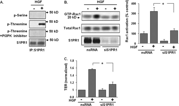
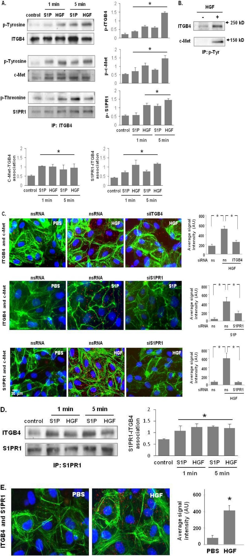
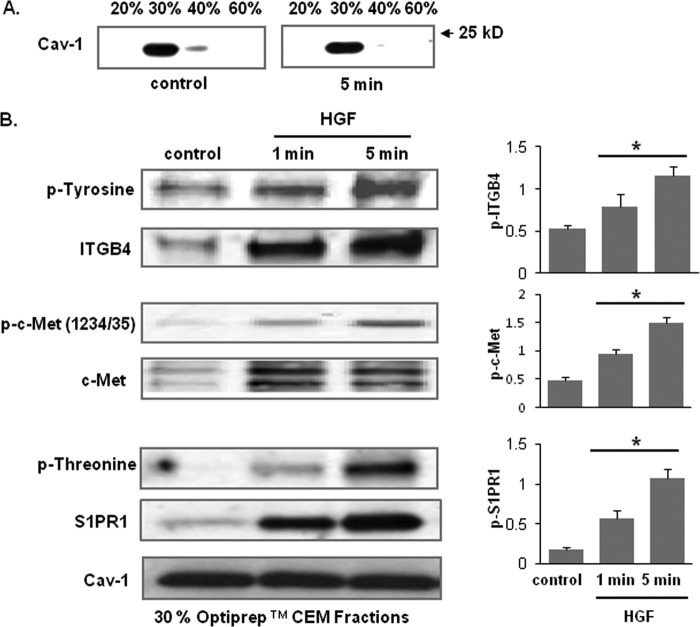
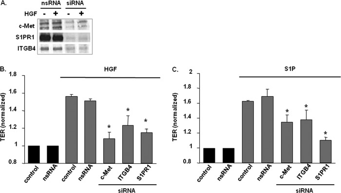
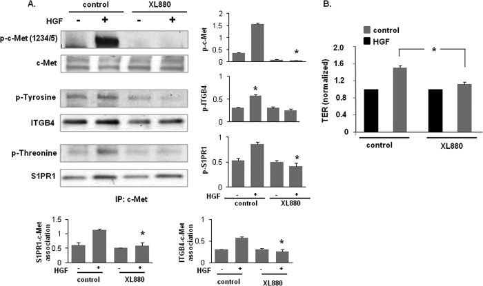
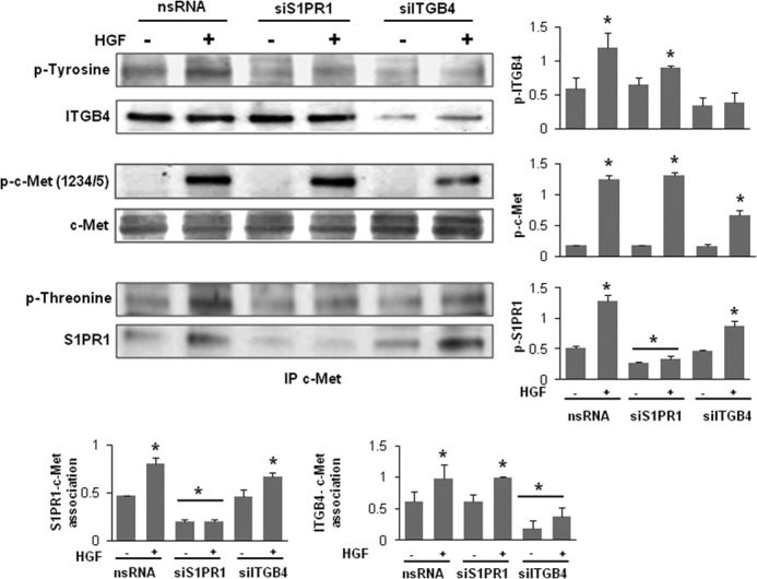
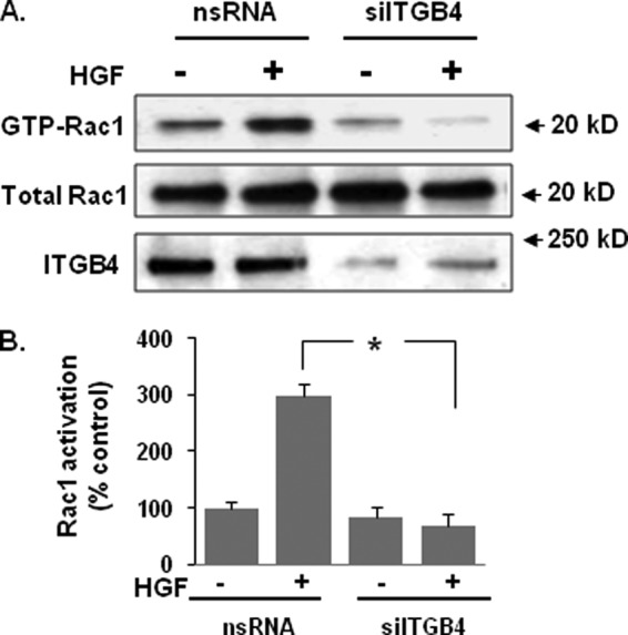
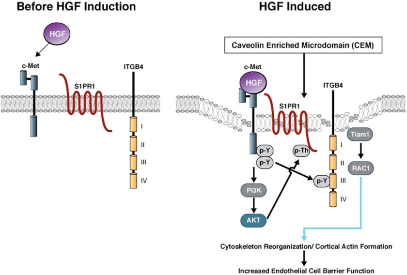
Vascular endothelial cell (EC) barrier integrity is critical to vessel homeostasis whereas barrier dysfunction is a key feature of inflammatory disorders and tumor angiogenesis. We previously reported that hepatocyte growth factor (HGF)-mediated increases in EC barrier integrity are signaled through a dynamic complex present in lipid rafts involving its receptor, c-Met. We extended these observations to confirm that S1PR1 (sphingosine 1-phosphate receptor 1) and integrin β4 (ITGB4) are essential participants in HGF-induced EC barrier enhancement. Immunoprecipitation experiments demonstrated HGF-mediated recruitment of c-Met, ITGB4 and S1PR1 to caveolin-enriched lipid rafts in human lung EC with direct interactions of c-Met with both S1PR1 and ITGB4 accompanied by c-Met-dependent S1PR1 and ITGB4 transactivation. Reduced S1PR1 expression (siRNA) attenuated both ITGB4 and Rac1 activation as well as c-Met/ITGB4 interaction and resulted in decreased transendothelial electrical resistance. Furthermore, reduced ITGB4 expression attenuated HGF-induced c-Met activation, c-Met/S1PR1 interaction, and effected decreases in S1P- and HGF-induced EC barrier enhancement. Finally, the c-Met inhibitor, XL880, suppressed HGF-induced c-Met activation as well as S1PR1 and ITGB4 transactivation. These results support a critical role for S1PR1 and ITGB4 transactivation as rate-limiting events in the transduction of HGF signals via a dynamic c-Met complex resulting in enhanced EC barrier integrity.


Figures

FIGURE 1.
FIGURE 1.
S1PR1 receptor transactivation is required for HGF-induced Rac1 activation and EC barrier enhancement. A, HPAEC were treated with a PI3K inhibitor (LY 294002, 10 μm, 1 h) prior to treatment with HGF (25 ng/ml, 5 min). Whole cell lysates were then immunoprecipitated with an anti-S1PR1 antibody and immunoblotted with an anti-phospho-serine, anti-phospho-threonine, or anti-S1PR1 antibody. HGF-induced S1PR1 threonine phosphorylation was abrogated by PI3K inhibition. B, in separate experiments, nonspecific (ns) siRNA or siRNA against S1PR1 were transfected into HPAEC prior to treatment with HGF (25 ng/ml, 5 min) and cell lysates then analyzed for acativated Rac1 (GTP-Rac1). Silencing of S1PR1 inhibited HGF-induced Rac1 activation. Densitometry analysis of Rac1 activation is expressed as a percentage relative to control cells (ns siRNA, untreated) (n = 3/group, *, p < 0.05). C, maximal TER responses measured in cells transfected with siRNA specific for S1PR1 and then treated with HGF (25 ng/ml) confirmed a marked reduction in HGF-induced TER increases compared with controls cells transfected with ns siRNA. (n = 3/condition, *, p < 0.05).
FIGURE 2.
FIGURE 2.
c-Met/S1PR1/ITGB4 complex modulation by HGF and S1P. A, HPAEC were treated with either S1P (1 μm) or HGF (25 ng/ml) for 1 and 5 min. Cell lysates were subjected to immunoprecipitation with an anti-ITGB4 antibody and tyrosine or threonine phosphorylation corresponding to the same molecular weight was determined via repeat immunoblotting on the same membrane (representative blots shown, densitometric data expressed as fold change phosphorylation relative to control and normalized to total protein, n = 3/group, *, p < 0.05 compared with untreated contols). B, in separate experiments, HPAEC were treated with HGF (25 ng/ml, 5 min) and immunoprecipitation performed with an antibody specific for tyrosine phosphorylation prior to immunoblotting for c-Met and ITGB4 (representative blots shown). C, associations of c-Met with ITGB4 induced by HGF (upper panels) and S1PR1 (middle panels), as well as HGF-induced associations between c-Met and S1PR1 were detected by in situ proximity ligation assay (red dots) in untreated HLMVEC as well as both cells transfected with specific siRNA (ITGB4 or S1PR1) and control cells (ns siRNA) prior to treatment with HGF (25 ng/ml, 5 min) or S1P (1 μm, 5 min). Average signal intensity for each condition was quantified as described under “Experimental Procedures” (n = 4/condition, *, p < 0.05). D, HPAEC were treated with either S1P (1 μm) or HGF (25 ng/ml) and lysates then immunoprecipitated with an anti-S1PR1 antibody followed by immunoblotting with an anti-ITGB4 or anti-S1PR1 antibody. Representative blots and densitometry of the association between S1PR1 and ITGB4 are shown. (n = 3/condition, *, p < 0.05 compared with untreated controls). E, interaction of S1PR1 and ITGB4 in response to HGF treatment (25 ng/ml, 5 min) was also as assessed by PLA in situ.
FIGURE 3.
FIGURE 3.
HGF recruits c-Met, ITGB4, and S1PR1 to lipid rafts. A, Optiprep fractions were isolated and subjected to immunoblotting with an anti-cav-1 antibody. The 30% Optiprep fraction represents the lipid rafts. B, HPAEC were treated with HGF (25 ng/ml) for 1 and 5 min and the 30% Optiprep fraction was isolated and subjected to immunoblotting with antibodies specific for c-Met, ITGB4, or S1PR1. Phosphorylation corresponding to the same molecular weight was determined via repeat immunoblotting on the same membrane with specific antibodies as shown (representative blots shown, densitometry data expressed as fold change phosphorylation relative to control and normalized to total protein, n = 3/condition, *, p < 0.05 compared with untreated controls).
FIGURE 4.
FIGURE 4.
Role of c-Met/S1PR1/ITGB4 complex components on HGF- and S1P-induced EC barrier enhancement. A, HPAEC were transfected with either nonspecific (ns) siRNA or siRNA specific for c-Met, S1PR1, or ITGB4 prior to treatment with HGF (25 ng/ml, 5 min). Cell lysates were then immunoblotted with anti-c-Met, anti-S1PR1 or anti-ITGB4 antibodies to confirm silencing. B and C, maximal TER responses after HGF (25 ng/ml) or S1P (1 μm) are shown for control cells and cells transfected with specific siRNA (n ≥ 5/condition, results normalized to control, untreated cells, *, p < 0.05 compared with control cells transfected with nsRNA and stimulated with HGF or S1P, respectively).
FIGURE 5.
FIGURE 5.
c-Met tyrosine kinase inhibition abrogates HGF-induced activation of S1PR1 and ITGB4 and EC barrier enhancement. A, HPAEC were pretreated with a c-Met inhibitor, XL880 (0.4 nmol/liter, 1 h) prior to HGF treatment (25 ng/ml, 5 min). Cell lysates were immunoprecipitated with an anti-c-Met antibody and then subjected to immunoblotting with antibodies specific for c-Met, ITGB4, or S1PR1. Phosphorylation corresponding to the same molecular weight was determined via repeat immunoblotting on the same membrane with specific antibodies as shown (representative blots shown, densitometry data expressed as fold change phosphorylation relative to control and normalized to total protein, n = 3/condition, *, p < 0.05 compared with HGF-treated control cells). B, maximal TER responses to HGF (25 ng/ml) were measured in cells pretreated with XL880 (0.4 nmol/liter, 1 h) and compared with untreated control cells (n ≥ 5/condition, *, p < 0.05).
FIGURE 6.
FIGURE 6.
Reductions in S1PR1 or ITGB4 expression inhibits c-Met/S1PR1/ITGB4 complex activation. HPAEC were transfected with nonspecific (ns) siRNA or siRNA specific for S1PR1 or ITGB4 prior to treatment with HGF (25 ng/ml, 5 min). Cells lysates were immunoprecipitated with an anti-c-Met antibody and then subjected to immunoblotting with antibodies specific for c-Met, ITGB4, or S1PR1. Phosphorylation corresponding to the same molecular weight was determined via repeat immunoblotting on the same membrane with specific antibodies as shown (representative blots shown, densitometry data expressed as fold change phosphorylation relative to control and normalized to total protein, n = 3/condition, *, p < 0.05 compared with untreated nsRNA controls).
FIGURE 7.
FIGURE 7.
HGF-induced Rac1 activation involves ITGB4-mediated signaling. A, HPAEC were transfected with nonspecific (ns) siRNA or ITGB4 siRNA prior to treatment with HGF (25 ng/ml, 5 min). Cell lysates were then prepared and analyzed for Rac1 activation (GTP-Rac1). Representative blots are shown. B, densitometry analysis of Rac1 activation is expressed as a percentage relative to control cells (ns siRNA, untreated) (n = 3/group, *, p < 0.05).
FIGURE 8.
FIGURE 8.
Model for HGF-mediated c-Met signalosome formation resulting in EC barrier enhancement. Based on our previous work and current study, an HGF-mediated, EC barrier-enhancing signalosome model is proposed. A constitutive c-Met/S1PR1/ITGB4 complex is present in lipid rafts of the plasma membrane. Treatment with HGF promotes further recruitment of complex components and strengthens the c-Met signalosome. Ligation of c-Met results in auto-activation and subsequent activation of key downstream targets such as PI3K and Akt, which drive phosphorylation of the S1PR1 cytoplasmic tail and its activation. Activated c-Met may also phosphorylate ITGB4, either directly or indirectly through c-Src, which leads to the activation of Rac1 through the adapter protein, Tiam1, resulting in cytoskeletal changes. We envisage the coordinated signals from the receptors in the c-Met complex bring about cytoskeletal reorganization leading to cortactin/actin interaction leading to EC barrier enhancement.

Similar articles

Cited by

References

    1. Singleton P. A., Salgia R., Moreno-Vinasco L., Moitra J., Sammani S., Mirzapoiazova T., Garcia J. G. (2007) CD44 regulates hepatocyte growth factor-mediated vascular integrity. Role of c-Met, Tiam1/Rac1, dynamin 2, and cortactin. J. Biol. Chem. 282, 30643–30657 - PubMed
    1. Garcia J. G., Liu F., Verin A. D., Birukova A., Dechert M. A., Gerthoffer W. T., Bamberg J. R., English D. (2001) Sphingosine 1-phosphate promotes endothelial cell barrier integrity by Edg-dependent cytoskeletal rearrangement. J. Clin. Invest. 108, 689–701 - PMC - PubMed
    1. Dudek S. M., Jacobson J. R., Chiang E. T., Birukov K. G., Wang P., Zhan X., Garcia J. G. (2004) Pulmonary endothelial cell barrier enhancement by sphingosine 1-phosphate: roles for cortactin and myosin light chain kinase. J. Biol. Chem. 279, 24692–24700 - PubMed
    1. Singleton P. A., Dudek S. M., Chiang E. T., Garcia J. G. (2005) Regulation of sphingosine 1-phosphate-induced endothelial cytoskeletal rearrangement and barrier enhancement by S1PR1 receptor, PI3 kinase, Tiam1/Rac1, and α-actinin. Faseb J. 19, 1646–1656 - PubMed
    1. Singleton P. A., Dudek S. M., Ma S. F., Garcia J. G. (2006) Transactivation of sphingosine 1-phosphate receptors is essential for vascular barrier regulation. Novel role for hyaluronan and CD44 receptor family. J. Biol. Chem. 281, 34381–34393 - PubMed

MeSH terms