. 2013 Mar;23(3):411-8.
doi: 10.1101/gr.145631.112. Epub 2012 Dec 3.

Nonallelic homologous recombination between retrotransposable elements is a driver of de novo unbalanced translocations

Affiliations

Nonallelic homologous recombination between retrotransposable elements is a driver of de novo unbalanced translocations

Caroline Robberecht et al. Genome Res. 2013 Mar.

Abstract

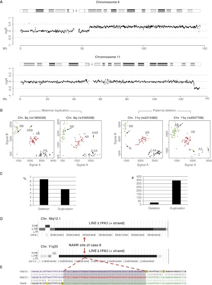
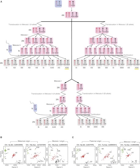
Large-scale analysis of balanced chromosomal translocation breakpoints has shown nonhomologous end joining and microhomology-mediated repair to be the main drivers of interchromosomal structural aberrations. Breakpoint sequences of de novo unbalanced translocations have not yet been investigated systematically. We analyzed 12 de novo unbalanced translocations and mapped the breakpoints in nine. Surprisingly, in contrast to balanced translocations, we identify nonallelic homologous recombination (NAHR) between (retro)transposable elements and especially long interspersed elements (LINEs) as the main mutational mechanism. This finding shows yet another involvement of (retro)transposons in genomic rearrangements and exposes a profoundly different mutational mechanism compared with balanced chromosomal translocations. Furthermore, we show the existence of compound maternal/paternal derivative chromosomes, reinforcing the hypothesis that human cleavage stage embryogenesis is a cradle of chromosomal rearrangements.


Figures

Figure 1.
Figure 1.
Determination of the origin and breakpoint sequence of the translocation in case 8. (A) SNP array intensity ratio plots. Log2 intensity ratio of test over reference of the affected chromosomes is plotted on the y-axis against the position from pter to qter on the x-axis. (Gray line) The average test over reference ratio per 10 SNP values. (B) SNP cluster plots of individual SNPs in case 8. Green, red, and black dots represent controls with a BB, AB, and AA genotype, respectively. The pink and blue dots represent the genotypes of the mother and father and the longer arrows point to them. The yellow dots indicate the genotype call of the index patient and are indicated by the shorter arrow. The SNP plots of parental homozygous SNPs demonstrate that the duplication (left two panels) is of maternal origin, while the deletion (right panel) is of paternal origin. (C) The graphs indicate the percentage (left) and total number (right) of informative SNPs found in the deleted and duplicated region of case 8. (D) The breakpoint junction of case 8 is located in 6-kb LINE L1PA3 elements of the same orientation (black bars) with 95% DNA sequence identity in the chromosome regions 8q12.1 (top) and 11q25 (bottom). (Red arrows) The location of the breakpoint junction determined by sequence analysis. (E) DNA sequence alignment of the PCR-amplified translocation junction fragment of case 8 (middle sequence). The breakpoint site was narrowed to a 53-bp segment (gray box) with 100% DNA sequence identity between chromosomes 11 (top) and 8 (bottom). (Purple nucleotides) Alignment with the chromosome 11 sequence; (green nucleotides) alignment with the chromosome 8 sequence; (yellow highlighted nucleotides) trans-morphic mismatches.
Figure 2.
Figure 2.
Resolving meiotic origin of de novo unbalanced translocations. (A) A schematic illustration of different segregants following a meiotic I or II de novo unbalanced translocation event. (Rose rectangles) Maternal gametes; (blue rectangles) paternal gametes; (gray rectangles) the zygote. In this figure, the translocation event occurs in the maternal gamete. Within those rectangles, the black and red colors represent maternal chromosomes, and dark gray and dark pink colors represent paternal chromosomes. To discriminate a meiosis I from a meiosis II event, maternal heterozygous SNPs and paternal homozygous SNPs are considered. The presence of ABB SNP-calls (underlined with yellow color) present in the duplicated region of the translocated chromosomes can only be the consequence of a premeiotic or a meiotic I event. (B) The SNP plots of parental homozygous SNPs demonstrate that both the deletion and duplication (left two panels) are of maternal origin, while the analysis of the maternal heterozygous and paternal homozygous SNPs demonstrates a premeiotic or meiosis I origin (right panel). (C) The SNP plots of parental homozygous SNPs demonstrate that both the deletion and duplication (left two panels) are of paternal origin, while the analysis of the paternal heterozygous and maternal homozygous SNPs demonstrates the premeiotic or meiosis I origin (right panel). Green, red, black, and gray dots represent controls with a BB, AB, AA, and NoCall genotype, respectively. The pink and blue dots represent the genotypes of the mother and father and the longer arrows point to them. The yellow dots indicate the genotype call of the index patient and are indicated by the shorter arrow. The SNP plots of parental homozygous SNPs demonstrate that the duplication (left two panels) is of maternal origin, while the deletion (right panel) is of paternal origin.

Similar articles

Cited by

References

    1. Ballif BC, Sulpizio SG, Lloyd RM, Minier SL, Theisen A, Bejjani BA, Shaffer LG 2007. The clinical utility of enhanced subtelomeric coverage in array CGH. Am J Med Genet A 143A: 1850–1857 - PubMed
    1. Bauters M, Van Esch H, Friez MJ, Boespflug-Tanguy O, Zenker M, Vianna-Morgante AM, Rosenberg C, Ignatius J, Raynaud M, Hollanders K, et al. 2008. Nonrecurrent MECP2 duplications mediated by genomic architecture-driven DNA breaks and break-induced replication repair. Genome Res 18: 847–858 - PMC - PubMed
    1. Belshaw R, Dawson AL, Woolven-Allen J, Redding J, Burt A, Tristem M 2005. Genomewide screening reveals high levels of insertional polymorphism in the human endogenous retrovirus family HERV-K(HML2): Implications for present-day activity. J Virol 79: 12507–12514 - PMC - PubMed
    1. Branzei D, Foiani M 2010. Maintaining genome stability at the replication fork. Nat Rev Mol Cell Biol 11: 208–219 - PubMed
    1. Burwinkel B, Kilimann MW 1998. Unequal homologous recombination between LINE-1 elements as a mutational mechanism in human genetic disease. J Mol Biol 277: 513–517 - PubMed

Publication types