. 2013 Nov;70(5):1360-8.
doi: 10.1002/mrm.24564. Epub 2012 Dec 4.

Motion and flow insensitive adiabatic T2 -preparation module for cardiac MR imaging at 3 Tesla

Affiliations

Motion and flow insensitive adiabatic T2 -preparation module for cardiac MR imaging at 3 Tesla

Elizabeth R Jenista et al. Magn Reson Med. 2013 Nov.

Abstract

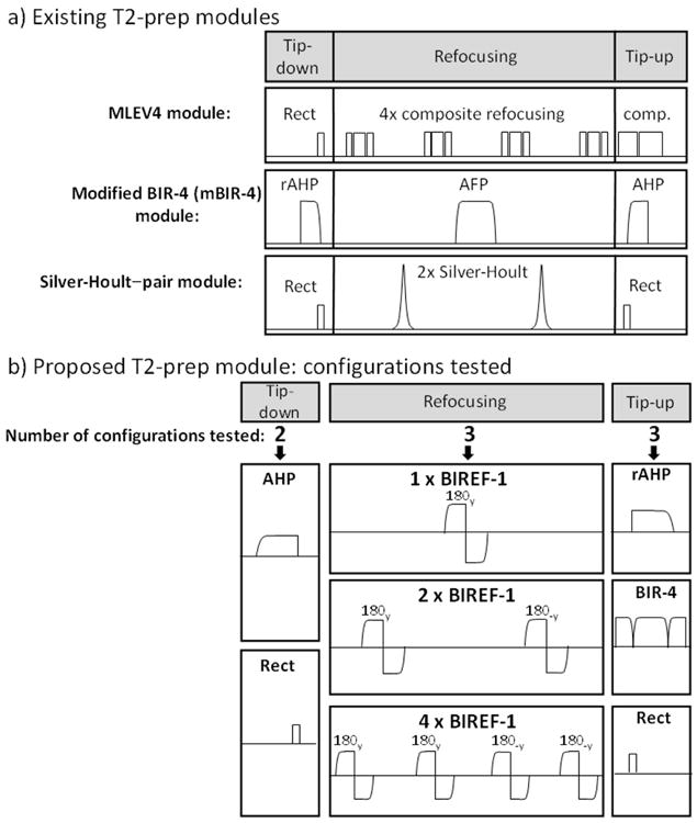
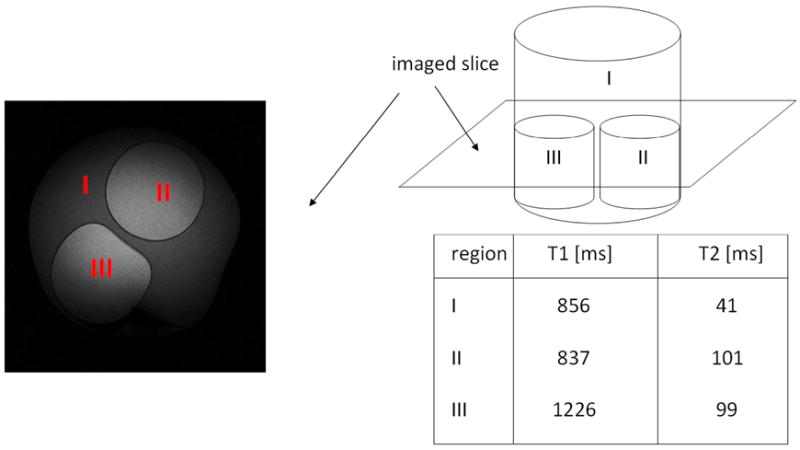
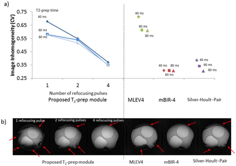
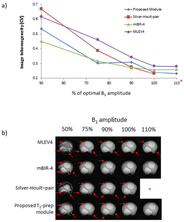
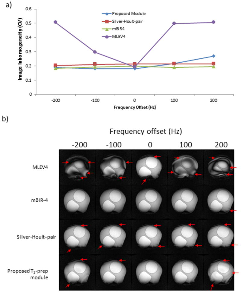
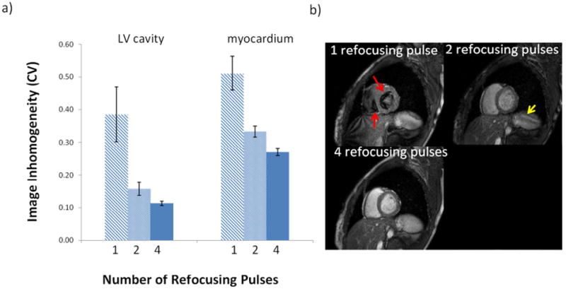
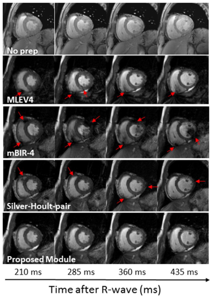
A versatile method for generating T2 -weighting is a T2 -preparation module, which has been used successfully for cardiac imaging at 1.5T. Although it has been applied at 3T, higher fields (B0 ≥ 3T) can degrade B0 and B1 homogeneity and result in nonuniform magnetization preparation. For cardiac imaging, blood flow and cardiac motion may further impair magnetization preparation. In this study, a novel T2 -preparation module containing multiple adiabatic B1 -insensitive refocusing pulses is introduced and compared with three previously described modules [(a) composite MLEV4, (b) modified BIR-4 (mBIR-4), and (c) Silver-Hoult-pair]. In the static phantom, the proposed module provided similar or better B0 and B1 insensitivity than the other modules. In human subjects (n = 21), quantitative measurement of image signal coefficient of variation, reflecting overall image inhomogeneity, was lower for the proposed module (0.10) than for MLEV4 (0.15, P < 0.0001), mBIR-4 (0.27, P < 0.0001), and Silver-Hoult-pair (0.14, P = 0.001) modules. Similarly, qualitative analysis revealed that the proposed module had the best image quality scores and ranking (both, P < 0.0001). In conclusion, we present a new T2 -preparation module, which is shown to be robust for cardiac imaging at 3T in comparison with existing methods.

Keywords: 3T; T2-weighting; adiabatic pulses; cardiac imaging.


Figures

Figure 1
Figure 1. Description of T2-preparation modules
a) Generically, T2-prep modules can be separated into tip-down, refocusing, and tip-up components. Three existing T2-prep modules are shown. The MLEV4 module uses a rectangular (Rect) tip-down pulse (90x), four composite refocusing pulses each consisting of (90x, 180y, 90x), MLEV phase cycling of the refocusing pulses as follows, [180y, 180y, 180-y, 180-y], and a composite tip-up pulse (comp) consisting of (270x, -360x). The mBIR-4 module uses a reverse adiabatic half passage (rAHP) tip-down, an adiabatic full passage (AFP) pulse, and an adiabatic half passage (AHP) tip-up. The Silver-Hoult–pair module uses Rect/Rect tip-down/tip-up pulses and two AFPs with sech/tanh modulation for refocusing. b) The different configurations tested for the proposed T2-prep module are shown. There were two options for tip-down (AHP or Rect), three for refocusing (1, 2 or 4 BIREF-1 pulses), and three for tip-up (AHP, BIR-4, Rect). When multiple BIREF-1 pulses were employed, MLEV phase cycling was used.
Figure 2
Figure 2. Phantom setup
A phantom was constructed out of Agarose gel and NiCl2, with three compartments with different combinations of T2 and T1 values.
Figure 3
Figure 3. The effect of the number of refocusing pulses
a) Left portion of the graph displays image inhomogeneity (coefficient of variation) versus number of refocusing pulses for the proposed T2-prep module with Rect/Rect tip-down/tip-up pulses. For all T2-prep times, more refocusing pulses improved image homogeneity (ie reduced coefficient of variation). The performance of MLEV4, mBIR-4 and Silver-Hoult–pair modules are shown on the right portion of the graph for comparison. The MLEV4 module had a relatively high level of inhomogeneity. b) Typical images with each module for a T2-prep time of 40 ms. Arrows show areas of inhomogeneity.
Figure 4
Figure 4. B1 Sensitivity
a) B1 amplitude was varied between 50% to 110% of the calibrated value for the tested modules (*Note that B1 amplitude at 110% could not be tested for the Silver-Hoult–pair module because peak amplitude exceeded scanner capability). T2-prep time was 60 ms for all modules, and the coefficient of variation (CV) is shown as a function of transmitter voltage. b) Typical images are shown. Red arrows identify regional inhomogeneities. The mBIR-4 and the proposed T2-prep module provided a larger range of insensitivity to B1 than the MLEV4 and Silver-Hoult–pair modules.
Figure 5
Figure 5. B0 sensitivity
a) Transmitter frequency was varied from -200 Hz to +200 Hz for the tested modules. T2-prep time was 60 ms for all modules, and the coefficient of variation (CV) is shown as a function of frequency offset. b) Typical images are shown. Red arrows point out areas of image inhomogeneity. The mBIR-4, Silver-Hoult-pair and proposed T2-prep modules were relatively insensitive to changes in B0 unlike the MLEV4 module.
Figure 6
Figure 6. Effect of the number of refocusing pulses in the proposed T2-prep module in human volunteers
a) The coefficient of variation in the LV cavity and myocardium averaged for all T2-prep times (40, 60, and 80 ms) in 4 subjects decreased with increasing number of refocusing pulses (p<0.0001 for both). b) Typical images from one volunteer (T2-prep time was 60 ms). With 1 refocusing pulse, there are significant artifacts due to motion and flow in the left and right ventricular cavities (red arrows point to signal voids). Signal void artifacts are mostly gone with 2 refocusing pulses, but the overall signal intensity is low and banding is still seen (yellow arrow). With 4 refocusing pulses, LV cavity signal is bright and homogeneous, and the myocardium is uniform.
Figure 7
Figure 7. Images acquired at different phases of the cardiac cycle
Images were acquired with incremental changes in the trigger delay (75 ms step size) using a T2-prep time of 60 ms for those with T2-prep modules. The top row shows images acquired without a T2-prep to show the difference in myocardial contrast. The horizontal axis represents the time from the R-wave to the center of k-space for the images. Regions of inhomogeneity are indicated with red arrows. The proposed module provided the most uniform tissue and blood preparation throughout the cardiac cycle of the four T2-prep modules that were tested.

References

    1. Botnar RM, Stuber M, Danias PG, Kissinger KV, Manning WJ. Improved coronary artery definition with T2-weighted, free-breathing, three-dimensional coronary MRA. Circulation. 1999;99(24):3139–3148. - PubMed
    1. Kellman P, Aletras AH, Mancini C, McVeigh ER, Arai AE. T2-prepared SSFP improves diagnostic confidence in edema imaging in acute myocardial infarction compared to turbo spin echo. Magnetic Resonance in Medicine. 2007;57(5):891–897. - PMC - PubMed
    1. Sparrow PJ, Kurian JB, Jones TR, Sivananthan MU. MR imaging of cardiac tumors. Radiographics. 2005;25(5):1255–1276. - PubMed
    1. Brittain JH, Hu BS, Wright GA, Meyer CH, Macovski A, Nishimura DG. Coronary Angiography with Magnetization-Prepared T2 Contrast. Magnetic Resonance in Medicine. 1995;33(5):689–696. - PubMed
    1. Wright GA, Nishimura DG, Macovski A. Flow-independent magnetic resonance projection angiography. Magnetic Resonance in Medicine. 1991;17(1):126–140. - PubMed

Publication types