. 2012 Dec 18;109(51):21110-5.
doi: 10.1073/pnas.1210804109. Epub 2012 Dec 3.

Possible role of pineal allopregnanolone in Purkinje cell survival

Affiliations

Possible role of pineal allopregnanolone in Purkinje cell survival

Shogo Haraguchi et al. Proc Natl Acad Sci U S A. .

Abstract

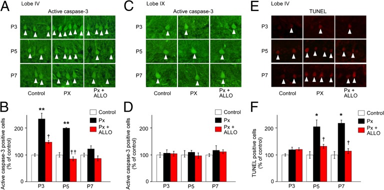
It is believed that neurosteroids are produced in the brain and other nervous systems. Here, we show that allopregnanolone (ALLO), a neurosteroid, is exceedingly produced in the pineal gland compared with the brain and that pineal ALLO acts on the Purkinje cell, a principal cerebellar neuron, to prevent apoptosis in the juvenile quail. We first demonstrated that the pineal gland is a major organ of neurosteroidogenesis. A series of experiments using molecular and biochemical techniques has further demonstrated that the pineal gland produces a variety of neurosteroids de novo from cholesterol in the juvenile quail. Importantly, ALLO was far more actively produced in the pineal gland than in the brain. Pinealectomy (Px) decreased ALLO concentration in the cerebellum and induced apoptosis of Purkinje cells, whereas administration of ALLO to Px quail chicks prevented apoptosis of Purkinje cells. We further found that Px significantly increased the number of Purkinje cells that expressed active caspase-3, a key protease in apoptotic pathway, and daily injection of ALLO to Px quail chicks decreased the number of Purkinje cells expressing active caspase-3. These results indicate that the neuroprotective effect of pineal ALLO is associated with the decrease in caspase-3 activity during the early stage of neuronal development. We thus provide evidence that the pineal gland is an important neurosteroidogenic organ and that pineal ALLO may be involved in Purkinje cell survival during development. This is an important function of the pineal gland in the formation of neuronal circuits in the developing cerebellum.


Conflict of interest statement

The authors declare no conflict of interest.

Figures

Fig. 1.
Fig. 1.
De novo PREG formation from cholesterol in the pineal gland. (A and B) RT-PCR analyses of StAR (A) and Cyp11a (B) mRNAs in the pineal gland of male quail chicks. Total RNA extracted from the tissue was reverse-transcribed with (+) or without (−) reverse transcriptase (RTase) followed by PCR amplification. (C) HPLC analysis of PREG formation from cholesterol in the pineal gland of male quail chicks. The pineal gland was incubated with [3H]cholesterol and homogenized after different incubation times, and then each extract was subjected to HPLC. The pineal gland with [3H]cholesterol was also incubated with aminoglutethimide. The arrowheads indicate the elution positions of the substrate cholesterol (open arrowhead) and its metabolite PREG (solid arrowhead). (D) GC-SIM analysis of the metabolite of nonradioactive cholesterol by the pineal gland of male quail chicks. GC-SIM was traced at m/z 298 for PREG as the metabolite of nonradioactive cholesterol. The arrowhead shows the peak corresponding to authentic PREG. (E) Western blot analysis of the pineal gland and extracts of COS-7 cells transfected with chicken Cyp11a cDNA with the anti-human P450scc antibody. Anti-human P450scc antibody was preadsorbed with chicken P450scc protein for control. (F) IHC of P450scc in the pineal gland. Arrowheads indicate immunoreactive cells. (G) IHC using P450scc antibody preadsorbed with a saturating concentration of chicken P450scc protein (10 μg/mL). (Scale bars, 10 μm.) Similar results were obtained in repeated experiments using three different samples.
Fig. 2.
Fig. 2.
Neurosteroid formation from PREG in the pineal gland. (A, D, and G) RT-PCR analyses of steroidogenic enzyme Cyp7b (A), Hsd3b (D), Srd5a (G), and Cyp17 (G) mRNAs in the pineal gland of male quail chicks. Total RNA was reverse-transcribed with (+) or without (−) RTase followed by PCR amplification. (B, E, and H) HPLC analyses of neurosteroid formation in the pineal gland of male quail chicks. (B) The pineal gland homogenates were incubated with [3H]PREG + NADPH, and the extracts were subjected to HPLC. The pineal gland homogenates with [3H]PREG were also incubated with ketoconazole. The arrowheads indicate elution positions of the substrate PREG (open arrowhead) and its metabolite 7α- and/or 7β-OH PREG (solid arrowhead). (E) The pineal gland homogenates were incubated with [3H]PREG + NAD+, and the extracts were subjected to HPLC. The pineal gland homogenates with [3H]PREG were also incubated with trilostane. The arrowheads indicate elution positions of the substrate PREG (open arrowhead) and its metabolite PROG (solid arrowhead). (H) The pineal gland homogenates were incubated with [3H]PROG + NADPH, and the extracts were subjected to HPLC. The pineal gland homogenates with [3H]PROG were also incubated with ketoconazole and finasteride. The arrowheads indicate elution positions of the substrate PROG (open arrowhead) and its metabolites 5α-DHP, ALLO and/or EPI and AD (solid arrowheads). (C, F, I, and J) GC-SIM analyses of the metabolites of nonradioactive PREG or PROG by the pineal gland of male quail chicks. GC-SIM was traced at m/z 386 for 7α- and 7β-OH PREG (C) and at m/z 510 for PROG (F), metabolites of nonradioactive PREG, or at m/z 514 for ALLO and EPI (I) and at m/z 482 for AD (J), metabolites of nonradioactive PROG. The arrowheads show the peaks corresponding to authentic 7α- and 7β-OH PREG (C), PROG (F), ALLO and EPI (I), and AD (J). Similar results were obtained in repeated experiments using three different samples.
Fig. 3.
Fig. 3.
ALLO and 7α-OH PREG are abundantly synthesized and released from the quail chick pineal gland. (A) Neurosteroids synthesized from PREG in the pineal gland of male quail chicks. The pineal glands were incubated with the substrate [3H]PREG, and the extracts were subjected to HPLC. The arrows indicate elution positions of the substrate PREG (open arrow) and its metabolites (solid arrows). (B) Comparison of the amount of neurosteroids synthesized from PREG in the pineal gland of male quail chicks by HPLC (n = 8). **P < 0.01 vs. 5α-DHP, AD, or T; †††P < 0.001 vs. PROG, 5α- and/or 5β-DHT, or E2. (C) Comparisons of 7α- and/or 7β-OH PREG synthesis and Cyp7b mRNA expression in the pineal gland of adults and chicks of both sexes (n = 8). **P < 0.01 vs. adult. (D) Comparisons of 7α- and/or 7β-OH PREG synthesis and Cyp7b mRNA expression among the pineal gland, cerebellum, and diencephalon of quail chicks of both sexes (n = 8). **P < 0.01 vs. cerebellum or diencephalon. (E) Comparisons of 7α- and 7β-OH PREG releases from the pineal gland, cerebellum, and diencephalon of male quail chicks by GC-MS (n = 6). ***P < 0.001 vs. cerebellum or diencephalon. (F) Comparisons of ALLO and/or EPI synthesis and Srd5a mRNA expression in the pineal gland of adults and chicks of both sexes (n = 8). **P < 0.01 vs. adult. (G) Comparisons of ALLO and/or EPI synthesis and Srd5a mRNA expression among the pineal gland, cerebellum, and diencephalon of quail chicks of both sexes (n = 8). ***P < 0.001 vs. cerebellum or diencephalon. (H) Comparisons of ALLO and EPI releases from the pineal gland, cerebellum, and diencephalon of male quail chicks by GC-MS (n = 6). **P < 0.01 vs. cerebellum or diencephalon. (I) Identified biosynthetic pathways of neurosteroids in the pineal gland. Each column and vertical line in BH represent the mean ± SEM.
Fig. 4.
Fig. 4.
Pineal ALLO saves Purkinje cells from cell death in Px quail chicks. Cerebella of control male quail chicks, Px at P2 male quail chicks, and Px at P2 male quail chicks treated with daily injection of 7α-OH PREG (30 ng/5 μL) or ALLO (30 ng/5 μL), or Px at P2 male quail chicks s.c. implanted with a silastic plate containing melatonin (10 mg/plate) from P2 to P7 were analyzed. (A) Number of Purkinje cells in the cerebellar lobes I–X at P21. Purkinje cell number is presented as the percentage of control (n = 12). *P < 0.05 or **P < 0.01 vs. control; P < 0.05 or ††P < 0.01 Px plus ALLO vs. Px. (B) Morphology of Purkinje cells in the cerebellar lobes IV and IX at P21. M, molecular layer; P, Purkinje cell layer. (Scale bars, 40 μm.) (C) Effects of Px and daily injection of ALLO on ALLO concentration in the cerebellum of male quail chicks at P7 (n = 12). **P < 0.01 vs. control; ††P < 0.01 Px plus ALLO vs. Px. (D) Comparisons of ALLO concentration among the pineal gland, rostral cerebellum, and caudal cerebellum in P2 male quail chicks (n = 7). **P < 0.01 vs. rostral cerebellum or caudal cerebellum. (E) Effects of Px on ALLO concentrations in the rostral and caudal cerebellum of male quail chicks (n = 7). *P < 0.05 vs. 0 h. (F) Contents of [3H]ALLO in the rostral and caudal cerebellum after injection of [3H]ALLO close to the pineal lumen of male quail chicks at P2. [3H]ALLO (20 pmol) was injected close to the pineal lumen, and the content of [3H]ALLO in the rostral and caudal cerebellum was counted in a liquid scintillation counter (n = 7). ★★★P < 0.001 vs. 0 h; ☆☆P < 0.01 or ☆☆☆P < 0.001 vs. 0 h; P < 0.05 rostral cerebellum vs. caudal cerebellum. Each column and vertical line in A and CF represent the mean ± SEM.
Fig. 5.
Fig. 5.
Neuroprotective effect of pineal ALLO is associated with the decrease in caspase-3 activity in Purkinje cells. (A and C) Purkinje cells expressing active caspase-3 in lobes IV (A) and IX (C) of control male quail chicks, Px male quail chicks, and Px plus ALLO male quail chicks at P3, P5, and P7. (B and D) Comparison of the number of Purkinje cells expressing active caspase-3 in lobes IV (B) and IX (D) among control male quail chicks, Px male quail chicks, and Px plus ALLO male quail chicks at P3, P5, and P7 (n = 12). **P < 0.01 vs. control; P < 0.05 or ††P < 0.01 Px plus ALLO vs. Px. (E) TUNEL-positive Purkinje cells in lobe IV of control male quail chicks, Px male quail chicks, and Px plus ALLO male quail chicks at P3, P5, and P7. (F) Comparison of the number of TUNEL-positive Purkinje cells in lobe IV among control male quail chicks, Px male quail chicks, and Px plus ALLO male quail chicks at P3, P5, and P7 (n = 12). *P < 0.05 vs. control; P < 0.05 Px plus ALLO vs. Px. Each column and vertical line in B, D, and F represent the mean ± SEM.

Similar articles

Cited by

References

    1. Kilic E, Hermann DM, Isenmann S, Bähr M. Effects of pinealectomy and melatonin on the retrograde degeneration of retinal ganglion cells in a novel model of intraorbital optic nerve transection in mice. J Pineal Res. 2002;32(2):106–111. - PubMed
    1. Tunç AT, et al. Neonatal pinealectomy induces Purkinje cell loss in the cerebellum of the chick: A stereological study. Brain Res. 2006;1067(1):95–102. - PubMed
    1. Baulieu EE. Neurosteroids: Of the nervous system, by the nervous system, for the nervous system. Recent Prog Horm Res. 1997;52:1–32. - PubMed
    1. Compagnone NA, Mellon SH. Neurosteroids: Biosynthesis and function of these novel neuromodulators. Front Neuroendocrinol. 2000;21(1):1–56. - PubMed
    1. Do Rego JL, et al. Neurosteroid biosynthesis: Enzymatic pathways and neuroendocrine regulation by neurotransmitters and neuropeptides. Front Neuroendocrinol. 2009;30(3):259–301. - PubMed

Publication types