. 2012 Dec 18;109(51):20913-8.
doi: 10.1073/pnas.1218792109. Epub 2012 Dec 3.

Out-of-register β-sheets suggest a pathway to toxic amyloid aggregates

Affiliations

Out-of-register β-sheets suggest a pathway to toxic amyloid aggregates

Cong Liu et al. Proc Natl Acad Sci U S A. .

Abstract

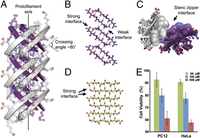
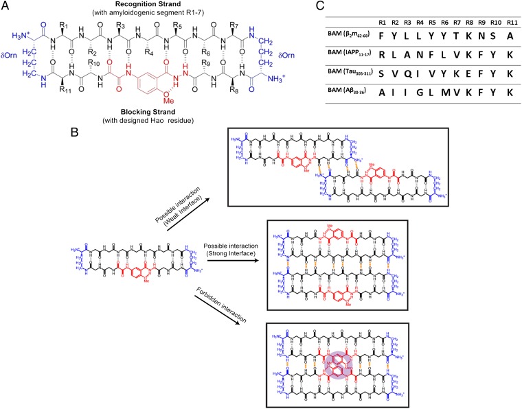
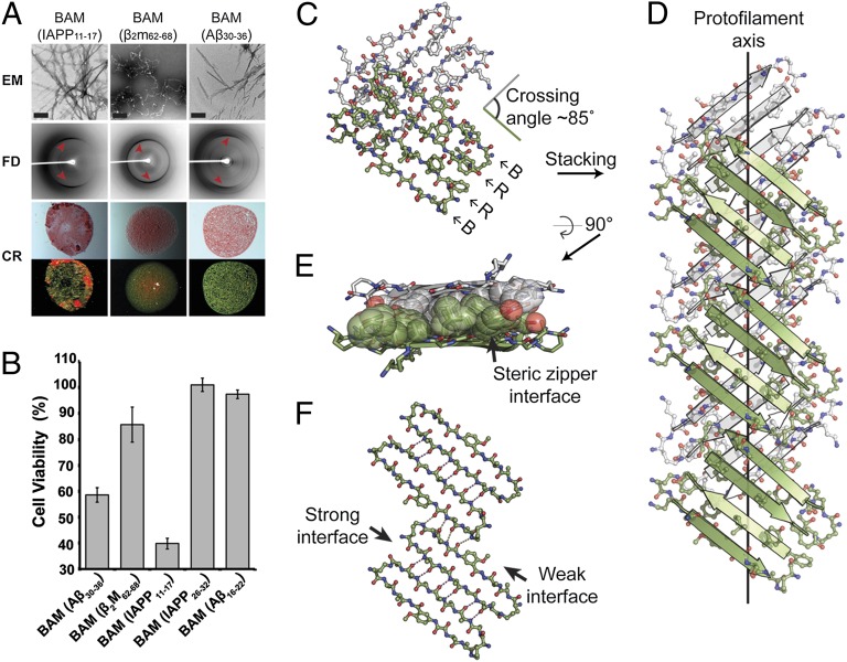
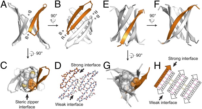
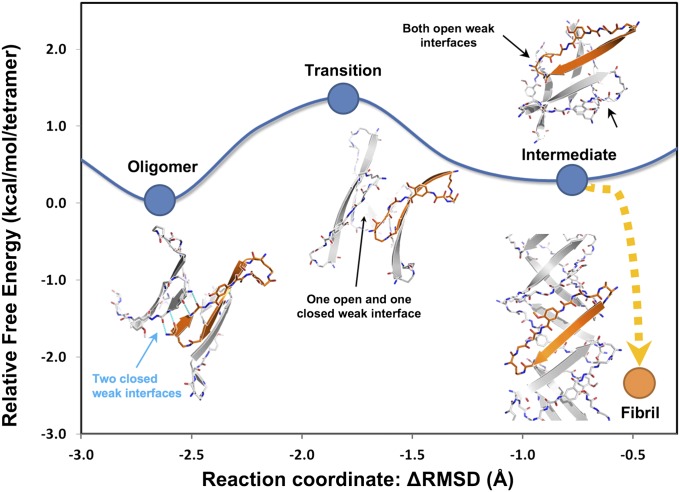
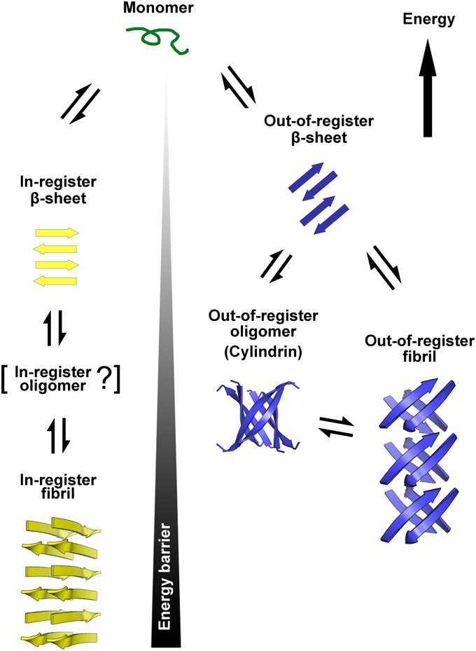
Although aberrant protein aggregation has been conclusively linked to dozens of devastating amyloid diseases, scientists remain puzzled about the molecular features that render amyloid fibrils or small oligomers toxic. Here, we report a previously unobserved type of amyloid fibril that tests as cytotoxic: one in which the strands of the contributing β-sheets are out of register. In all amyloid fibrils previously characterized at the molecular level, only in-register β-sheets have been observed, in which each strand makes its full complement of hydrogen bonds with the strands above and below it in the fibril. In out-of-register sheets, strands are sheared relative to one another, leaving dangling hydrogen bonds. Based on this finding, we designed out-of-register β-sheet amyloid mimics, which form both cylindrin-like oligomers and fibrils, and these mimics are cytotoxic. Structural and energetic considerations suggest that out-of-register fibrils can readily convert to toxic cylindrins. We propose that out-of-register β-sheets and their related cylindrins are part of a toxic amyloid pathway, which is distinct from the more energetically favored in-register amyloid pathway.


Conflict of interest statement

The authors declare no conflict of interest.

Figures

Fig. 1.
Fig. 1.
Crystal structure of out-of-register, antiparallel protofilament formed by the amyloidogenic peptide KDWSFY (β2m58–63) and its fibril toxicity. (A) View perpendicular to the protofilament axis. The two β-sheets are colored in purple and white. Side chains are shown in ball-and-stick representation. The protofilament axis is denoted by a black line. Note that the β-strands are antiparallel, out-of-register, and not perpendicular to the protofilament axis. The crossing angle between the β-strands of adjacent β-sheets is ∼80°. (B) The geometry of the back (purple) out-of-register β-sheet. For clarity, side chains are omitted. Hydrogen bonds are shown by dotted lines. (C) View down the protofilament axis showing the dry steric zipper interface between the pair of β-sheets. Side chains contributing to this interface are shown in space-filling representation. (D) The geometry of a single in-register antiparallel β-sheet from the KLVFFA crystal structure (Protein Data Bank ID code 2Y2A). (E) Assessment of cytotoxicity of KDWSFY (β2m58–63) fibril samples by MTT-based cell viability assay. KDWSFY (β2m58–63) fibrils show toxicity to both PC12 and HeLa cell lines in a dose-dependent manner. Error bars represent 1 SD (n = 4).
Fig. 2.
Fig. 2.
Structure and interaction of BAMs. (A) Structure of BAMs. The macrocyclic ring is connected by two ornithines colored in blue. The Hao residue, which can support intramolecular but not intermolecular hydrogen bonding, is colored in red. All natural amino acids are colored black. In BAM, the recognition strand (upper strand) contains 7 natural aa residues that are replaced with the various amyloidogenic segments in this study. The blocking strand (lower strand) contains 4 natural aa residues to facilitate the folding of the β-conformation and increase the solubility of the molecule. R10 is sometimes replaced with 4-bromo phenylalanine for phase determination of crystal structures. (B) Three types of potential BAM interactions are shown. (Top) Between blocking strands out of register, four intermolecular hydrogen bonds (colored in orange) can be formed. (Middle) Between recognition strands in register, eight intermolecular hydrogen bonds can be formed. (Lower) Between blocking strands in register, six intermolecular hydrogen bonds can be formed. However, the clash between Hao residues precludes this interaction. (C) Sequences of four different BAMs for out-of-register assembly studies.
Fig. 3.
Fig. 3.
Characterization of fibril-forming BAMs showing their amyloid-like character. (A) Negatively stained transmission electron micrographs (EM), fibril diffraction (FD), and Congo red staining (CR) of fibrils formed by BAM (IAPP11–17), BAM (β2m62–68), and BAM (Aβ30–36). Black bars in the EM images represent 200 nm. Meridional reflections corresponding to about 4.7 Å are highlighted by red arrowheads in FD images. CR of BAM fibril pellets shows apple-green birefringence. Pictures of the same view taken under normal (upper row) and polarized light (lower row) are shown. (B) MTT-based cytotoxicity assays of BAMs with mammalian cell lines PC12. Fibril-forming BAMs (left three) show significant cytotoxicity (P < 0.01) compared with nonfibril-forming BAMs [BAM (IAPP26–32) and BAM (Aβ16–22) as determined by one-sided Student t test] (Table S3). Error bars represent 1 SD calculated from four replicates. The concentration of each BAM is 50 μM (monomer equivalence). (C) The asymmetric unit of the BAM (β2m62–68) crystal. Two dimers of macrocycles are shown in green and white. Recognition (R) and blocking strands (B) are labeled. The crossing angle between the green and white strands is ∼85°. (D) View perpendicular to the protofilament axis. Note that the strands are not perpendicular to the protofilament axis and the lack of registration between dimers within a single sheet. (E) A view 90° rotated from D looking down the protofilament axis shows the steric zipper interface. Side chains contributing to the interface are shown in space-filling representation. For clarity, other side chains are omitted. (F) Geometry of a single sheet showing the out-of-register nature of the weak interface. Only backbone atoms are shown. Hydrogen bonds are shown by dotted lines.
Fig. 4.
Fig. 4.
Comparison of two cylindrin-like oligomers: crystal structures of BAM (Aβ30–36) and K11V (αB crystallin68–162). (A) The crystal structure of the fibril-forming BAM (Aβ30–36) in cartoon form shows a cylindrin-like oligomer containing four macrocycles, one of which is colored in orange. The view is oriented to show a weak interface between the blocking strands (B). (B) A view 90° rotated from A showing a strong interface between two recognition strands (R). (C) Another view 90° rotated from A showing the steric zipper-like interface in the center of the barrel. Side chains contributing to this interface are shown in space-filling representation. (D) Geometry of the cylindrin-like tetramer. Only backbone atoms are shown. Hydrogen bonds are shown by dotted lines. (E) Cartoon representation of cylindrin structure of K11V (Protein Data Bank ID code 3SGO) from αB crystallin. The view is oriented to show a weak interface analogous to A. (F) A view 90° rotated from E showing a strong interface analogous to B. (G) Another view 90° rotated from E showing the center of the cylindrin. Side chains contributing to the steric zipper-like core are shown in space-filling representation. (H) Geometry of K11V cylindrin. Pairs of hydrogen bonds between corresponding residues are shown by magenta (for strong interface) and black (for weak interface) dotted lines. Notice the out-of-register weak interfaces in both D and H.
Fig. 5.
Fig. 5.
Free energy landscape associated with the out-of-register aggregation pathway of BAM (Aβ30–36). The Gibbs free energy landscape of the structural conversion from the cylindrin-like, out-of-register oligomer state to out-of-register transition state to out-of-register intermediate state was explored by MD simulation with explicit water molecules. Representative structural models from the MD trajectories of each state are shown. The reaction coordinate (x axis) is indicated by Δrmsd from the coordinates of the starting and ending states (cylindrin structure and the pair of β-sheets structure). Over the process of the conversion, the hydrogen bonds in the strong interface are maintained. The transition of the conversion is initiated by disrupting the weak interface as shown in the transition state. As the cylindrin unrolls, the weak interface further dissociates (intermediate state), and the interface within the barrel is rearranged to the out-of-register fibril state. When two cylindrin-like oligomers were simulated to form an out-of-register fibril, the free energy dropped by ∼3 kcal/mol-of-tetramer (equivalent to ∼5 RT at 300 K), suggesting a facile conversion between out-of-register oligomers and fibrils. Furthermore, a small free energy difference between the out-of-register oligomer and fibril states indicates that both are relatively stable, consistent with their detection in solution and in crystals. In contrast, the free energy of the KDWSFY out-of-register oligomer is ∼20 kcal/mol-of-monomer higher than the KDWSFY out-of-register fibril (Fig. S3), consistent with our observation that only out-of-register fibrils could be detected in solution and crystals.
Fig. 6.
Fig. 6.
Schematic diagram of two distinct amyloid aggregation pathways. Two distinct pathways branch from monomers to fibrils: one through in-register β-strands (yellow) and the other through out-of register β-strands (blue). The in-register pathway features an in-register β-sheet that serves as the fundamental element, perhaps passing through an in-register intermediate oligomer that has not yet been characterized at the atomic level and then into the in-register fibril, which is widely observed and structurally characterized. The out-of-register pathway features an out-register β-sheet, out-of-register oligomer, and out-of-register fibril. The out-of-register oligomer and out-of-register fibril are structurally related by a common out-of-register β-sheet, suggesting easy interconversion between the two states. A high energy barrier requiring rearrangement of entire hydrogen-bonding networks separates the in-register and out-of-register β-sheet aggregation pathways. Because of their unsatisfied hydrogen bonds, out-of-register fibrils are generally less stable than in-register fibrils, and the energy differences separating out-of-register oligomers and fibrils from monomers are smaller than the corresponding differences for in-register fibrils. Hence, accumulation of in-register fibrils is common, but accumulation of out-of-register fibrils is rare.

References

    1. Eisenberg D, Jucker M. The amyloid state of proteins in human diseases. Cell. 2012;148(6):1188–1203. - PMC - PubMed
    1. Chiti F, Dobson CM. Protein misfolding, functional amyloid, and human disease. Annu Rev Biochem. 2006;75:333–366. - PubMed
    1. Härd T, Lendel C. Inhibition of amyloid formation. J Mol Biol. 2012;421(4–5):441–465. - PubMed
    1. Eichner T, Radford SE. A diversity of assembly mechanisms of a generic amyloid fold. Mol Cell. 2011;43(1):8–18. - PubMed
    1. Ma B, Nussinov R. Selective molecular recognition in amyloid growth and transmission and cross-species barriers. J Mol Biol. 2012;421(2–3):172–184. - PMC - PubMed

Publication types

LinkOut - more resources