. 2012 Dec 18;109(51):20907-12.
doi: 10.1073/pnas.1211872110. Epub 2012 Dec 3.

Complete fiber structures of complex trimeric autotransporter adhesins conserved in enterobacteria

Affiliations

Complete fiber structures of complex trimeric autotransporter adhesins conserved in enterobacteria

Marcus D Hartmann et al. Proc Natl Acad Sci U S A. .

Abstract

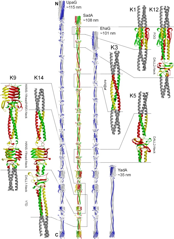
Trimeric autotransporter adhesins (TAAs) are modular, highly repetitive surface proteins that mediate adhesion to host cells in a broad range of Gram-negative pathogens. Although their sizes may differ by more than one order of magnitude, they all follow the same basic head-stalk-anchor architecture, where the head mediates adhesion and autoagglutination, the stalk projects the head from the bacterial surface, and the anchor provides the export function and attaches the adhesin to the bacterial outer membrane after export is complete. In complex adhesins, head and stalk domains may alternate several times before the anchor is reached. Despite extensive sequence divergence, the structures of TAA domains are highly constrained, due to the tight interleaving of their constituent polypeptide chains. We have therefore taken a "domain dictionary" approach to characterize representatives for each domain type by X-ray crystallography and use these structures to reconstruct complete TAA fibers. With SadA from Salmonella enterica, EhaG from enteropathogenic Escherichia coli (EHEC), and UpaG from uropathogenic E. coli (UPEC), we present three representative structures of a complex adhesin that occur in a conserved genomic context in Enterobacteria and is essential in the infection process of uropathogenic E. coli. Our work proves the applicability of the dictionary approach to understanding the structure of a class of proteins that are otherwise poorly tractable by high-resolution methods and provides a basis for the rapid and detailed annotation of newly identified TAAs.


Conflict of interest statement

The authors declare no conflict of interest.

Figures

Fig. 1.
Fig. 1.
Electron microscopy of SadA. Transmission (A and C) and scanning electron micrographs (B and D) of E. coli Top10 carrying an overexpression vector for Salmonella typhimurium SadA (C and D) or an empty vector as control (A and B). Cells were grown in liquid culture and immobilized on polylysine-coated coverslips. In A and C, cells were labeled after immobilization with an antibody raised against SadA and affinity purified against SadAK9 and chemically fixated by using glutaraldehyde. (Scale bars: 1 µm.)
Fig. 2.
Fig. 2.
Crystal structures of fragments and reconstructed full fibers of SadA, UpaG, and EhaG. The three chains of the SadA trimers are colored individually; the coiled-coil adaptors fused to the fragments are shown in gray. Only one of the two structures obtained for SadAK9 (K9cfI) is shown. The reconstructed fibers are compared with a prototypical simple trimeric adhesin, YadA. Indicated lengths are measured on the atomic coordinates of the models and include the membrane anchor.
Fig. 3.
Fig. 3.
Neck domains in context with different upstream domains. (AC) Neck domains in SadA in context with different upstream domains. One chain of each trimer is colored individually. The neck (thick lines) forms a continuous β-sheet with the last β-strand of the respective upstream domain (transparently thick). Structurally invariant water molecules are in black. (A) Long neck following a DALL2 domain in SadAK5. (B) Short neck following a Ylhead domain in SadAK14. (C) Short neck following a HIM3 domain in SadAK9. (D) Superposition of nine known neck structures as indicated in H, solved in context with their native upstream domain, colored by trimer. The insertion sequence in the neck of Hia (1S7M) was cut for clarity. (E) Top view of D. (F) Head insert motifs HIM2 and HIM3 fold tightly around their respective neck: Superimposition of B and C (HIM3) with a HIM2-neck domain, highlighting the head insert motif (thick lines). (G) Close up of the β-layer in the neck of SadAK5, highlighting the hydrogen bonding network that involves the central water molecule. (H) Sequence alignment of neck sequences, highlighting the central β-layer residue (mostly valine) from the DAVN consensus sequence.
Fig. 4.
Fig. 4.
The DALL domain. (A) Structure of the DALL1 domain in SadAK14 together with the downstream neck domain. One chain is colored yellow; the DALL1 domain is drawn in thick lines. The central water molecule of the DALL1 and the water molecules interacting mostly with the yellow chain are drawn in red, the central water of the neck in black, and others in gray. Notably, the N-terminal coiled coil is kinked with respect to the trimer axis of the DALL1-neck tandem. (B) Close-up of the β-sheet on one face of the DALL1-neck tandem. Compared with DALL2 in E, the β-sheet of the DALL1 is invaded by bridging water molecules. (C) Top view of the DALL1 domain, highlighting the β-layer interactions. Although the backbone interactions of the central β-layer residues are almost undisturbed, the coordination of the central water molecule is asymmetric because of the kink of the coiled coil. (D) Structure of the DALL2 domain in SadAK12 together with the downstream neck domain. One chain is colored green; the DALL2 domain is drawn in thick. The central water molecule and the water interacting with the green chain are shown in red, the central water molecule of the neck in black, and others in gray. Superposed in thin black is the structure of the DALL2-neck tandem in SadAK5. (E) Close-up of the continuous β-sheet formed between the DALL2 and neck domain. (F) Top view of the DALL2 domain in SadAK12, highlighting the interchain interaction between the conserved Tryptophan and Histidine and the β-layer interactions. (G) Superposition of the DALL1 structure of SadAK14 and the DALL2 structure of SadAK12 on the C-terminal halves. Because of the waters invading the β-sheet in DALL1, the spacing of the β-strands differs between the two structures. The N-terminal coiled-coil of DALL1 in SadAK14 is 10° kinked with respect to the symmetric DALL2 in SadAK12. (H) Sequence alignment of DALL1 and DALL2 domains, highlighting the central β-layer residues and the conserved residues in interchain interactions.
Fig. 5.
Fig. 5.
β-Layers as universal adaptors. The structure of β-layers in three different contexts, highlighting the β-interactions of the three chains around the central water molecule. (A) Side and top views are shown for the β-layer in a DALL domain (Left; DALL2 from SadAK12) leading from alpha to beta, a β-layer in a neck domain (Right; from SadAK12), leading from beta to alpha, and a β-layer separating two coiled-coil segments, leading form alpha to alpha, from structure 2BA2 (Center). B and C show a superposition of the three different β-layers in side and top view, respectively. The superposition is based on the central β-layer residues and the central water molecule. With a total of 8 DALL and 12 neck domains, the whole SadA fiber comprises 20 β-layers.
Fig. 6.
Fig. 6.
The HANS domain. (A) Side view of the last HANS domain in SadA from structure K9cfI, second in the alignment in G. The dashed line indicates where the N-terminal coiled coil breaks into a second, distorted segment. (B) Top view of the same HANS domain, showing the side chains of the second β-strand. (C and D) Superposition of all structures of the last HANS domain of SadA (K9cfI black, K9cfII gray, K14 light blue) with the HANS from 3LAA (salmon) on the KYFHANS consensus motif, illustrating the conformational flexibility of the N-terminal helical segment. C shows a side view of a superposition of all individual chains, and D shows the top view of the full trimers. (E and F) Superposition of two different conformations of the last HANS domain in SadA (from K9cfI and from K9cfII) onto the coiled coil above the distorted segment (indicated by the dashed line). Between the two conformations, the C-terminal Ylhead is rotated by 25° with respect to the N-terminal coiled-coil. (G) Sequence alignment of HANS domains.
Fig. 7.
Fig. 7.
FGG domain and NxYTD motif. Side (A) and top (C) view of the FGG domain. The β-hairpins inserted between the coiled-coil segments fold around the bundle like a collar and cause a 120° rotation of the helical bundle. (B) Directly at the junction of the two coiled-coil segments, the N-terminal helices appear to pass seamlessly into to the C-terminal helices. (D) Sequence alignment of FGG domains. (E and F) Side and top view of the NxYTD motif in SadAK14, highlighting hydrogen bonding interactions and the invariant water network. Before reaching the anchor, many TAAs contain a right-handed coiled-coil segment in the stalk that contains a YTD sequence motif at the transition to the final left-handed segment; as described recently for YadA (20), this motif forms an elaborate network of hydrogen bonds: The threonines of the three chains form hydrogen bonds with each other in the core via their hydroxyl groups, whereas the tyrosines and aspartates form interchain hydrogen bonds around the bundle. Careful analysis however shows that the extent of the network is even larger. In more than half of all occurrences, YTD is preceded by an asparagine two residues before the tyrosine, yielding the motif NxYTD. These asparagines, together with the backbone oxygen of the residue preceding them and the threonines, coordinate three water molecules in the core of the bundle. Inspection of the YadA structures and their experimental data reveals that the three water molecules are also present in the two NxYTD motifs in YadA, albeit not modeled consistently.

Similar articles

Cited by

References

    1. Gerlach RG, Hensel M. Protein secretion systems and adhesins: The molecular armory of Gram-negative pathogens. Int J Med Microbiol. 2007;297(6):401–415. - PubMed
    1. Hoiczyk E, Roggenkamp A, Reichenbecher M, Lupas A, Heesemann J. Structure and sequence analysis of Yersinia YadA and Moraxella UspAs reveal a novel class of adhesins. EMBO J. 2000;19(22):5989–5999. - PMC - PubMed
    1. Linke D, Riess T, Autenrieth IB, Lupas A, Kempf VA. Trimeric autotransporter adhesins: Variable structure, common function. Trends Microbiol. 2006;14(6):264–270. - PubMed
    1. Leo JC, Grin I, Linke D. Type V secretion: Mechanism(s) of autotransport through the bacterial outer membrane. Philos Trans R Soc Lond B Biol Sci. 2012;367(1592):1088–1101. - PMC - PubMed
    1. Bölin I, Norlander L, Wolf-Watz H. Temperature-inducible outer membrane protein of Yersinia pseudotuberculosis and Yersinia enterocolitica is associated with the virulence plasmid. Infect Immun. 1982;37(2):506–512. - PMC - PubMed

Publication types

MeSH terms

LinkOut - more resources