. 2012 Dec 18;109(51):20820-5.
doi: 10.1073/pnas.1214753109. Epub 2012 Dec 3.

F-actin buckling coordinates contractility and severing in a biomimetic actomyosin cortex

Affiliations

F-actin buckling coordinates contractility and severing in a biomimetic actomyosin cortex

Michael P Murrell et al. Proc Natl Acad Sci U S A. .

Abstract

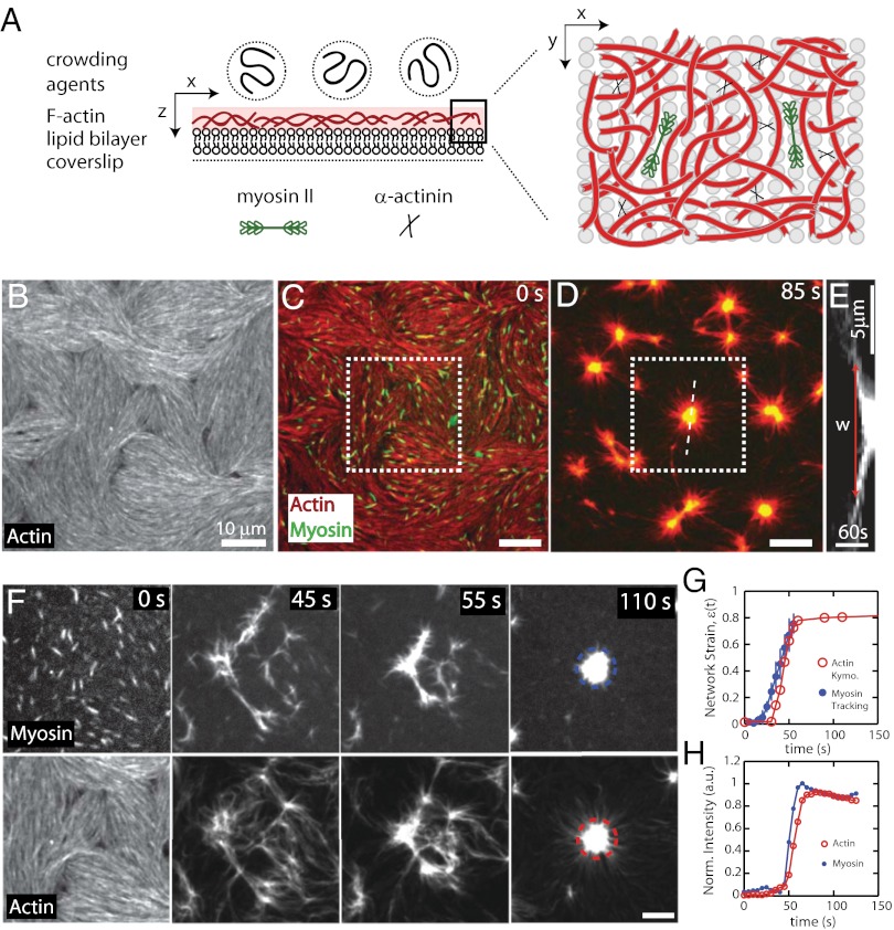
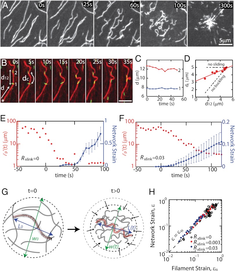
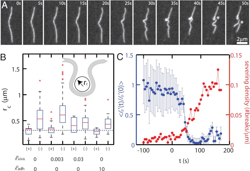
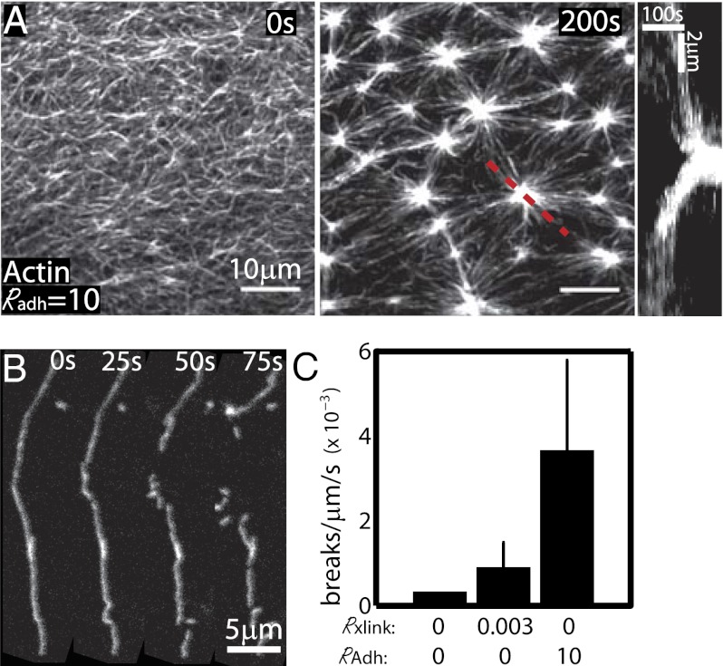
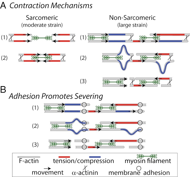
Here we develop a minimal model of the cell actomyosin cortex by forming a quasi-2D cross-linked filamentous actin (F-actin) network adhered to a model cell membrane and contracted by myosin thick filaments. Myosin motors generate both compressive and tensile stresses on F-actin and consequently induce large bending fluctuations, which reduces their effective persistence length to <1 μm. Over a large range of conditions, we show the extent of network contraction corresponds exactly to the extent of individual F-actin shortening via buckling. This demonstrates an essential role of buckling in breaking the symmetry between tensile and compressive stresses to facilitate mesoscale network contraction of up to 80% strain. Portions of buckled F-actin with a radius of curvature ~300 nm are prone to severing and thus compressive stresses mechanically coordinate contractility with F-actin severing, the initial step of F-actin turnover. Finally, the F-actin curvature acquired by myosin-induced stresses can be further constrained by adhesion of the network to a membrane, accelerating filament severing but inhibiting the long-range transmission of the stresses necessary for network contractility. Thus, the extent of membrane adhesion can regulate the coupling between network contraction and F-actin severing. These data demonstrate the essential role of the nonlinear response of F-actin to compressive stresses in potentiating both myosin-mediated contractility and filament severing. This may serve as a general mechanism to mechanically coordinate contractility and cortical dynamics across diverse actomyosin assemblies in smooth muscle and nonmuscle cells.


Conflict of interest statement

The authors declare no conflict of interest.

Figures

Fig. 1.
Fig. 1.
Reconstitution of a model contractile actomyosin cortex. (A) Schematic illustration of reconstituted system. F-actin is crowded to the surface of a supported lipid bilayer (SLB) after which cross-linking proteins and skeletal muscle myosin II filaments are added. Data in B–H are from 1 μM F-actin (100% labeled) crowded to the surface of 91% EPC/9% nickel-tagged lipid (NTA) (mol/mol) SLB with Rxlink = 0 and RAdh = 0. (B) Alexa-568–labeled F-actin at the SLB surface. (C and D) F-actin (red) and myosin II (green) (C) immediately (0 s) after myosin thick filament formation and (D) 85 s after myosin filaments assembled (t = 0 s). (E) Kymograph of the actin over time taken at the white dotted line in D with the width of the contractile zone w indicated. (F) Magnified images of myosin and F-actin from square region indicated in C during contraction. Myosin filaments appear at 0 s. (G) Contractile strain of the actin network from the kymograph in E. (H) Average normalized myosin and actin fluorescence intensity within the center of contraction, indicated in F.
Fig. 2.
Fig. 2.
Myosin-induced F-actin buckling occurs concomitantly with contraction. (A) Images of individual F-actin (1–2% labeled) during contraction of sample with Rxlink = 0 and RAdh = 0 (Movie S10). Myosin thick filaments form at 0 s. Data in B–D are from sample with Rxlink = 0.003 and RAdh = 0 (Movie S11). (B) Myosin (green) translocating along F-actin (red) where d is the distance of myosin from F-actin barbed end (*), dc is length of F-actin between myosin punctae, and d12 is the Euclidean distance between myosin thick filaments. (C) Distance d of myosin punctae from the barbed end of F-actin over time. (D) dc as a function of d12, with dashed lines indicating the results for changes expected due to buckling, with no actomyosin sliding (no sliding), and those for relative sliding, with no F-actin buckling (no buckling). (E and F) Persistence length, lp′ (red) and network strain (blue) for (E) Rxlink = 0 (RAdh = 0) (Movie S10) and (F) Rxlink = 0.03 (RAdh = 0) (Movie S9). The lp′ data are for a single F-actin, and the network strain reflects the average of four myosin pairs in E and five in F, each within a single experiment. (G) Schematic indicating measurement of filament compressive strain, determined by tracking changes in end-to-end length of single filaments, and network contractile strain, determined by changes in the size of the network. (H) Network strain ɛ as a function of filament strain ɛfil during contraction for Rxlink = 0, 0.003, and 0.03 (RAdh = 0). Dashed line indicates ɛ = ɛfil. The data are the average of four myosin pairs and nine filaments for Rxlink = 0, seven myosin pairs and six filaments for Rxlink = 0.003, and five myosin pairs and seven filaments for Rxlink = 0.03, each for a single experiment.
Fig. 3.
Fig. 3.
Filament buckling at high curvature induces severing. (A) F-actin images during contraction of a sparsely labeled network (Rxlink = 0, RAdh = 0). (B) Box plot of the filament radii of curvature rc measured preceding a severing event (+) or not (−). Dashed line indicates 300 nm. The sample sizes for the different conditions are as follows: Rxlink = 0/Radh = 0 (Nsever = 14, Nstable = 58), Rxlink = 0.003/Radh = 0 (Nsever = 22, Nstable = 123), Rxlink = 0.03/Radh = 0 (Nsever = 4, Nstable = 58), and Rxlink = 0/RAdh = 10 (Nsever = 10, Nstable = 21). Nsever is the number of measurements of rc taken that sever in the following time point. Nstable is the number of rc measurements, selected at random, that do not sever in the next time point. Data reflect eight independent experiments. (Inset) Schematic of radius of curvature. (C) Persistence length normalized to initial value before contraction (blue) and mean severing density as a function of time (red) for Rxlink = 0.003/RAdh = 0. The data reflect the curvature of 5 filaments (lp′) and the severing (Nsever) of 102 filaments from a single experiment.
Fig. 4.
Fig. 4.
Membrane adhesion modulates mechanically induced severing. (A) F-actin images in RAdh = 10, Rxlink = 0. (Left) Precontraction, (Middle) postcontraction, and (Right) a kymograph of actin intensity for red dashed line. (B) Individual F-actin during contraction of a sparsely labeled network with RAdh = 10, Rxlink = 0. (C) Severing rates for Rxlink = 0/Radh = 0 (Nfil = 48, Nexpt = 1), Rxlink = 0.003 (Nfil = 204, Nexpt = 4), and Radh = 10 (Nfil = 133, Nexpt = 3). Nfil is the total number of filaments observed through a total of Nexp independent experiments.
Fig. 5.
Fig. 5.
Schematics of contraction mechanisms. (A) In sarcomeres, myosin filaments (green) are segregated toward pointed ends of F-actin and cross-links (black “x”) are at the barbed end. This configuration permits solely tensile forces (red) and, thus, translocation of actin filaments (black arrows), resulting in contraction. In disordered, nonsarcomeric actomyosin bundles and networks that lack segregation of motors and cross-links, actomyosin interactions result in both tension and compression (blue). Compressive stresses are relieved through filament buckling and severing, keeping only tensile forces and, thus, driving contraction. (B) Cross-links to the membrane (open circles) spatially constrain stresses generated by motors and thus promote severing and prevent long-range transmission of tensile stresses. Numbers delineate sequential steps in each process.

Similar articles

Cited by

References

    1. Bray D, White JG. Cortical flow in animal cells. Science. 1988;239(4842):883–888. - PubMed
    1. Guha M, Zhou M, Wang YL. Cortical actin turnover during cytokinesis requires myosin II. Curr Biol. 2005;15(8):732–736. - PubMed
    1. Medeiros NA, Burnette DT, Forscher P. Myosin II functions in actin-bundle turnover in neuronal growth cones. Nat Cell Biol. 2006;8(3):215–226. - PubMed
    1. Wilson CA, et al. Myosin II contributes to cell-scale actin network treadmilling through network disassembly. Nature. 2010;465(7296):373–377. - PMC - PubMed
    1. Vicente-Manzanares M, Ma X, Adelstein RS, Horwitz AR. Non-muscle myosin II takes centre stage in cell adhesion and migration. Nat Rev Mol Cell Biol. 2009;10(11):778–790. - PMC - PubMed

Publication types