. 2012 Dec 18;109(51):21146-51.
doi: 10.1073/pnas.1210789109. Epub 2012 Dec 4.

Arabidopsis lateral organ boundaries negatively regulates brassinosteroid accumulation to limit growth in organ boundaries

Affiliations

Arabidopsis lateral organ boundaries negatively regulates brassinosteroid accumulation to limit growth in organ boundaries

Elizabeth M Bell et al. Proc Natl Acad Sci U S A. .

Abstract

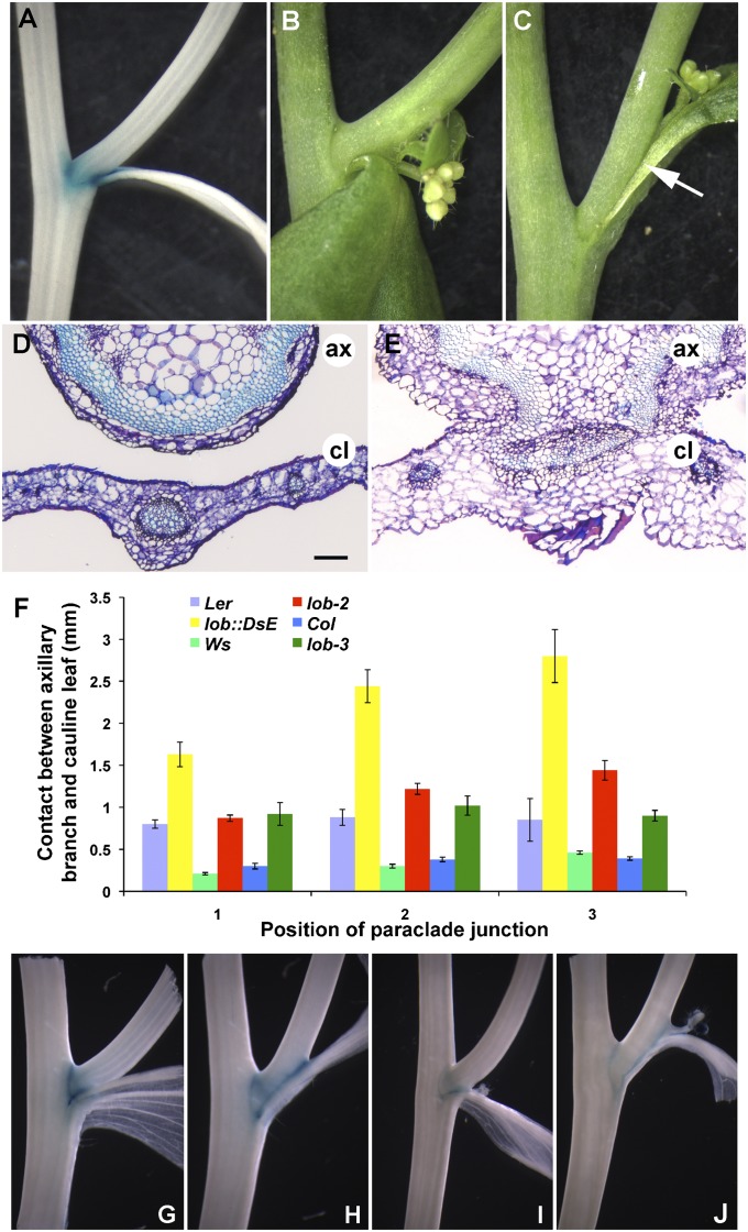
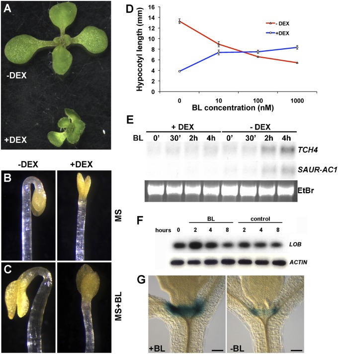
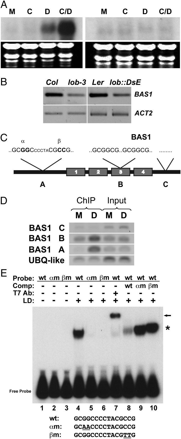
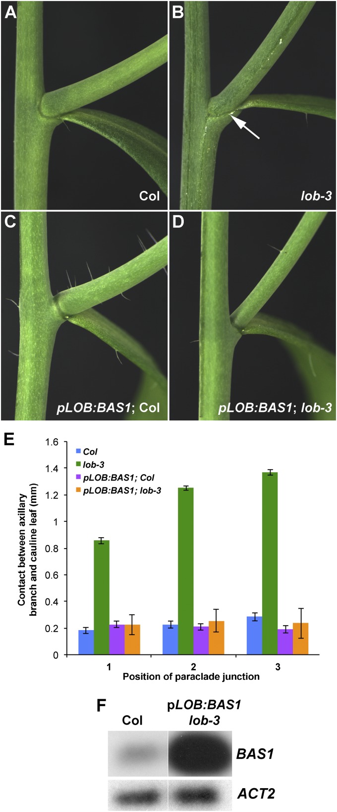
Leaves and flowers begin life as outgrowths from the edges of shoot apical meristems. Stem cell divisions in the meristem center replenish cells that are incorporated into organ primordia at the meristem periphery and leave the meristem. Organ boundaries, regions of limited growth that separate forming organs from the meristem, serve to isolate these two domains and are critical for coordination of organogenesis and meristem maintenance. Boundary formation and maintenance are poorly understood processes, despite the identification of a number of boundary-specific transcription factors. Here we provide genetic and biochemical evidence that the Arabidopsis thaliana transcription factor lateral organ boundaries (LOB) negatively regulates accumulation of the plant steroid hormone brassinosteroid (BR) in organ boundaries. We found that ectopic expression of LOB results in reduced BR responses. We identified BAS1, which encodes a BR-inactivating enzyme, as a direct target of LOB transcriptional activation. Loss-of-function lob mutants exhibit organ fusions, and this phenotype is suppressed by expression of BAS1 under the LOB promoter, indicating that BR hyperaccumulation contributes to the lob mutant phenotype. In addition, LOB expression is BR regulated; therefore, LOB and BR form a feedback loop to modulate local BR accumulation in organ boundaries to limit growth in the boundary domain.


Conflict of interest statement

The authors declare no conflict of interest.

Figures

Fig. 1.
Fig. 1.
Mutations in LOB result in organ fusion. (A) GUS activity in pLOB:GUS between the main stem and axillary stem and between the axillary stem and cauline leaf. (B and C) Paraclade junction between main stem, axillary stem, and cauline leaf in wild-type (B) and lob::DsE (C) plants. The axillary stem and cauline leaf are separated in wild type and fused in lob::DsE (white arrow). (D and E) Cross sections through junction between axillary stem (ax) and cauline leaf (cl) in wild type (D) and lob::DsE (E). (F) Length of fused region in wild-type Landsberg erecta, lob::DsE, wild-type Wassilewskija, lob-2, wild-type Columbia, and lob-3. Position 1 corresponds to lowest cauline leaf axil on stem. SEs are indicated; n ≥ 11 for positions 1 and 2, and n ≥ 5 for position 3 (not all plants have three paraclade junctions). (G–J) GUS expression in boundary marker lines ET4016 (11) (G and H) and GT185 (34) (I and J) in wild type (G and I) and lob (H and J). ET4016 reports expression of LOF1 (11) and GT185 reports expression of ORGAN BOUNDARY1 (13). Expression of both markers is extended throughout the fused region in lob mutants, indicating an expansion or overgrowth of the boundary domain. (Scale bar in D, 100 μm for D and E.)
Fig. 2.
Fig. 2.
Ectopic LOB expression disrupts brassinosteroid responses. (A) 35S:LOB-GR plants are dwarfed when grown on 3 μM DEX. (B and C) Dark-grown, 4-d-old 35S:LOB-GR seedlings produce an apical hook when grown on MS medium in the absence of DEX (−DEX) and lack an apical hook when grown in the presence of 3 μM DEX (+DEX). Apical hook formation is not restored by addition of 100 nM epi-brassinolide in the medium (C; MS+BL). (D) Hypocotyl lengths of 35S:LOB-GR seedlings grown in the dark on increasing concentrations of epi-brassinolide (BL) in the presence or absence of 3 μM DEX. SEs (n ≥ 15) are indicated. (E) Northern blot analyses of TCH4 and SAUR-AC1 transcripts. Seven-day-old 35S:LOB-GR seedlings were pretreated overnight in the presence or absence of 3 μM DEX and then incubated in the presence of 1 μM epi-brassinolide (BL) for the indicated times. (F) RT-PCR analysis of LOB transcript levels in 7-d-old wild-type seedlings following treatment with 1 μM epi-brassinolide (BL) for 2, 4, or 8 h. RT-PCR products were detected by blotting and probing with gene-specific probes, following either 15 (LOB) or 12 cycles (ACTIN) of amplification. (G) GUS activity in pLOB:GUS:LOB-3′IGR seedlings after 3-h incubation in liquid MS supplemented with (Left) or without (Right) 1 μM BL. (Scale bar in G and H, 100 μm.)
Fig. 3.
Fig. 3.
BAS1 is a direct target of LOB. (A) Northern blot analyses of BAS1 transcript levels in 35S:LOB-GR (Left) and Columbia wild-type (Right) 8-d-old seedlings following 4-h mock (M), cycloheximide (C), DEX (D), or cycloheximide plus DEX (C/D) treatment. (B) RT-PCR analysis of BAS1 transcript levels in dissected cauline leaf-axillary stem junctions of Col, lob-3, Ler, and lob::DsE. RT-PCR products were detected by blotting and probing with gene-specific probes, following either 15 (BAS1) or 12 cycles (ACT2) of amplification. (C) Cartoon of the genomic structure of BAS1 showing the locations of LBD motifs and regions tested for enrichment after ChIP. BAS1 A contained two partial LBD sites separated by five nucleotides, 306 bp upstream of the ATG. BAS1 B contained two full LBD sites separated by 46 nucleotides, 1,700 bp downstream of the ATG in the third exon. The BAS1 C region contained no LBD sites and was 2,900 bp downstream of the ATG. (D) PCR products were amplified from DNA obtained before (Input) and after (ChIP) collection of specific LOB-DNA complexes by a LOB 1° antibody. DEX (D) and mock-treated (M) 35S:LOB-GR plants were used in ChIP experiments. BAS1 A and B regions were amplified for 27 cycles, and BAS1 C was amplified for 38 cycles. Thirty cycles of amplification were performed with the control gene UBQ-LIKE (At3g26980). (E) The LOB domain (LD) of LOB was incubated with a 133-bp radiolabeled probe generated from the BAS1 A region and separated on a native polyacrylamide gel. Probes contained unmodified LBD motifs (wt; lanes 1, 4, and 7–10), a mutation in the 5′-most motif in which the central GG residues were mutated to AA (αm; lanes 2 and 5), or a mutation in the 3′-most motif in which the central GG residues were mutated to AA (βm; lanes 3 and 6). Inclusion of T7 Ab against the tag on LD results in a supershift, demonstrating that recombinant LD protein bound the wild-type probe (lane 7). An excess of cold wild-type αm or βm DNA was used in competition experiments to demonstrate specificity of binding (lanes 8–10). The sequence of the central motif in the wt and mutant probes is shown.
Fig. 4.
Fig. 4.
Expression of BAS1 under the LOB promoter suppresses fusion in the lob mutant. (A–D) Cauline leaf/axillary stem junctions of Columbia wild type (A), lob-3 (B), transgenic pLOB:BAS1 in Col (C), and pLOB:BAS1 in lob-3 (D). Arrow in B indicates fused region. (E) Length of fused region in Col, lob-3, pLOB:BAS1 Col, and pLOB:BAS1 lob-3. Position 1 corresponds to lowest cauline leaf axil on stem. Expression of pLOB:BAS1 suppresses the fusion in lob-3. SEs (n ≥ 10) are indicated. (F) RT-PCR analysis of BAS1 transcript levels in isolated paraclade junctions of Col and pLOB:BAS1 lob-3 plants. RT-PCR products were detected by blotting and probing with gene-specific probes, following either 15 (BAS1) or 12 cycles (ACT2) of amplification.

Comment in

References

    1. Barton MK. Twenty years on: The inner workings of the shoot apical meristem, a developmental dynamo. Dev Biol. 2010;341(1):95–113. - PubMed
    1. Hussey G. Cell division and expansion and resultant tissue tensions in shoot apex during formation of a leaf primordium in tomato. J Exp Bot. 1971;22:702–714.
    1. Breuil-Broyer S, et al. High-resolution boundary analysis during Arabidopsis thaliana flower development. Plant J. 2004;38(1):182–192. - PubMed
    1. Rast MI, Simon R. The meristem-to-organ boundary: More than an extremity of anything. Curr Opin Genet Dev. 2008;18(4):287–294. - PubMed
    1. Aida M, Ishida T, Fukaki H, Fujisawa H, Tasaka M. Genes involved in organ separation in Arabidopsis: An analysis of the cup-shaped cotyledon mutant. Plant Cell. 1997;9(6):841–857. - PMC - PubMed

Publication types

Associated data