

BEST1 sequence variants in Italian patients with vitelliform macular dystrophy
- PMID: 23213274
- PMCID: PMC3513188
BEST1 sequence variants in Italian patients with vitelliform macular dystrophy
Abstract
Purpose: To analyze the spectrum of sequence variants in the BEST1 gene in a group of Italian patients affected by Best vitelliform macular dystrophy (VMD).
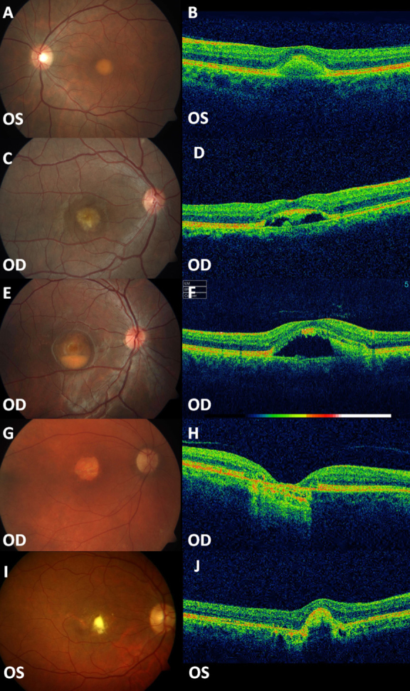
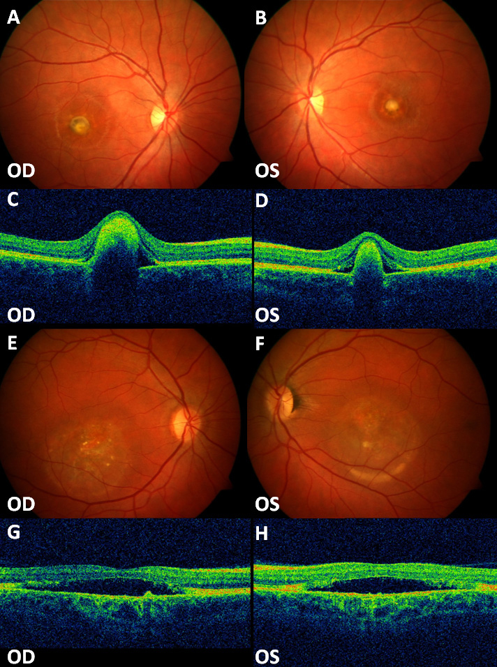
Methods: Thirty Italian patients with a diagnosis of VMD and 20 clinically healthy relatives were recruited. They belonged to 19 Italian families predominantly originating from central Italy. They received a standard ophthalmologic examination, OCT scan, and electrophysiological tests (ERG and EOG). Fluorescein and ICG angiographies and fundus autofluorescence imaging were performed in selected cases. DNA samples were analyzed for sequence variants of the BEST1 gene by direct sequencing techniques.
Results: Nine missense variants and one deletion were found in the affected patients; each patient carried one mutation. Five variants [c.73C>T (p.Arg25Trp), c.652C>T (p.Arg218Cys), c.652C>G (p.Arg218Gly), c.728C>T (p.Ala243Val), c.893T>C (p.Phe298Ser)] have already been described in literature while another five variants [c.217A>C (p.Ile73Leu), c.239T>G (p.Phe80Cys), c.883_885del (p.Ile295del), c.907G>A (p.Asp303Asn), c.911A>G (p.Asp304Gly)] had not previously been reported. Affected patients, sometimes even from the same family, occasionally showed variable phenotypes. One heterozygous variant was also found in five clinically healthy relatives with normal fundus, visual acuity and ERG but with abnormal EOG.
Conclusions: Ten variants in the BEST1 gene were detected in a group of individuals with clinically apparent VMD, and in some clinically normal individuals with an abnormal EOG. The high prevalence of novel variants and the frequent report of a specific variant (p.Arg25Trp) that has rarely been described in other ethnic groups suggests a distribution of BEST1 variants peculiar to Italian VMD patients.
Figures


References
-
- Gass JDM. Best’s Disease. in: Stereoscopic Atlas of Macular Disease. Diagnosis and Treatment. Mosby. St.Louis-London-Philadelphia-Sydney-Toronto, 1997, pp.304–313.
-
- Deutman AF, Hoyng CB. Macular Dystrophies. in: Retina. Ryan SJ Ed. Mosby. St.Louis-London-Philadelphia-Sydney-Toronto, 2001, pp. 1210–1257.
-
- Souied E, Kaplan J, Coscas G, Soubrane G. Les dystrophies maculaires. J Fr Ophtalmol. 2003;26:743–62. - PubMed
-
- Best F. Uber eine hereditare Maculaaffektion. Beitrag zur Vererbungslehre. Z Augenheilk. 1905;13:199–212.
-
- Andrade RE, Farah ME, Costa RA. Photodynamic therapy with verteporfin for subfoveal choroidal neovascularization in Best disease. Am J Ophthalmol. 2003;136:1179–81. - PubMed
MeSH terms
Substances
LinkOut - more resources
Full Text Sources