. 2012 Jan 15;1(1):19-29.
doi: 10.1242/bio.2011013. Epub 2011 Oct 24.

Transcription factor NF-Y is involved in differentiation of R7 photoreceptor cell in Drosophila

Affiliations

Transcription factor NF-Y is involved in differentiation of R7 photoreceptor cell in Drosophila

Yasuhide Yoshioka et al. Biol Open. .

Abstract

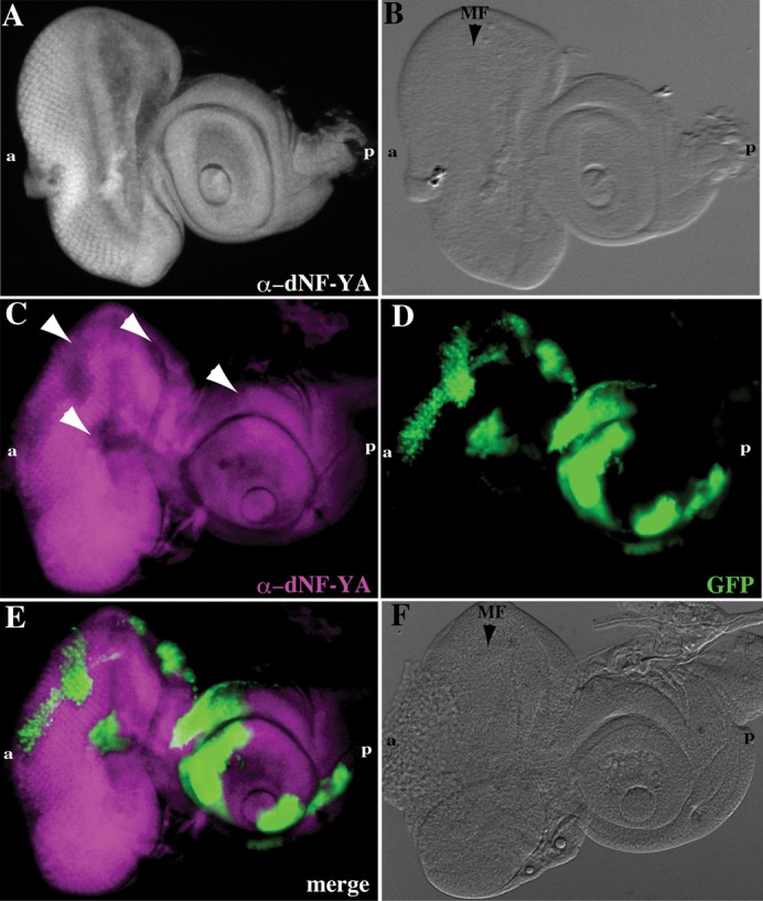
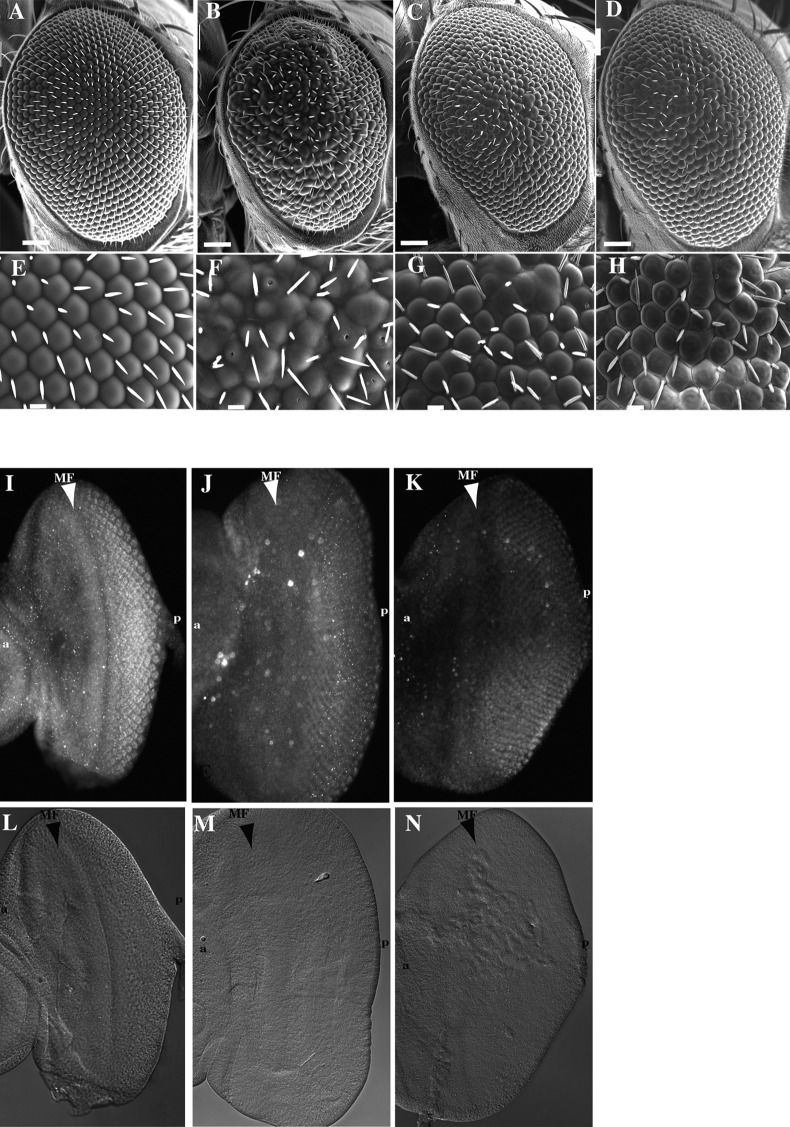
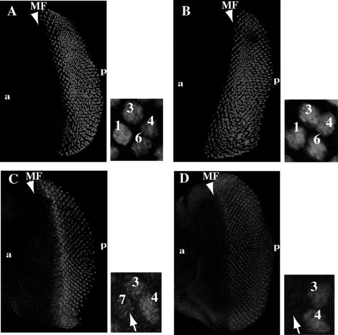
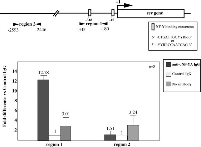
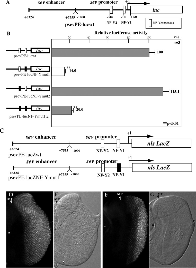
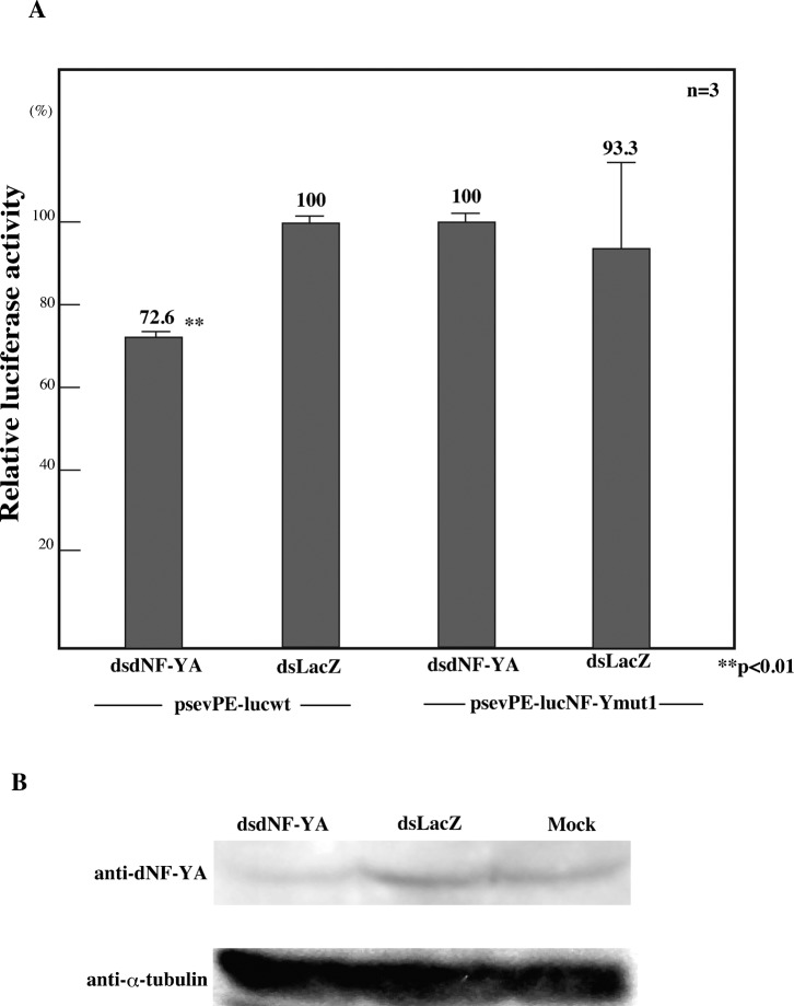
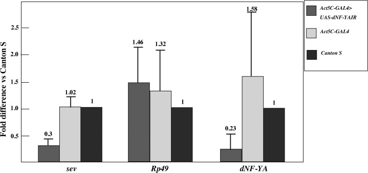
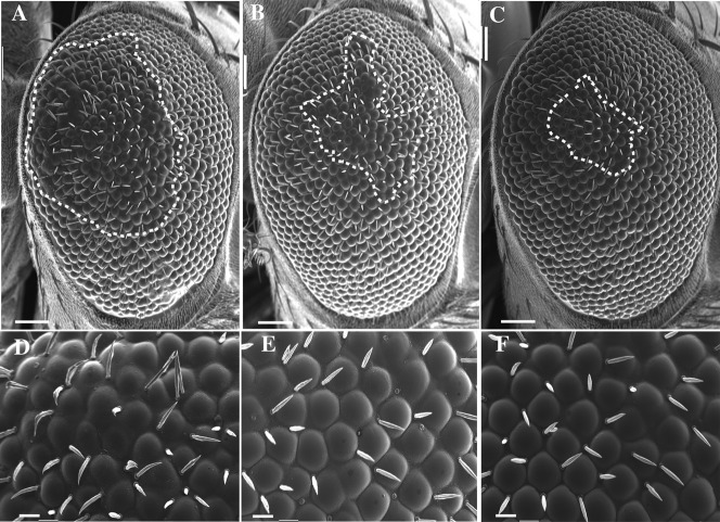
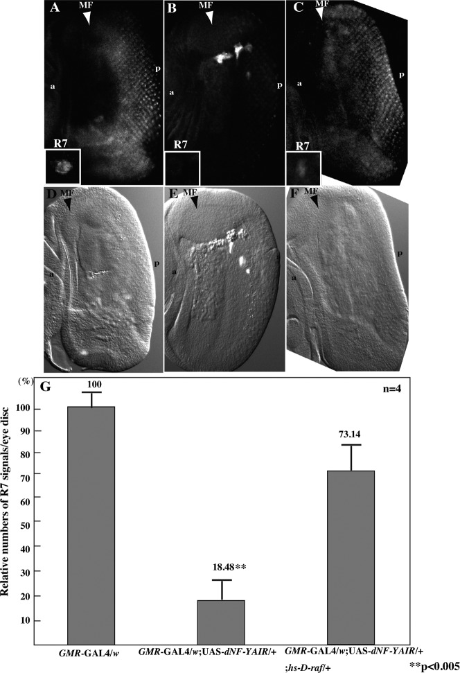
The CCAAT motif-binding factor NF-Y consists of three different subunits, NF-YA, NF-YB and NF-YC. Knockdown of Drosophila NF-YA (dNF-YA) in eye discs with GMR-GAL4 and UAS-dNF-YAIR resulted in a rough eye phenotype and monitoring of differentiation of photoreceptor cells by LacZ expression in seven up-LacZ and deadpan-lacZ enhancer trap lines revealed associated loss of R7 photoreceptor signals. In line with differentiation of R7 being regulated by the sevenless (sev) gene and the MAPK cascade, the rough eye phenotype and loss of R7 signals in dNF-YA-knockdown flies were rescued by expression of the sev gene, or the D-raf gene, a downstream component of the MAPK cascade. The sev gene promoter contains two dNF-Y-binding consensus sequences which play positive roles in promoter activity. In chromatin immunoprecipitation assays with anti-dNF-YA antibody and S2 cells, the sev gene promoter region containing the NF-Y consensus was effectively amplified in immunoprecipitates from transgenic flies by polymerase chain reaction, indicating that dNF-Y is necessary for appropriate sev expression and involved in R7 photoreceptor cell development.

Keywords: MAPK; NF-Y; R7 photoreceptor; chromatin immunoprecipitation; sevenless.


Figures

Fig. 1.
Fig. 1.. Expression of dNF-YAdsRNA reduces dNF-YA levels in eye-antennal imaginal discs.
(A, B) Expression pattern in eye imaginal discs. (A) Immunostaining of eye discs with anti-dNF-YA IgG. (B) Nomarski image of the wing disc shown in panel A. (C–F) Flip out experiments. In the dNF-YA RNAi clone area, levels of dNF-YA signals are reduced (white arrowheads). (C) Immunostaining of an eye-antennal disc with anti-dNF-YA IgG. (D) Cells expressing dNF-YAdsRNA are marked with GFP (Green). (E) Merged image of anti-dNF-YA and GFP signals. (F) Nomarski image of the eye disc. a, anterior; p, posterior; MF, morphogenetic furrow.
Fig. 2.
Fig. 2.. Knockdown of dNF-YA induced a rough eye phenotype.
(A–H) Scanning electron micrographs of an adult compound eye. (I–K) Immunostaining of the eye imaginal discs with anti-dNF-YA antibody. (L–N) Nomarski images of the eye discs shown in panels I to K. (A, E, I, L) GMR-GAL4/w. (B, F, J, M) GMR-GAL4/w; UAS-dNF-YAIR231-399/+ (strain 67). (C, G, K, N) GMR-GAL4/w; UAS-dNF-YAIR63-228/+ (strain 81). (D, H) GMR-GAL4/w; UAS-dNF-YAIR231-399/+; UAS-HA-dNF-YA/+. Scale bars are for 50 µm in (A) to (D) and 10 µm in (E) to (H). The arrowhead indicates morphogenetic furrow (MF). a, anterior; p, posterior.
Fig. 3.
Fig. 3.. Expression of dNF-YAdsRNA inhibits R7 development in eye imaginal discs.
R3, R4, R1, R6 photoreceptor cells are marked by the AE127 (svp-lacZ) enhancer trap line and the R3, R4, R7 photoreceptor cells are marked by the P82 (deadpan-lacZ) enhancer trap line. (A) GMR-GAL4/w; svp-lacZ/+. (B) GMR-GAL4/w; UAS-NF-YAIR/+; svp-lacZ/+. (C) GMR-GAL4/w; deadpan-lacZ/+. (D) GMR-GAL4/w; UAS-NF-YAIR/deadpan-lacZ. The insets illustrate ommatidia. The arrow indicates the position of an R7 cell. a, anterior; p, posterior; MF, morphogenetic furrow.
Fig. 4.
Fig. 4.. Examination of NF-YA-binding in the 5′-flanking region of the sev gene by chromatin immunoprecipitation.
(A) Schematic illustration of NF-Y consensus in the 5′-flanking region of the sev gene. The transcription initiation site is indicated by the arrow and designated as +1. Arrowheads show the positions of primers used for the chromatin immunoprecipitation assays for two genomic regions (regions 1 and region 2). The boxes indicate NF-Y-binding consensus sites located at −10 and −318. (B) Chromatin immunoprecipitation for two genomic regions of the sev gene. The data shown are derived from quantitative real-time PCR analysis of two genomic regions, 1 and 2, of the sev gene shown in panel A. Chromatin from S2 cells was immunoprecipitated with either anti-dNF-YA IgG or control rabbit IgG. The fold difference values are for anti-dNF-YA immunoprecipitated samples (shown as anti-dNF-YA IgG column) compared to the corresponding control rabbit IgG immunoprecipitated samples (control IgG column) defined as 1. A sample without antibody treatment was also included as a negative control (no antibody column). Mean values with standard deviations from three independent immunoprecipitations are shown.
Fig. 5.
Fig. 5.. Roles of NF-Y consensus sequences in promoter activity of the sev gene in S2 cells and in eye imaginal discs.
(A) Schematic features of the sev promoter-enhancer-luciferase fusion plasmid, psevPE-lucwt, are illustrated. NF-Y consensus 1 and 2 are indicated by open boxes. (B) The psevPE-lucwt and its base-substituted derivatives, psevPE-lucNF-Ymut1, psevPE-lucNF-Ymut2 and psevPE-lucNF-Ymut1,2 are shown, with mutated CCAAT indicated by closed boxes. Plasmids were transfected into S2 cells and promoter activities measured 48 h thereafter. Luciferase activity was normalized to Renilla luciferase activity and expressed relative to that of psevPE-lucwt. The mean values with standard deviations from four independent transfections are shown. The P-value by Welch's t-test is also given. (C) Schematic features of the sev promoter-enhancer-lacZ fusion plasmids, psevPE-lacZwt and its base-substituted derivative, psevPE-lacZNF-Ymut1, are shown. (D) Expression of lacZ in the eye disc from the transgenic fly carrying the wild type sev promoter-enhancer-lacZ fusion gene. (E) Nomarski image of the eye disc in panel D. (F) Expression of lacZ in the eye disc from the transgenic fly carrying the mutant type sev promoter-enhancer-lacZ fusion gene. (G) Nomarski image of the eye disc shown in panel F. a, anterior; p, posterior; MF, morphogenetic furrow.
Fig. 6.
Fig. 6.. Effects of dNF-YAdsRNA treatment on sev gene promoter activity in S2 cells.
(A) Four days after treatment with dNF-YAdsRNA (dsdNF-YA) or LacZdsRNA (dsLacZ), S2 cells were transfected with 0.5 µg each of psevPE-lucwt or psevPE-lucNF-Ymut1. Promoter activity was measured at 48 h thereafter. The luciferase activity was normalized to Renilla luciferase activity. The luciferase activity relative to that of LacZdsRNA treated cells is shown. Mean activities with standard deviations from three independent transfections are shown, with the P-value by Welch's t-test. (B) Western immunoblot analysis of cells treated with dNF-YAdsRNA (dsdNF-YA), LacZdsRNA (dsLacZ) or no dsRNA (Mock). Proteins were probed with anti-dNF-YA antibody and anti-α-tubulin antibody.
Fig. 7.
Fig. 7.. Knockdown of dNF-YA reduces sev mRNA levels in third instar larvae.
dNF-YA mRNA and sev mRNA levels were measured by quantitative RT-PCR. mRNA for Rp49 was used as a negative control. Fold differences against the amplification with RNA samples from Canton S are shown with standard deviations from three independent preparations of RNA.
Fig. 8.
Fig. 8.. Scanning electron micrographs of adult compound eyes.
Expression of sev or D-raf suppressed the rough eye phenotype induced by knockdown of dNF-YA. (A, D) GMR -GAL4/w;UAS-dNF-YAIR231-399/+. (B, E) GMR-GAL4/hs-sev;UAS-dNF-YAIR/+;. (C, F) GMR-GAL4/w; UAS-dNF-YAIR231-399/+;hs-D-raf /+. Scale bars are for 50 µm in (A) to (C) and for 10 µm in (D) to (F). The rough areas of the compound eyes are marked with dot lines.
Fig. 9.
Fig. 9.. Expression of sev or D-raf suppresses reduction of R7 signals induced by knockdown of dNF-YA.
(A, B and C) R7 photoreceptor cells are marked by B38 (klingon-lacZ) enhancer trap line. (A) GMR-GAL4/w;B38/+. (B) GMR-GAL4/w; UAS-dNF-YAIR231-399/+; B38/+. (C) GMR-GAL4/w;UAS-dNF-YAIR231-399/+;B38/hs-Draf. (D, E and F) Nomarski images of the eye discs shown in panels A, B and C, respectively. The insets illustrate R7 cells. a, anterior; p, posterior; MF, morphogenetic furrow. (G) Summary of the numbers of R7 signals in the eye imaginal disc of GMR-GAL4/w;B38/+, GMR-GAL4/w; UAS-dNF-YAIR231-399/+; B38/+ and GMR-GAL4/w;UAS-dNF-YAIR231-399/+;B38/hs-Draf flies. Mean activities with standard deviations from four discs are shown, with the P-value by Welch's t-test.

Similar articles

Cited by

References

    1. Banerjee U., Renfranz P. J., Pollok J. A., Benzer S. (1987). Molecular characterization and expression of sevenless, a gene involved inneuronal pattern formation in the Drosophila eye. Cell 49, 281–291 10.1016/0092-8674(87)90569-1 - DOI - PubMed
    1. Basile V., Mantovani R., Imbriano C. (2005). DNA Damage Promotes Histone Deacetylase 4 Nuclear Localization and Repression of G2/M Promoters, via p53 C-terminal Lysines. J Biol Chem. 281, 2347–2357 10.1074/jbc.M507712200 - DOI - PubMed
    1. Basler K., Hafen E. (1988). Sevenless and Drosophila eye development: a tyrosine kinase controls cell fate. Trends Genet 4, 74–79 10.1016/0168-9525(88)90044-3 - DOI - PubMed
    1. Basler K., Siegrist P., Hafen E. (1989). The spatial and temporal expression pattern of sevenless is exclusively controlled by gene-internal elements. The EMBO J. 8, 2381–2386 - PMC - PubMed
    1. Bhattacharya A., Deng J. M., Zhang Z., Behringer R., Crombrugghe B., Marty S. N. (2003). The B subunit of the CCAAT box binding transcription factor complex (CBF/NF-Y) is essential for early mouse development and cell proliferation. Cancer res. 63, 8167–8172 - PubMed