. 2012 Oct 15;1(10):994-1005.
doi: 10.1242/bio.20122154. Epub 2012 Aug 20.

Drosophila sosie functions with β(H)-Spectrin and actin organizers in cell migration, epithelial morphogenesis and cortical stability

Affiliations

Drosophila sosie functions with β(H)-Spectrin and actin organizers in cell migration, epithelial morphogenesis and cortical stability

Olivier Urwyler et al. Biol Open. .

Abstract

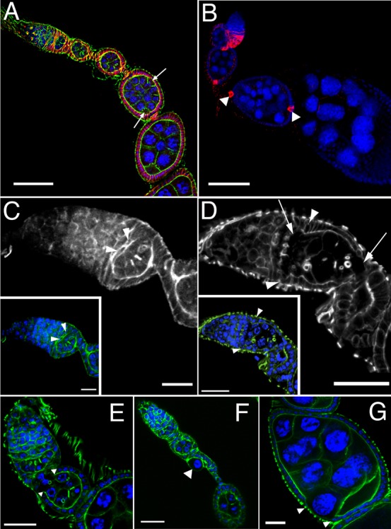
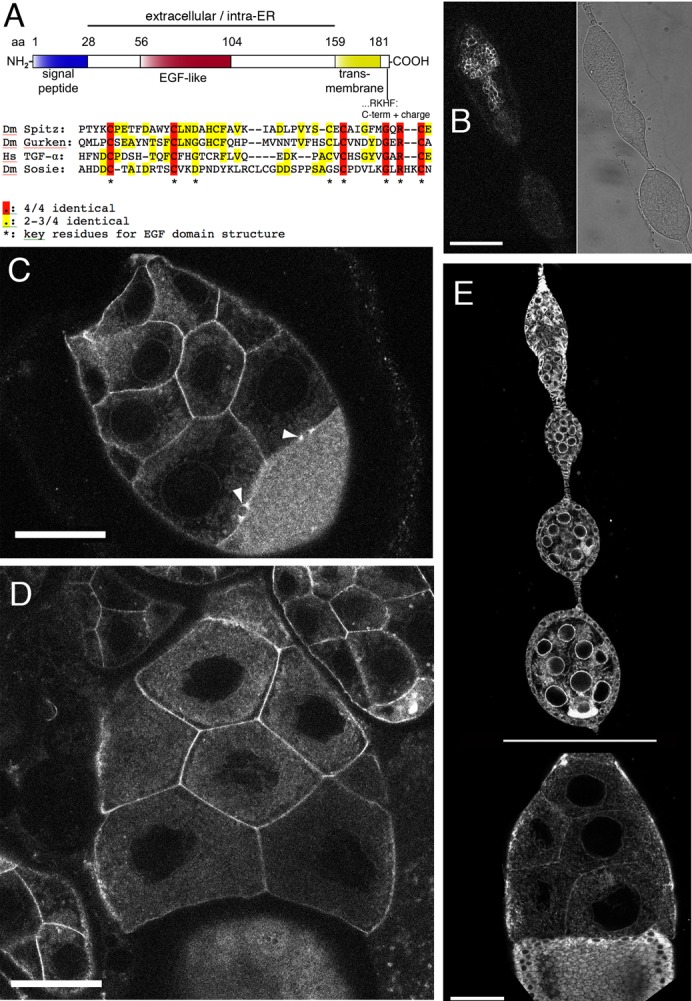
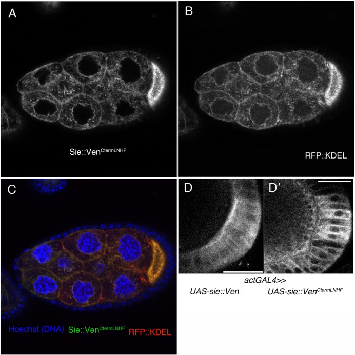
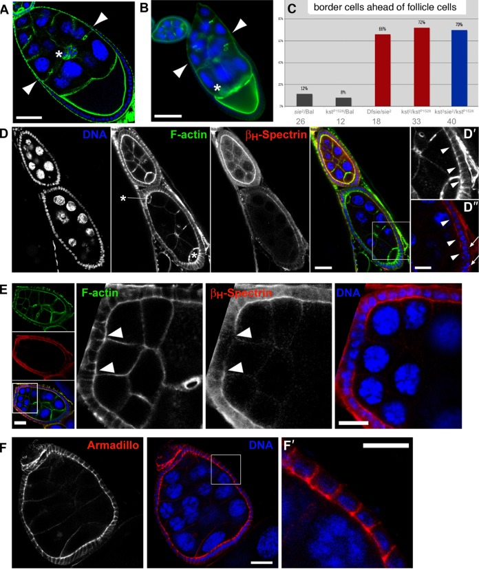
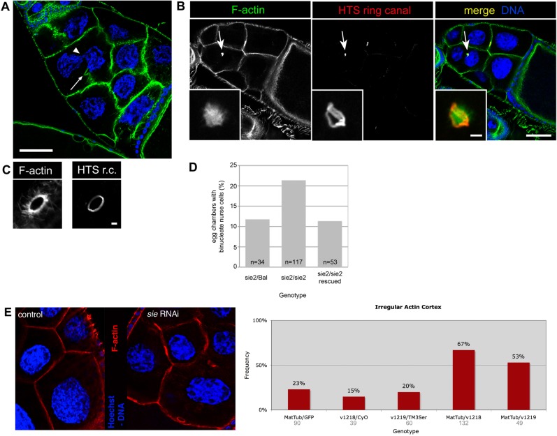
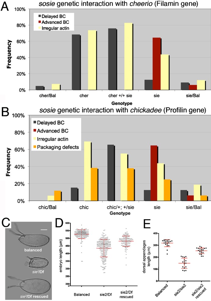
Morphogenesis in multicellular organisms requires the careful coordination of cytoskeletal elements, dynamic regulation of cell adhesion and extensive cell migration. sosie (sie) is a novel gene required in various morphogenesis processes in Drosophila oogenesis. Lack of sie interferes with normal egg chamber packaging, maintenance of epithelial integrity and control of follicle cell migration, indicating that sie is involved in controlling epithelial integrity and cell migration. For these functions sie is required both in the germ line and in the soma. Consistent with this, Sosie localizes to plasma membranes in the germ line and in the somatic follicle cells and is predicted to present an EGF-like domain on the extracellular side. Two positively charged residues, C-terminal to the predicted transmembrane domain (on the cytoplasmic side), are required for normal plasma membrane localization of Sosie. Because sie also contributes to normal cortical localization of β(H)-Spectrin, it appears that cortical β(H)-Spectrin mediates some of the functions of sosie. sie also interacts with the genes coding for the actin organizers Filamin and Profilin and, in the absence of sie function, F-actin is less well organized and nurse cells frequently fuse.

Keywords: Cell migration; Cortical organization and stability; Drosophila oogenesis; Epithelia; Morphogenesis; Spectrin.


Conflict of interest statement

Competing interests: The authors have no competing interests to declare.

Figures

Fig. 1.
Fig. 1.. sosie genetics and compound egg chamber phenotype.
(A) The sosie locus and its mutants (adapted from Flybase: http://flybase.org). Exons are shown as solid boxes, connecting lines represent introns. Start codons (ATG) of two different predicted sosie open reading frames (ORF) RA and RB, respectively (encoding PA and PB, respectively) are indicated as vertical bars. They are in the same frame ending with TAG. RA is the major transcript in adult females, but there is also evidence for a third, minor transcript, RC, that initiates 1.6 kb downstream of RB, encodes the small PB ORF and is mainly expressed in pupae and adult males. Transposon insertions are shown as triangles. sie1 and sie2 are the Exelixis PiggyBac insertions c03947 and f00514, respectively. sie3 is the Minos transposon insertion MB03846, which was used to create deletions sie4 and sie5 by imprecise excision (indicated as horizontal lines). The predicted sosie/CG13636-RB mRNA splice variant (top) with the ATGPB start codon in its fourth exon could not be detected by RT-PCR from ovarian extracts, and only one expressed sequence tag (EST) was listed on http://flybase.org for CG13636-RB at the time when 12 ESTs were found for CG13636-RA. The positions and orientations of the primers used for the RT-PCR shown in D are also indicated (a–d). The neighbor gene CG3368 is encoded on the opposite strand. (B) Compound egg chamber in a sie1/Df(3R)Exel6200 ovary. Two focal planes of the same egg chamber are shown and the inset is a magnification of the oocyte at the posterior of the compound egg chamber, revealing the presence of four ring canals. Blue: DNA. Green: filamentous actin. Red: Egalitarian (oocyte marker). The nurse cells are easily recognizable by their big polyploid nuclei (blue). Note that the egg chamber contains more than 16 germ line cells and two oocytes. Scale bar represents 20 µm. Posterior is to the right. (C) Compound egg chambers at later developmental stages in a sie2/sie2 ovary. The upper one contains even more than twice the normal complement of germ line cells and appears as a long tube of fused germ line cysts that are not separated by somatic follicle cells. Oocytes are labeled (oo). Blue: DNA. Green: F-actin. Scale bar represents 100 µm. (D) Expression of polyadenylated sie mRNA in heterozygous or homozygous sie mutant ovaries. Reverse transcription (RT) reactions were primed with oligo-dT. Lanes labeled with “−” are controls where the reverse transcriptase was omitted from the RT. a, b, c and d indicates the primers that were used for the individual PCRs (primer positions are shown in A). Products amplified with primer pair a/b were loaded on a separate agarose gel as indicated by the vertical separator line. Rearrangements of lanes within a gel are indicated by dashed vertical lines. The DNA molecular weight standard is shown on both sides.
Fig. 2.
Fig. 2.. sosie functions in encapsulating the germ line cysts in the germarium.
DNA is shown in blue and F-actin in green in all colored panels. (A) Compound egg chamber containing 2 germ line cysts at an early developmental stage in a sie4/sie4 ovariole. Its oocytes are indicated by arrows. The compound egg chamber is flanked by a normal stage 3 egg chamber anteriorly, and by a normal stage 6 egg chamber posteriorly, indicating that it is a stage 4–5 egg chamber. Adducin-related protein/Hts staining is shown in red. (B) FasIII immunostaining (red) of a sie1/Df(3R)Exel6200 ovariole. Arrowheads indicate proper restriction of FasIII signal to polar follicle cells after developmental stage 3 in a compound egg chamber. (C) F-actin staining in a wild-type germarium reveals follicle cells that extend F-actin-rich centripetal processes and migrate around the anterior border of a germ line cyst to encapsulate it (arrowheads). (D) sie5/Df(3R)Exel6200 germarium. As revealed by F-actin staining, follicle cells fail to extend processes and to migrate around germ line cells (arrowheads), leading to an open continuum of germ line cells from germarial region 2 to 3 (arrows). (E) Open follicle cell epithelium at the anterior end of a budding sie4/sie4 egg chamber. Arrowheads indicate the borders of the epithelium. (F) Occasionally, single germ line cells that were not packaged into egg chambers can be observed. The arrowhead indicates such a polyploid pseudo-nurse cell in a sie4/sie4 ovariole. (G) A nurse cell appearing at the surface of a stage 7 sie4/sie4 egg chamber due to a gap in the follicle cell epithelium (arrowheads indicate its border). Anterior is top left and scale bars represent 50 µm in A,B, 10 µm in C, and 20 µm in D–G.
Fig. 3.
Fig. 3.. Sosie protein structure and localization.
(A) Schematic representation of the Drosophila melanogaster (Dm) Sosie protein (Fig. 1A, isoform PA). Computationally predicted domains are indicated. aa: amino acid. NH2: N-terminus. COOH: C-terminus. EGF: epidermal growth factor. ER: endoplasmic reticulum. Hs: Homo sapiens. Another program predicts the signal peptide from aa12–33 and the transmembrane domain from aa159–180 (http://phobius.sbc.su.se). (B–D) Distribution of Sosie::Venus in germ line cells (the UAS construct was driven by nanosGAL4). Scale bars represent 50 µm. (B) Germarium. Left panel, Sosie::Venus fluorescence, accumulating at cell membranes. Right panel, transmitted light image. (C) Stage 8 egg chamber. Note the strong Sosie::Venus signal on nurse cell membranes and on two ring canals between nurse cells and the oocyte (arrowheads). The uniformly distributed signal in the oocyte (bottom right) is likely to stem from yolk autofluorescence. (D) Stage 10 egg chamber. Sosie::Venus signal accumulates much stronger on plasma membranes than eYFP-ER (see E). (E) Appearance of the ER in the germ line and somatic cells of the ovary, visualized with an ER-targeted eYFP. The horizontal line indicates that the stage 10 egg chamber (bottom) was recorded in a different image. Note the prominent accumulation of signal around the nurse cell nuclei and at the oocyte posterior during early oogenesis. Scale bar represents 50 µm.
Fig. 4.
Fig. 4.. C-terminal RK residues are required for Sosie plasma membrane localization.
(A–C) Tangential section through a stage 9 egg chamber expressing Sosie::Venus with an altered C-terminal sequence and the ER marker KDEL::RFP both from an UAS promoter. Expression was driven by the germ line specific matTubGAL4 driver. (A) Expression of Sosie::Venus with an altered C-terminal sequence (…LNHF instead of …RKHF) fails to produce a preferential plasma membrane signal. Instead, the signal resembles an ER staining. (B) Expression of the ER marker RFP:: KDEL. (C) Overlay of A and B showing also Hoechst staining for DNA (blue). Large nuclei are nurse cell nuclei, small ones are follicle cell nuclei. (D) Driven with the general driver actGAL4, wild type Sosie::Venus produces a signal that is enriched at the apical plasma membrane in follicle cells (D), while the C-term mutant Sosie::VenusCtermLNHF produces a cytoplasmic or ER signal (D′). Scale bars represent 20 µm.
Fig. 5.
Fig. 5.. Sie–kst/βH-Spectrin interaction, BC/FC migration and maintenance of epithelial structures.
(A,B) DNA is in blue and filamentous F-actin in green. Scale bars represent 50 µm. (A) Wild type stage 9 egg chamber with coordinated migration of BC (asterisk) and outer FC (arrowheads mark most anterior ones). (B) Stage 9 sie5/Df(3R)Exel6200 egg chamber with BC (asterisk) that have already reached the oocyte while the most anterior outer FC (arrowheads) have not even completed half of their migration. (C) Quantification of mis-coordination between outer FC and BC migration in heterozygous, hemizygous and compound heterozygous sosie and karst mutants. See text for details. Bal stands for balancer chromosome, which is wild type for sie and kst. Dfsie is a small deficiency that removes sie. Numbers below the genotypes indicate the number of stage 8 to 9 egg chambers counted for each genotype. Having only one functional copy of sie in a kst background did not further enhance the frequency of this phenotype. (D) Disruption of apical βH-Spectrin localization in follicle cells of a sie4/sie4 compound egg chamber. Asterisks indicate the position of the two oocytes. D′,D″ show a magnified view of the area boxed in the merge panel. Arrowheads point to the apical sides of follicle cells that have lost βH-Spectrin signal (D″), while the appearance of F-actin (D′) seems normal. Arrows in D″ point to cytoplasmic puncta of βH-Spectrin signal. Scale bars represent 20 µm in the main panels and 10 µm in D′,D″. (E) Anterior follicle cells of a sie2/sie2 compound egg chamber have lost apical βH-Spectrin signal. Left panels show an overview of the egg chamber. Note the difference in size and ploidy of the nurse cells that originated from two different cystoblasts. The boxed area denotes the region that is magnified in the panels on the right. In these, arrowheads point to the border between follicle cells that show normal apical accumulation of βH-Spectrin signal and those in which apical βH-Spectrin signal becomes virtually undetectable. Scale bars represent 20 µm in the overview panels and 10 µm in the panels showing a magnified view. (F) Adherens junctions appear normal and are precisely located in an apico-lateral position in a sie2/sie2 compound egg chamber as assayed by immunostaining for Armadillo/β-catenin. F′ shows a magnification of the area boxed in the middle panel. Scale bars represent 20 µm in F and 10 µm in F′, respectively.
Fig. 6.
Fig. 6.. Loss of sosie function leads to degeneration of actin structures in older egg chambers.
(A) sie4/sie4 stage 10 egg chamber. Arrow points to a degenerated F-actin cortex between two nurse cells. In this binucleate nurse cell two nuclei appear to “kiss” one another (arrowhead). Scale bar represents 50 µm. (B) Stage 10 sie2/sie2 egg chamber with a binucleate nurse cell containing a degenerating ring canal (arrow). Insets show a magnified view of another example of a degenerating ring canal. Note the amorphous F-actin staining and the, although distorted, still ring shaped structure for the inner rim component Adducin-like/Hu-Li Tai Shao r.c. Scale bar represents 50 µm in the main panel and 2.5 µm in the inset. (C) Morphology of a wild-type ring canal revealed by staining for F-actin and HTS r.c. Note the very regular ring form and the actin filaments extending from the ring canal (“actin basket”) (Nicolas et al., 2009). Scale bar represents 2.5 µm. (D) Quantification of the binucleate nurse cell phenotype. n: number of stage 10 egg chambers counted. Full genotypes: w; +/SM1; sie2/TM3 Sb (“sie2/Bal”). w; +/SM1; sie2/sie2 (“sie2/sie2”). w; att58A[gsie+ w+]/+; sie2/sie2 (“sie2/sie2 rescued”). (E) Germ line knock down of sie affects cortical F-actin. matTubGAL4 driving UAS-GFP as “control” and a representative example of the sie RNAi constructs that was scored as defective. F-actin is visualized in red and DNA in blue. Additional controls were the two RNAi lines (v1218, v1219) over the balancers (CyO, TM3Ser). MatTub/v1218: sie RNAi construct v1218 driven by the matTubGal4 driver (right panel). MatTub/v1219: sie RNAi construct v1219 driven by the matTubGal4 driver.
Fig. 7.
Fig. 7.. Interactions between sie and actin organizers.
(A,B) sie acts as a second site non-complementer for the actin organizers cher and chic. (A) Enhanced loss of coordination of BC/FC migration and of cortical F-actin integrity in double heterozygous cheerio and sie4 mutants. Egg chambers analyzed are from stage 3 to 10. Number of egg chambers from left to right: n = 41, 19, 29, 48, 33. (B) Enhancement of the chic phenotypes by lack of one copy of sie. Cortical F-actin defects, packaging defects and defects in coordination of BC/FC migration are as strong in the double heterozygous chic/+; +/sie4 as they are in homozygous chic mutants. Egg chambers analyzed ranged from stage 3 to 10. Number of egg chambers inspected: 17, 13, 29, 48 and 16 (from left to right). (C–E) Smaller eggs and eggshell defects in sie mutants. “Balanced” means sie2 or Df3R(niki-sie) over a TM3 Sb balancer. “Df” is Df3R(niki-sie). “Rescued” means that in addition to being mutant for sie, the female flies had a genomic sie+ rescue construct (supplementary material Fig. S2A) in the landing platform att58A on the second chromosome. (C,D) Some of the eggs laid by sie mutant mothers are short (“dumpless” phenotype) and have rudimentary and broadened dorsal appendages. This phenotype can be rescued by a genomic sosie transgene. Note that only a fraction of eggs laid by sosie mutant mothers is shorter than normal, and that this fraction has approximately the same size as stage 10 egg chambers that show the F-actin degeneration phenotype (Fig. 6A). Also note that the y-axis starts at 200 µm in D. (C,E) Phenotype (C) and quantitative analysis (E) of the dorsal appendages of eggs laid by sie2 homozygous mutant mothers. (D,E) each data-point represents one embryo, and bars indicate mean and standard deviations. Scale bar in C represents 100 µm.

Similar articles

Cited by

References

    1. Baum B., Perrimon N. (2001). Spatial control of the actin cytoskeleton in Drosophila epithelial cells. Nat. Cell Biol. 3, 883–890 10.1038/ncb1001-883 - DOI - PubMed
    1. Bischof J., Maeda R. K., Hediger M., Karch F., Basler K. (2007). An optimized transgenesis system for Drosophila using germ-line-specific phiC31 integrases. Proc. Natl. Acad. Sci. USA 104, 3312–3317 10.1073/pnas.0611511104 - DOI - PMC - PubMed
    1. Bogdanov M., Xie J., Dowhan W. (2009). Lipid-protein interactions drive membrane protein topogenesis in accordance with the positive inside rule. J. Biol. Chem. 284, 9637–9641 10.1074/jbc.R800081200 - DOI - PMC - PubMed
    1. Brand A. H., Perrimon N. (1993). Targeted gene expression as a means of altering cell fates and generating dominant phenotypes. Development 118, 401–415. - PubMed
    1. Conder R., Yu H., Zahedi B., Harden N. (2007). The serine/threonine kinase dPak is required for polarized assembly of F-actin bundles and apical-basal polarity in the Drosophila follicular epithelium. Dev. Biol. 305, 470–482 10.1016/j.ydbio.2007.02.034 - DOI - PubMed