. 2012 Nov 15;1(11):1083-93.
doi: 10.1242/bio.20121396. Epub 2012 Aug 28.

Lhx1 in the proximal region of the optic vesicle permits neural retina development in the chicken

Affiliations

Lhx1 in the proximal region of the optic vesicle permits neural retina development in the chicken

Takumi Kawaue et al. Biol Open. .

Abstract

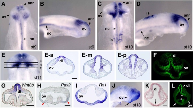
How the eye forms has been one of the fundamental issues in developmental biology. The retinal anlage first appears as the optic vesicle (OV) evaginating from the forebrain. Subsequently, its distal portion invaginates to form the two-walled optic cup, which develops into the outer pigmented and inner neurosensory layers of the retina. Recent work has shown that this optic-cup morphogenesis proceeds as a self-organizing activity without any extrinsic molecules. However, intrinsic factors that regulate this process have not been elucidated. Here we show that a LIM-homeobox gene, Lhx1, normally expressed in the proximal region of the nascent OV, induces a second neurosensory retina formation from the outer pigmented retina when overexpressed in the chicken OV. Lhx2, another LIM-homeobox gene supposed to be involved in early OV formation, could not substitute this function of Lhx1, while Lhx5, closely related to Lhx1, could replace it. Conversely, knockdown of Lhx1 expression by RNA interference resulted in the formation of a small or pigmented vesicle. These results suggest that the proximal region demarcated by Lhx1 expression permits OV development, eventually dividing the two retinal domains.

Keywords: Chicken; Eye development; LIM-homeobox; Lhx1; Lhx5; Neural retina; Optic vesicle.


Conflict of interest statement

Competing interests: The authors have no competing interests to declare.

Figures

Fig. 1.
Fig. 1.. Lhx1 is expressed in the proximal region of the OV.
Whole mount in situ hybridization (WISH) of embryos at Hamburger's and Hamilton's stages 9 (A,B), stage 10 (C,D), stage 11 (E), and stage 13 (J). (A,C,E) Dorsal views. (B,D,J) Lateral views. Transverse sections cut after WISH are shown in (E-a,E-m,E-p). At the middle OV level (E-m), Lhx1 is distinctly expressed in the proximal region of the OV and dorsal diencephalon (di). Outside these regions, Lhx1 is also expressed in the anterior neural ridge (anr) and notochord (nc). (F,L) Immunohistochemistry of Lhx1/Lhx5 protein. Transverse sections at the OV level of stage 11 (F), and stage 13 (L). (G–I) Expression patterns of Wnt8b (G), Pax2 (H), and Rx1 (I) at stage 11. Transverse sections at the OV level are shown for comparison. (JL) At stage 13, Lhx1 is expressed in the dorsal diencephalon and by a subset of cell in the ventral diencephalon (arrow in J–L), but not in the presumptive neural retina (arrowhead in L). Scale bars: 100 µm.
Fig. 2.
Fig. 2.. Lhx1 overexpression induces a second NR formation from the presumptive RPE.
(A,B) In the EGFP-overexpressing control eye, Lhx1 is not expressed at stage 15/16 (24 hours after electroporation). (C,D) In the Lhx1-overexpressing eye, Lhx1 mRNA is strongly detected in the protruding region, as indicated by arrowheads and intense EGFP fluorescence. (E,F) In the EGFP-overexpressing control eye, Pax6 is expressed in the optic cup and lens vesicle. (G,H) In the Lhx1-overexpressing eye, Pax6 is ectopically expressed in the protruding region of the optic cup, as indicated by arrowheads and intense EGFP fluorescence. (I,J) Hematoxylin-Eosin staining at stage 24 (60 hours after electroporation). Control EGFP-overexpressing (I) and Lhx1-overexpressing retina (J), where the outer layer of the optic cup is partly thickened (arrow). (K,L) Immunostaining for β3-tubulin (red), which is localized on the vitread side of the neural retina. Beta3-tubulin is localized to the thickened epithelium (arrow in L) in a mirror-image symmetrical pattern when compared to the authentic retina. (M,N) Immunostaining for phosphohistone-H3 (ph-H3; red), which marks mitotic cells on the apical side of the neuroepithelium. Phosphohistone-H3 is localized to the thickened epithelium (arrow in N) in a mirror-image symmetrical pattern when compared to the authentic retina. In (K–N), EGFP fluorescence is shown in green, and nuclei were stained with DAPI (blue). (O,P) Immunostaining for a neurofilament 3A10 antigen (green), which is an early neuronal differentiation marker. In this experiment, a bicistronic vector, RFP-2A-Lhx1/pCAGGS (P) or a mock vector, RFP-2A/pCAGGS (O) was electroporated. Within 48 hours after electroporation, only RFP-positive cells (red) in the outer layer of the OV are thickened to form rosette that express 3A10 antigen. (P′–P′″) Single-channel confocal images of (P). NR, neural retina; RPE, retinal pigment epithelium; and vit, vitreous. Scale bars: 100 µm.
Fig. 3.
Fig. 3.. Immunostaining for neural differentiation makers of the stage 29 optic cup.
Immunoreactive signals are shown in green or red. Control (A,C,E,G,I,K) and Lhx1-overexpressing eyes (B,D,F,H,J,L). In these experiments, a bicistronic vector of pCAGGS-RFP-2A-Lhx1 was used. (A,B) 3A10, (C,D) N-cadherin, (E,F) HuC/D RNA-binding protein, (G,H) Islet1, (I,J) visinin (calcium-binding protein), and (K,L) AP2α. N-cadherin is localized to the neuroepithelium, visinin is localized to future cone photoreceptors, and others are localized to early retinal ganglion cells, and AP2α is localized to differentiating amacrine cells. Note that induced neural retina (iNR) is thicker than the authentic NR in this experimental condition. Scale bar: 100 µm.
Fig. 4.
Fig. 4.. Lhx1 overexpression activates NR specification genes and suppresses RPE specification genes in the presumptive RPE.
Expression of Rx1 (A,A′,H,H′), Six3 (B,B′,I,I′), Six6 (C,C′,J,J′), Chx10 (D,D′,K,K′), Sox2 (E,E′,L,L′), Otx2 (F,F′,M,M′), and Mitf (G,G′,N,N′). Frontal sections were shown. Eyes were examined at stage 15/16 (24 hours post-electroporation). Contralateral (A–G′) and Lhx1-overexpressing (H–N′) eyes are shown. (A′–N′) High magnification of (A–N), respectively. Arrows in (H′–N′) indicates Lhx1-overexpressing cells, as confirmed morphologically as thickened epithelial areas. Results were obtained by in situ hybridization, except for Sox2 by immunostaining. Scale bars: 100 µm.
Fig. 5.
Fig. 5.. Lhx1 overexpression induces ectopic Fgf gene expressions.
(A–E′) Expression of Fgf19 (A,A′,C,C′) and EGFP fluorescence (B,B′) was examined at 24 hours post- electroporation. (A′,C′) High magnification of (A,C), respectively. Lhx1-overexpressing (A,B,A′,B′) and contralateral (C,C′) eyes. (D) Section of the optic cup after Lhx1-overexpression, showing ectopic induction of Fgf19. (E,E′) Expression of Fgf8 was examined by in situ hybridization in the eye overexpressing Lhx1 at 48 hours post-electroporation. Control (E), and in (E′) arrowhead shows ectopic induction of Fgf8 in the center of the second NR. Scale bar: 50 µm.
Fig. 6.
Fig. 6.. Differential effects of Lhx2 and Lhx5 on the OV and genetic interactions between Lhx1 and Lhx5.
(A) Phylogenetic tree of chicken LIM-homeodomain (LIM-HD) factors. The LIM-HD factors are depicted by the NJ (neighbor-joining) method. Chicken LMO3 was used as an outgroup. The scale bar is calibrated in substitutions per site. Numbers show bootstrap confidence values. Amino acid sequences used in the tree construction were deduced from the nucleotide sequences listed in supplementary material Table S2. (B–D) Lhx2 is expressed in the forebrain and whole OV at stage 11. Dorsal (B) and lateral (C) views. (D) Transverse section of the OV. (E–H) EGFP fluorescence images at 24 hours post-electroporation. Overexpression of Lhx2 (F) did not induce transformation of the outer optic cup to the NR, whereas overexpression of Lhx5 (G) or Lhx1 (H) induced a protrusion of the OV. (I,J) Histology of an Lhx5-overexpressing retina at stage 24. A second NR transformed from the outer layer of the optic cup. (K,L) In situ hybridization of an Lhx5-overexpressing retina at stage 21. Arrows show activation of Chx10 (K) and suppression of Otx2 (L) in the developing second NR. (M–Q) Whole mount in situ hybridization (WISH) of embryos at Hamburger's and Hamilton's stages 9 (M,N), stage 10 (O,P), and stage 11 (Q). (M,O,Q) Dorsal views. (N,P) Lateral views. Transverse sections cut after WISH are shown in (Q-a,Q-m,Q-p). At the middle OV level (Q-m), Lhx5 is distinctly expressed in the proximal region of the OV, while it is expressed in the ventral diencephalon at the posterior OV level (Q-p). (R–T) EGFP fluorescence (R) and in situ hybridization of Lhx1 (S,T) at 24 hours post-electroporation. Lhx1 expression is ectopically induced in the protruding outer layer of the optic cup after Lhx5 overexpression (arrowhead in S,T). Scale bars: 100 µm.
Fig. 7.
Fig. 7.. Lhx1-RNAi reduces the eye size.
(A–L) Lhx1-RNAi resulted in a small eye phenotype at stage 24. Electroporated right eyes are shown in the left two columns, and contralateral left eyes are shown in the right column. RFP fluorescence images are shown in the middle column. All images are shown in the same magnification. (M) Percentage of embryos with small eye phenotypes, Class I to Class III (see Materials and Methods), at stage 24. Scale bar: 300 µm.
Fig. 8.
Fig. 8.. Lhx1-RNAi perturbs NR formation.
(A–E) Hematoxylin-Eosin staining at stage 24. (B) Magnified view of the posterior retina in (A); distinct pigmentation is observed on the basal side of the single-layered RPE. (C) Small eye phenotype after Lhx1-RNAi, in which a pigmented vesicle (pv) forms and lens (le) development is perturbed. (D) The optic epithelium abutting the lens. (E) Posterior optic epithelium with distinct polarized pigmentation. (F–M) Expression of Chx10, Otx2, Mitf, and Mmp115. Eyes were examined at 24 hours (F,G,J,K), 36 hours (H,L) or 60 hours post-electroporation (I,M). In situ hybridization was performed under the same conditions for controls and Lhx1-RNAi, depending on the probes. (J) Chx10 expression in the inner layer of the optic cup is reduced (arrowhead). (K) The Otx2 expression domain is expanded to the inner layer of the optic cup (arrowhead). (L) Mitf expression is detected in the inner layer as well as outer layer of the optic cup. (M) Mmp115 expression is detected in the dorsal epithelium of the OV (arrowheads) and abutting the lens vesicle (arrow). The axis of the eye is depicted as follows: A, anterior; D, dorsal; P, posterior; V, ventral; nr, neural retina; and rpe, retinal pigmented epithelium. Scale bars, 100 µm (A,C), 10 µm (B,D,E).

References

    1. Bhatia B., Singhal S., Tadman D. N., Khaw P. T., Limb G. A. (2011). SOX2 is required for adult human muller stem cell survival and maintenance of progenicity in vitro. Invest. Ophthalmol. Vis. Sci. 52, 136–145 10.1167/iovs.10-5208 - DOI - PubMed
    1. Chow R. L., Lang R. A. (2001). Early eye development in vertebrates. Annu. Rev. Cell Dev. Biol. 17, 255–296 10.1146/annurev.cellbio.17.1.255 - DOI - PubMed
    1. Crossley P. H., Martinez S., Ohkubo Y., Rubenstein J. L. (2001). Coordinate expression of Fgf8, Otx2, Bmp4, and Shh in the rostral prosencephalon during development of the telencephalic and optic vesicles. Neuroscience 108, 183–206 10.1016/S0306-4522(01)00411-0 - DOI - PubMed
    1. Das R. M., Van Hateren N. J., Howell G. R., Farrell E. R., Bangs F. K., Porteous V. C., Manning E. M., McGrew M. J., Ohyama K., Sacco M. A.et al. (2006). A robust system for RNA interference in the chicken using a modified microRNA operon. Dev. Biol. 294, 554–563 10.1016/j.ydbio.2006.02.020 - DOI - PubMed
    1. Eiraku M., Takata N., Ishibashi H., Kawada M., Sakakura E., Okuda S., Sekiguchi K., Adachi T., Sasai Y. (2011). Self-organizing optic-cup morphogenesis in three-dimensional culture. Nature 472, 51–56 10.1038/nature09941 - DOI - PubMed