. 2012 Nov 15;1(11):1161-8.
doi: 10.1242/bio.20122824. Epub 2012 Sep 20.

A role for Rap2 in recycling the extended conformation of LFA-1 during T cell migration

Affiliations

A role for Rap2 in recycling the extended conformation of LFA-1 during T cell migration

Paula Stanley et al. Biol Open. .

Abstract

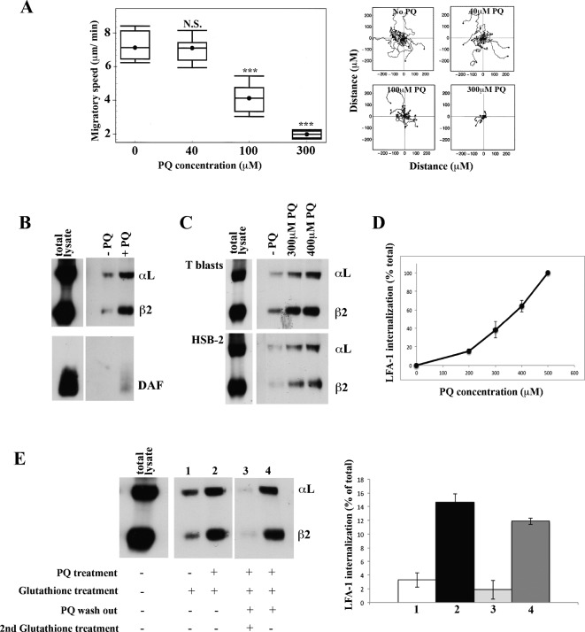
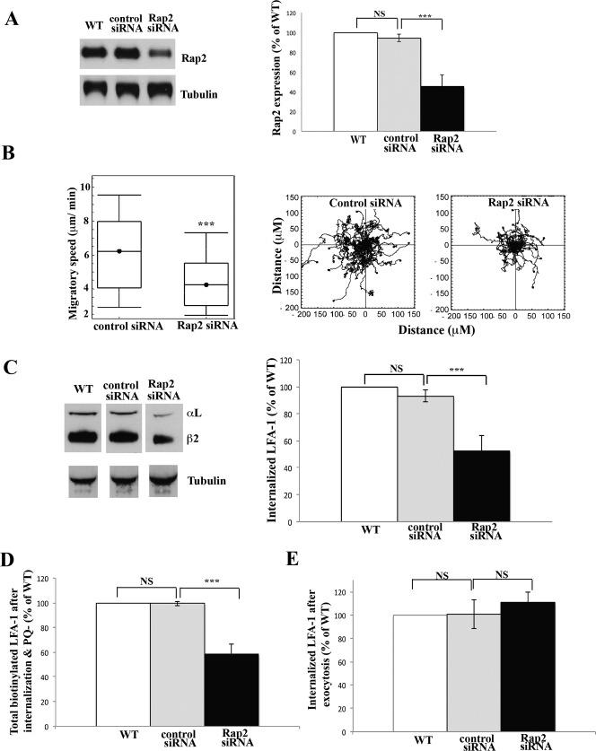
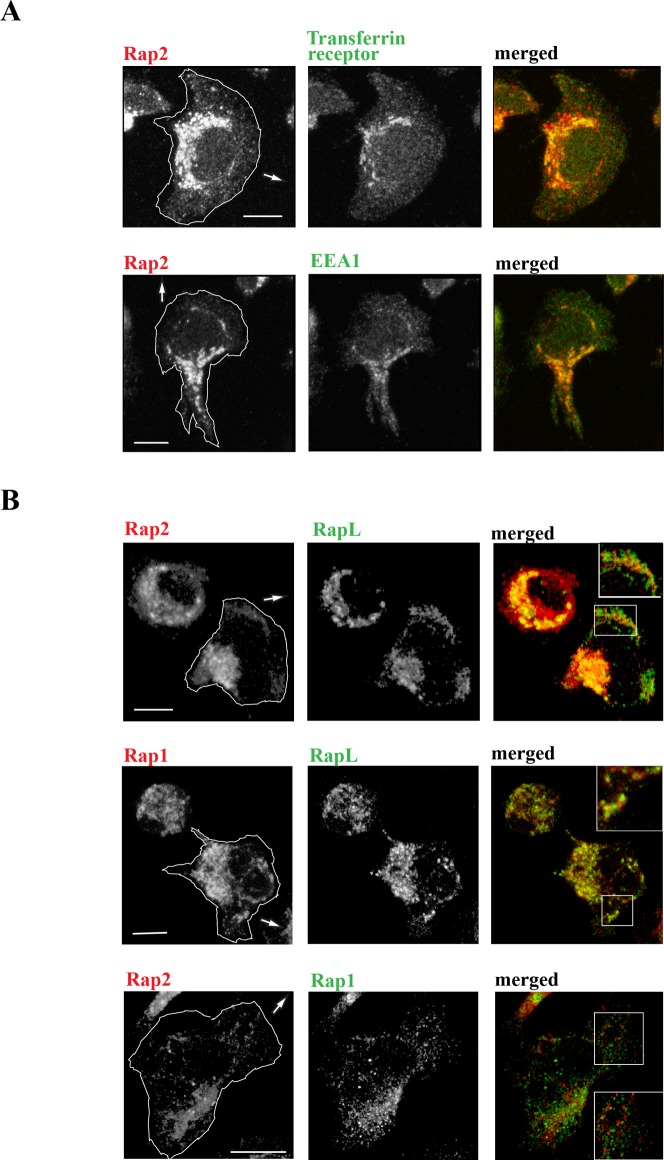
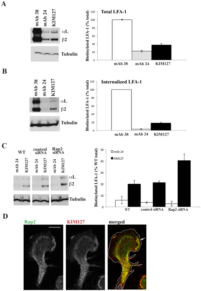
T lymphocytes make use of their major integrin LFA-1 to migrate on surfaces that express ICAM-1 such as blood vessels and inflamed tissue sites. How the adhesions are turned over in order to supply traction for this migration has not been extensively investigated. By following the fate of biotinylated membrane LFA-1 on T lymphocytes, we show in this study that LFA-1 internalization and re-exposure on the plasma membrane are linked to migration. Previously we demonstrated the GTPase Rap2 to be a regulator of LFA-1-mediated migration. SiRNA knockdown of this GTPase inhibits both LFA-1 internalization and also its ability to be re-exposed, indicating that Rap2 participates in recycling of LFA-1 and influences its complete endocytosis-exocytosis cycle. Confocal microscopy images reveal that the intracellular distribution of Rap2 overlaps with endosomal recycling vesicles. Although the homologous GTPase Rap1 is also found on intracellular vesicles and associated with LFA-1 activation, these two homologous GTPases do not co-localize. Little is known about the conformation of the LFA-1 that is recycled. We show that the extended form of LFA-1 is internalized and in Rap2 siRNA-treated T lymphocytes the trafficking of this LFA-1 conformation is disrupted resulting in its intracellular accumulation. Thus LFA-1-mediated migration of T lymphocytes requires Rap2-expressing vesicles to recycle the extended form of LFA-1 that we have previously found to control migration at the leading edge.

Keywords: Integrin; LFA-1; Migration; Rap2; Recycling.


Conflict of interest statement

Competing interests: The authors have no competing interests to declare.

Figures

Fig. 1.
Fig. 1.. LFA-1 is internalized and re-expressed by T cells migrating on ICAM-1.
(A) Speed of T lymphoblasts treated with increasing concentrations of primaquine (PQ) migrating on immobilized ICAM-1 showing cell trajectories tracked by video microscopy. Mean speed ± s.e.m. of three independent experiments (left) and the migratory tracks in a typical experiment (right) are shown; n = 20 cells per condition, ***P<0.001. (B) Western blots of immunoprecipitated internalized LFA-1 and DAF following biotinylation of T lymphoblast surface membranes followed by 30 min migration on ICAM-1: total cell lysate immunoprecipitated for LFA-1 and DAF (sample diluted 2×) and internalized protein ± 300 µM PQ revealed by removal of biotin from cell membrane receptors with glutathione. (C) Western blots comparing internalized LFA-1 in T lymphoblasts and HSB2 T cell line: total biotinylated LFA-1 (total lysate at 2× dilution) and similar amounts of internalized LFA-1 ± PQ. (D) Internalized T cell LFA-1 after 30 min on ICAM-1 in the presence of increasing amounts of PQ ± s.d. from n = 3 experiments. (E) Total biotinylated LFA-1 (total lysate at 2× dilution). Lanes 1 and 2: LFA-1 internalized after 30 min ± 300 µM PQ. LFA-1 re-exposure on the membrane following PQ washout is demonstrated by lack of intracellular LFA-1 in lane 3 (treated with glutathione to remove detection of membrane LFA-1) and lane 4 (no glutathione treatment allowing membrane and intracellular LFA-1 to be detected). Left: typical experiment. Right: mean ± s.d. of n = 3 experiments.
Fig. 2.
Fig. 2.. Rap2 regulates internalization of T cell LFA-1 during migration.
(A) HSB2 T cells were either not electroporated as control WT or electroporated with control or Rap2 siRNAs. Western blots were probed for Rap2 after 72 h and α-tubulin as a loading sample control. Left: typical experiment showing Rap2 siRNA knockdown compared with non-electroporated (WT) or control siRNA-treated T cell controls. Right: quantification of siRNA knockdown compared with controls, mean ± s.d. of n = 5 experiments, ***P<0.001. (B) HSB2 T cells electroporated with control or Rap2 siRNAs migrating on ICAM-1 for 30 min. Left: mean speed ± s.d. of n = 3 experiments. Right: migratory tracks of individual cells; n = 40 cells per condition, ***P<0.001. (C) Biotinylated LFA-1 internalization + PQ in WT, control siRNA- and Rap2 siRNA-treated HSB2 T cells. LFA-1 internalization following Rap2 siRNA compared with controls, showing (left) Western blot of a typical experiment and (right) quantification of 3 experiments, mean ± s.d. ***P<0.001. (D) Using T cells treated as in C, total biotinylated LFA-1 in control and Rap2 siRNA-treated T cells is shown following PQ washout and a 20 min incubation. No glutathione treatment allows assessment of internal and re-exposed LFA-1; mean ± s.d. of 3 experiments, ***P<0.001. (E) Re-expression of LFA-1 in control and Rap2 siRNA-treated T cells following PQ washout and 20 min incubation. LFA-1 remaining inside the cells revealed following removal of glutathione sensitive membrane LFA-1; mean ± s.d. of 3 experiments, ***P<0.001.
Fig. 3.
Fig. 3.. Expression of Rap2, Rap1 and recycling vesicles in migrating T lymphoblasts.
(A) Confocal microscopy images showing the co-distribution of Rap2 with intracellular vesicle markers EEA1 and transferrin receptor at the interface with ICAM-1. (B) Rap2 and Rap1 each co-localize with effector RAPL, but there is a lack of overlap between these two GTPases; insets show detail at leading edge. Arrows show direction of T cell migration. Scale bar = 5 µm.
Fig. 4.
Fig. 4.. Rap2 regulates internalization of the extended conformation of LFA-1.
MAbs specific for pan-LFA-1 (38), high affinity (24) and extended (KIM127) LFA-1 used to immunoprecipitate (A) total LFA-1 and (B) internalized LFA-1 from T cells. (A,B) Left: typical western blot experiment. Right: mean ± s.d. of n = 4 experiments. (C) Internalization of extended but not high affinity conformations of LFA-1 are increased following Rap2 siRNA knockdown compared with control siRNA and WT treatment of T cells. Left: typical western blot experiment. Right: mean ± s.d. of n = 3 experiments. (D) Confocal microscopy image showing overlap of Rap2 and KIM127-expressing LFA-1 concentrated in the lamella with some lamellipodial distribution in the direction of T lymphoblast migration indicated by an arrow. Scale bar = 5 µm.

Similar articles

Cited by

References

    1. Arjonen A., Alanko J., Veltel S., Ivaska J. (2012). Distinct recycling of active and inactive β1 integrins. Traffic 13, 610–625 10.1111/j.1600-0854.2012.01327.x - DOI - PMC - PubMed
    1. Bivona T. G., Wiener H. H., Ahearn I. M., Silletti J., Chiu V. K., Philips M. R. (2004). Rap1 up-regulation and activation on plasma membrane regulates T cell adhesion. J. Cell Biol. 164, 461–470 10.1083/jcb.200311093 - DOI - PMC - PubMed
    1. Bolte S., Cordelières F. P. (2006). A guided tour into subcellular colocalization analysis in light microscopy. J. Microsc. 224, 213–232 10.1111/j.1365-2818.2006.01706.x - DOI - PubMed
    1. Caswell P. T., Vadrevu S., Norman J. C. (2009). Integrins: masters and slaves of endocytic transport. Nat. Rev. Mol. Cell Biol. 10, 843–853 10.1038/nrm2799 - DOI - PubMed
    1. Christoforidis S., McBride H. M., Burgoyne R. D., Zerial M. (1999). The Rab5 effector EEA1 is a core component of endosome docking. Nature 397, 621–625 10.1038/17618 - DOI - PubMed