. 2012 Feb 15;1(2):82-91.
doi: 10.1242/bio.2011018. Epub 2011 Nov 3.

The CUL3-KLHL18 ligase regulates mitotic entry and ubiquitylates Aurora-A

Affiliations

The CUL3-KLHL18 ligase regulates mitotic entry and ubiquitylates Aurora-A

Saili Moghe et al. Biol Open. .

Abstract

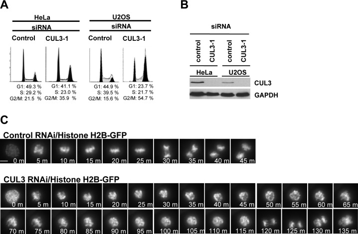
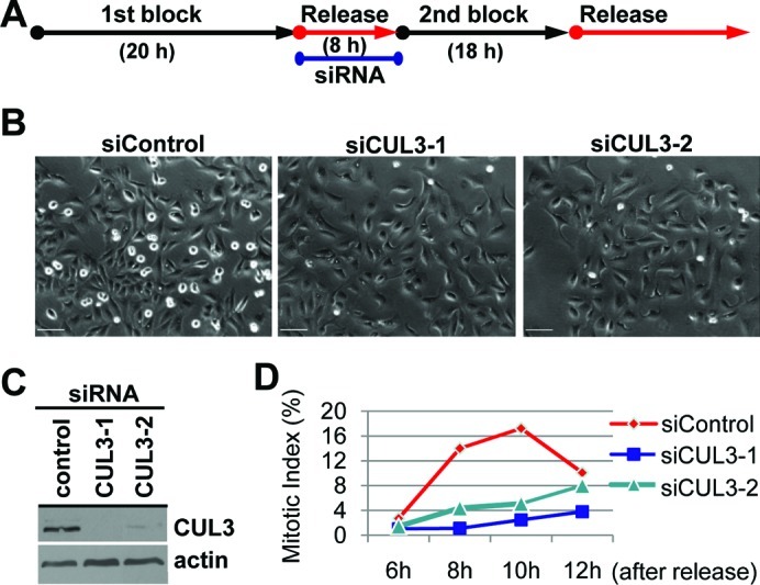
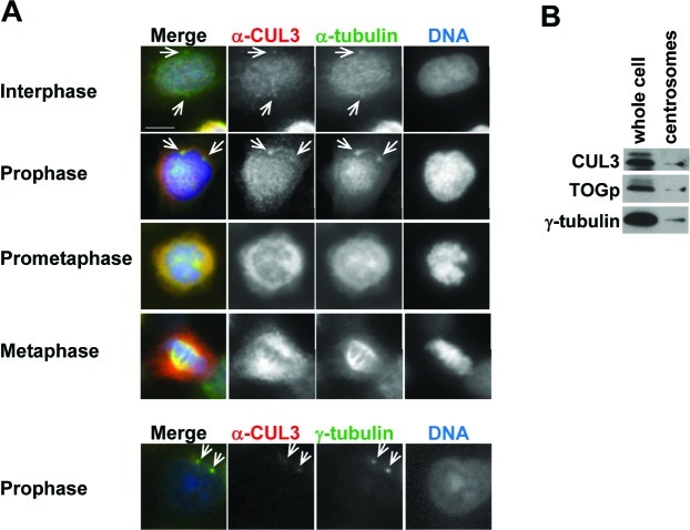
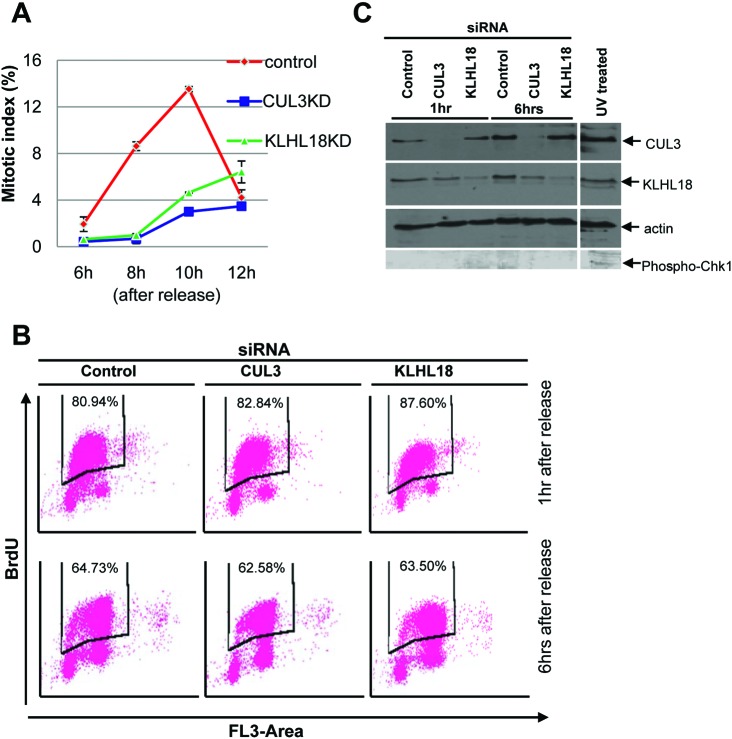
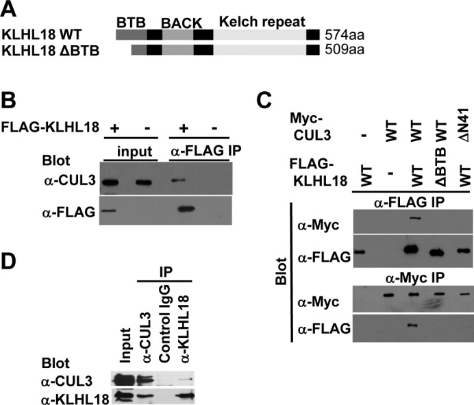
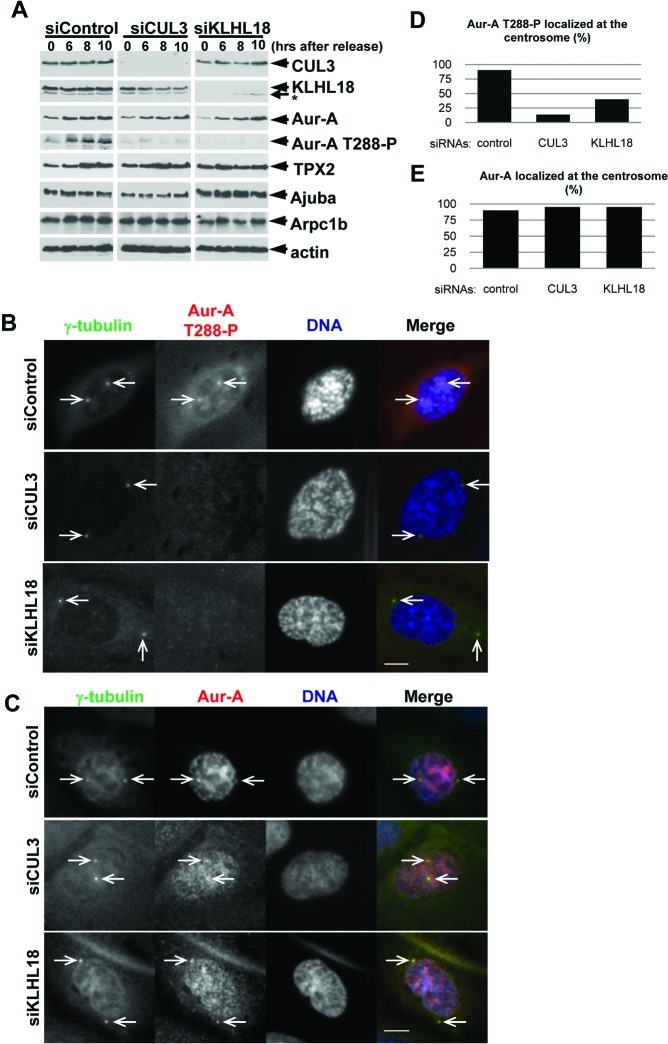
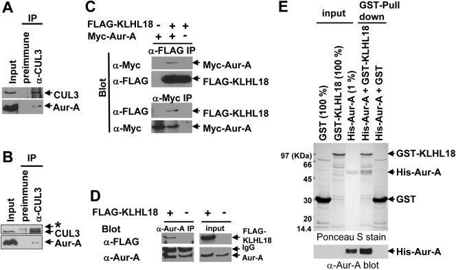
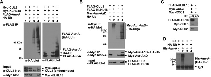
The cullin-RING family of ubiquitin ligases regulates diverse cellular functions, such as cell cycle control, via ubiquitylation of specific substrates. CUL3 targets its substrates through BTB proteins. Here we show that depletion of CUL3 and the BTB protein KLHL18 causes a delay in mitotic entry. Centrosomal activation of Aurora-A, a kinase whose activity is required for entry into mitosis, is also delayed in depleted cells. Moreover, we identify Aurora-A as a KLHL18-interacting partner. Overexpression of KLHL18 and CUL3 promotes Aurora-A ubiquitylation in vivo, and the CUL3-KLHL18-ROC1 ligase ubiquitylates Aurora-A in vitro. Our study reveals that the CUL3-KLHL18 ligase is required for timely entry into mitosis, as well as for the activation of Aurora-A at centrosomes. We propose that the CUL3-KLHL18 ligase regulates mitotic entry through an Aurora-A-dependent pathway.

Keywords: Aurora-A; BTB; CUL3; POZ; mitotic entry; ubiquitin.


Figures

Fig. 1.
Fig. 1.. CUL3 functions in mitotic progression.
(A) Asynchronous HeLa and U2OS cells were transfected with indicated siRNAs. 72 hours after transfection, cell cycle profile was analyzed by flow cytometry, and (B) cell lysate was analyzed by western blotting (WB) with anti-CUL3 or anti-GAPDH (control). (C) Histone H2B-GFP stably expressing HeLa cells were transfected with either control or CUL3 siRNAs. 48h after transfection, cells were analyzed by time-lapse antibodies fluorescence microscopy. One representative cell for control and CUL3 RNAi is shown from chromosome condensation (0 min). Scale bars, 10 μm.
Fig. 2.
Fig. 2.. CUL3 functions in mitotic entry.
(A) A schematic protocol for cell synchronization (by double thymidine block) and RNAi. (B, C, D) U2OS cells were synchronized and transfected as indicated and subjected to the protocol shown in (A) with indicated siRNAs. (B) Phase pictures of indicated siRNA transfected cells at 8 hours after release. Scale bars, 100 μm. (C) When released from 2nd thymidine block, cells were lysed and subjected to WB with indicated antibodies. (D) Cells were fixed and stained with DAPI at the indicated times after release. Over 500 cells were analyzed and mitotic index was determined.
Fig. 3.
Fig. 3.. CUL3 localizes at centrosomes and the mitotic spindle
(A) U2OS cells were fixed with paraformaldehyde (top panels) or methanol (bottom panels) and immunostained with indicated antibodies. DNA was counter-stained by DAPI. Representative pictures of cells in late G2 phase (Interphase), prophase, prometaphase, and metaphase are shown. White arrowheads indicate colocalization of α-tubulin or γ-tubulin and CUL3 in interphase and propahse. Scale bars, 10 μm. (B) Whole cell extract and purified centrosomes from U2OS cells were subjected to WB with indicated antibodies.
Fig. 4.
Fig. 4.. CUL3 and KLHL18 knockdown cells have delayed mitotic entry but progress through S phase normally
(A) U2OS cells were synchronized and transfected with indicated siRNAs as shown in Fig. 2A. Cells were fixed and stained with DAPI at the indicated times after release. Over 500 cells were analyzed and the mitotic index was determined, data are represented as mean ± SD. (B, C) U2OS cells were synchronized and transfected as indicated in Fig. 2A. (B) At the indicated times following release cells were pulse labeled with 10uM BrdU for 2 hours and stained with anti-BrdU-FITC and 7-AAD, and analyzed by flow cytometry. The percentages of incorporated BrdU are indicated. (C) Lysates from the indicated time points after release were analyzed by WB with indicated antibodies. Lysates from U2OS cells treated with UV were used as a positive control for phospho-Chk1 blot.
Fig. 5.
Fig. 5.. KLHL18 interacts with CUL3.
(A) Schematic figure of wild type and mutant of KLHL18. (B) FLAG-tagged KLHL18 expressing plasmids were transfected to HeLa cells and treated with 100 ng/mL Nocodazole for 12 hours before lysis. FLAG KLHL18 and endogenous CUL3 interaction was examined by immunoprecipitation (IP) and WB with indicated antibodies. (C) Wild type and mutant KLHL18 and CUL3 expressing plasmids were transfected to HeLa cells. Interactions were examined by IP-WB analysis with indicated antibodies. (D) Endogenous CUL3 and KLHL18 interaction in U2OS cells was examined by IP-WB with indicated antibodies
Fig. 6.
Fig. 6.. The CUL3-KLHL18 ligase is involved in Aurora-A activation at the centrosomes
U2OS cells were synchronized and transfected with indicated siRNAs according to the protocol in Fig. 2A. (A) Cells were lysed at the indicated times after release and examined by WB with indicated antibodies. (B, C) Cells were fixed at 10 hours after release and immunostained with indicated antibodies. DNA was counter-stained by DAPI. Representative pictures of cells in prophase are shown. White arrowheads indicate colocalization of γ-tubulin and Aurora-A T288-P or γ-tubulin and Aurora-A. Scale bars, 10 μm. (D, E) Colocalization of γ-tubulin and Aurora-A T288-P (D) and γ-tubulin and Aurora-A (E) in more than 50 of each siRNA transfected prophase cells were quantified.
Fig. 7.
Fig. 7.. KLHL18 interacts with Aurora-A directly.
(A) U2OS cells treated with 100 ng/mL Nocodazole for 12 hours were lysed and examined by IP-WB with indicated antibodies. (B) CSF-arrested Xenopus egg extracts were examined by IP-WB analysis with indicated antibodies. Asterisk shows non-specific band. (C) FLAG-tagged KLHL18 and Myc-tagged Aurora-A (Myc-Aur-A) expressing plasmids were co-transfected to 293T cells and their interactions were determined by IP-WB analysis with indicated antibodies. (D) FLAG-tagged KLHL18 expressing plasmid was transfected to 293T cells and the FLAG-KLHL18 and endogenous Aurora-A interaction was determined by IP-WB with indicated antibodies. (E) Bacterially expressed and purified GST fused KLHL18 or GST and 6x Histidine fused (His-) Aurora-A were incubated in vitro as indicated and their interactions were determined by GST-pull down assay. The input and GST-pull down reactions were separated by SDS-PAGE, transferred to a nitrocellulose membrane and examined by Ponceau S staining (upper panel). His-Aurora-A was examined by WB with anti-Aurora-A antibody as well (bottom panel).
Fig. 8.
Fig. 8.. The CUL3-KLHL18 ligase ubiquitylates Aurora-A in vivo and in vitro.
(A, B) FLAG-tagged Aurora-A (A) or Myc-tagged Aurora-A deleted C-terminal D-box (Aur-AΔD) (B) were cotransfected with Myc-tagged or FLAG-tagged CUL3, Myc-tagged or FLAG-tagged KLHL18, and HA-tagged Ubiquitin expressing plasmids to 293T cells as indicated. Twenty four hours after transfection, cells were lysed with a 1% SDS containing lysis buffer and boiled for 15 min. Denatured lysates were then diluted with 0.5% NP-40 lysis buffer and IP with anti-FLAG (A) or anti-Myc (B) antibody. The washed immunoprecipitates and the total cell lysate (input) were resolved by SDS-PAGE, followed by WB with indicated antibodies. (C) CUL3-KLHL18-ROC1 complexes were prepared from triply transfected 293T cells by IP using anti-FLAG antibody and used as the source of E3 ligase. (D) Bacterially expressed and purified His-tagged Aurora-A (His-Aur-A) (Fig. 6E) was incubated with the KLHL18-CUL3-ROC1 E3 complex (C), E1, E2, HA-tagged ubiquitin (HA-Ub), and ATP. The reactions were immunoprecipitated with anti-HA antibody and blotted with anti-Aurora-A antibody to examine Aurora-A ubiquitylation.

Similar articles

Cited by

References

    1. Bennett E. J., Rush J., Gygi S. P., Harper J. W. (2010). Dynamics of cullin-RING ubiquitin ligase network revealed by systematic quantitative proteomics. Cell 143, 951–965 10.1016/j.cell.2010.11.017 - DOI - PMC - PubMed
    1. Cassimeris L., Morabito J. (2004). TOGp, the human homolog of XMAP215/Dis1, is required for centrosome integrity, spindle pole organization and bipolar spindle assembly. Mol. Biol. Cell 15, 1580–1590 10.1091/mbc.E03-07-0544 - DOI - PMC - PubMed
    1. Crane R., Kloepfer A., Ruderman J. V. (2004). Requirements for the destruction of human Aurora-A. J. Cell Sci. 117, 5975–5983 10.1242/jcs.01418 - DOI - PubMed
    1. Cummings C. M., Bentley C. A., Perdue S. A., Baas P. W., Singer J. D. (2009). The Cul3/Klhdc5 E3 ligase regulates p60/katanin and is required for normal mitosis in mammalian cells. J. Biol. Chem. 284, 11663–11675 10.1074/jbc.M809374200 - DOI - PMC - PubMed
    1. De Luca M., Lavia P., Guarguaglini G. (2006). A functional interplay between Aurora-A, Plk1 and TPX2 at spindle poles: Plk1 controls centrosomal localization of Aurora-A and TPX2 spindle association. Cell Cycle 5, 296–303 10.4161/cc.5.3.2392 - DOI - PubMed