. 2012 Feb 15;1(2):109-19.
doi: 10.1242/bio.2011023. Epub 2011 Nov 18.

Wnt signalling antagonizes stress granule assembly through a Dishevelled-dependent mechanism

Affiliations

Wnt signalling antagonizes stress granule assembly through a Dishevelled-dependent mechanism

Pabitra K Sahoo et al. Biol Open. .

Abstract

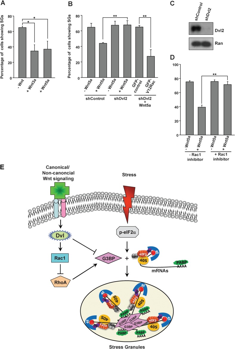
Cells often respond to diverse environmental stresses by inducing stress granules (SGs) as an adaptive mechanism. SGs are generally assembled as a result of aggregation of mRNAs stalled in a translational pre-initiation complex, mediated by a set of RNA-binding proteins such as G3BP and TIA-1. SGs may serve as triage centres for storage, translation re-initiation or degradation of specific mRNAs. However, the mechanism involved in the modulation of their assembly/disassembly is unclear. Here we report that Wnt signalling negatively regulates SG assembly through Dishevelled (Dvl), a cytoplasmic Wnt effector. Overexpression of Dvl2, an isoform of Dvl, leads to impairment of SG assembly through a DEP domain dependent mechanism. Intriguingly, the Dvl2 mutant K446M, which corresponds to an analogous mutation in Drosophila Dishevelled DEP domain (dsh(1)) that results in defective PCP pathway, fails to antagonize SG assembly. Furthermore, we show that Dvl2 exerts the antagonistic effect on SG assembly through a mechanism involving Rac1-mediated inhibition of RhoA. Dvl2 interacts with G3BP, a downstream component of Ras signalling involved in SG assembly, and functional analysis suggests a model wherein the Dvl-Rac1-RhoA axis regulates G3BP's SG-nucleating activity. Collectively, these results define an antagonistic effect of Wnt signalling on SG assembly, and reveal a novel role for Wnt/Dvl pathway in the modulation of mRNA functions.

Keywords: Dvl; G3BP; RNA; Stress granule; Wnt.


Figures

Fig. 1.
Fig. 1.. Dvl2 disassembles SGs in a DEP-dependent manner.
(A) NIH3T3 cells expressing indicated proteins were treated with sodium arsenite and immunostained for overexpressed proteins with HA antibodies (green) and for SGs with FXR1 antibodies (red). DNA was stained with Hoechst-33342 dye (blue). HA-firefly luciferase (Fluc) was used as the control. Scale bars: 10 µm. (B) NIH 3T3 cells were transfected with HA-Fluc or HA-Dvl2 and were subjected to arsenite treatment before immunostaining with HA antibodies (green) for detecting overexpressed proteins (green) and with Dcp1a antibodies (red) for identifying the P bodies. (C) Schematic representation of mouse Dvl2 constructs used in this study, and the ability of them to localize to and/or disassemble SGs upon overexpression. Amino acid numbers are as indicated. Deleted regions are shown as doted lines. ND denotes, not determined. (D) Cells were transfected with indicated mutant Dvl2 constructs and were immunostained with indicated antibodies after sodium arsenite treatment. (E) The graph depicts number of NIH3T3 cells transfected with indicated constructs displaying SG assembly after sodium arsenite treatment. Data represents average of three independent experiments. Error bars indicate ±SD. Data was analyzed by Student's t test (*P<0.05, **P<0.005).
Fig. 2.
Fig. 2.. Dvl2 promotes SG disassembly via Rac1-mediated inhibition of RhoA.
(A, upper panel) NIH3T3 cells were transfected with GFP-control vector or GFP-tagged version of constitutively-active Rac1 (V12Rac1), dominant-negative Rac1 (N17Rac1), constitutively-active RhoA (V14RhoA) or dominant-negative RhoA (N19RhoA) construct (green) and analyzed for SG assembly using HuR as the SG marker (red). DNA was stained in blue. (A, lower panel) NIH3T3 cells were transfected with GFP-V12Rac1 or GFP-N17Rac1 (green) with myc-V14RhoA or myc-N19RhoA (blue) as indicated, and analyzed for SG assembly using HuR (red). (B) The graph depicts number of NIH3T3 cells transfected with indicated constructs displaying SG assembly after sodium arsenite treatment. (C) NIH3T3 cells were co-transfected with HA-Dvl2 (blue) and GFP-tagged version of Rac1 or RhoA constructs (green), as indicated. Cells were analyzed for the SG assembly using HuR (red) as the marker. Scale bars: 10 µm. (D) Quantitative representation of SG assembly from experiments described in C. (E) NIH3T3 cells were treated with control (siControl), Rac1 (siRac1)- or RhoA (siRhoA)-specific siRNA and the cell lysate was analyzed for indicated proteins by Western blotting. α-tubulin was used as a loading control. (F) Quantitative depiction of SG assembly in control (siControl) or RhoA-specific siRNA (siRhoA) transfected cells. (G) NIH3T3 cells were transfected with control (siControl), Rac1-specific siRNA (siRac1) alone or with HA-Dvl2 as indicated, and analyzed for the number of transfected cells showing SG assembly after arsenite treatment. Data (in B, D, F and G) represents average of three independent experiments. Error bars indicate ±SD. Data was analyzed by Student's t test (**P<0.001, N.S.; non significant).
Fig. 3.
Fig. 3.. Dvl2 interacts with G3BP.
(A) HeLa S3 cells were transfected with GFP control or GFP-Dvl2 constructs and were subjected to arsenite treatment (0.2 mM, 30 min), and analyzed by western blotting (WB) with indicated antibodies. (B) HEK293T cells were lysed in the presence of RNasin or RNase A, and were subjected to immunoprecipitation (IP) using rabbit-IgG (control IP) or Dvl2-specific antibodies (Dvl2 IP). The immunoprecipitates were probed with indicated antibodies. (C) HEK293T cells were co-transfected with GFP or GFP-tagged Drosophila G3BP (GFP-G3BP) and HA-Dvl2 fragments as indicated. IP with GFP-specific antibody was performed and analyzed for the presence of Dvl2 fragments by Western blotting (WB). *indicates non-specific band. (D) Cells expressing GFP (control) or GFP-G3BP and indicated HA-Dvl2 deletion constructs were subjected to immunoprecipitation with GFP antibody and assayed for the presence of Dvl2 proteins by Western blotting with HA antibody. (E) Schematic representation of human G3BP (hG3BP) constructs used in this study. Different domains and phosphorylation-defective (S149A) and phospho-mimetic (S149E) mutants are indicated (for details, see (Tourriere et al., 2003). (F) HEK293T cells were co-transfected with HA-Dvl2 and indicated hG3BP constructs. Immunoprecipitation (IP) was performed using GFP-specific antibody and analyzed by Western blotting (WB) using indicated antibodies. (G & H) GFP-G3BP (green) was co-transfected with full length HA-Dvl2 (G, red) or indicated deletion mutants (H, red), and the cells were stained with HA-specific antibodies. (I) Cells were co-transfected with HA-Dvl2 (red) construct and indicated GFP-G3BP deletion mutants (green). DNA is stained in blue. Scale bars: 10 µm.
Fig. 4.
Fig. 4.. Dvl2 inhibits G3BP's ability to induce SGs.
(A) NIH3T3 cells were transfected with GFP-G3BP (green) alone or with HA-Dvl2 (blue) and stained for the SG marker HuR (red) in the absence (A) or presence (B) of sodium arsenite. Arrows indicate cytoplasmic puncta positive for GFP-G3BP and HuR. Arrowheads indicate puncta positive for GFP-G3BP and HA-Dvl2. Scale bars: 10 µm. (C) NIH3T3 cells were transfected with indicated constructs and subjected to arsenite treatment (0.5 mM, 30 min). Percentage of transfected cells displaying SGs, as identified by G3BP antibodies, was scored. (D) Quantitative data for experiments described in B. (E) Quantitative representation of cells showing SG assembly as assessed by HuR staining after transfection with indicated constructs. All the quantitative data represent average of three independent experiments. Error bars indicate ±SD. Data was analyzed by Student's t test (**P<0.001).
Fig. 5.
Fig. 5.. Wnt signalling antagonizes SG assembly.
(A) NIH3T3 cells were treated with Wnt3a or Wnt5a at a final concentration of 100 ng/ml for 13.5 h. Later, sodium arsenite (0.2 mM final concentration) was added to the medium for 30 min, and cells were stained for an SG marker, HuR. (B) NIH3T3 cells transfected with control (shControl) or Dvl2-shRNA (shDvl2) and GFP control or GFP-V12Rac1 were untreated (−) or treated (+) with Wnt5a as indicated and subjected to arsenite stress. SG assembly was analyzed by immunostaining with an SG specific marker (HuR). (C) Control or Dvl2-specific shRNA construct was transfected into NIH3T3 cells and analyzed for depletion of Dvl2 through Western blotting using affinity purified rabbit anti-Dvl2 antibodies. Ran was used as the loading control. (D) NIH3T3 cells were untreated (−) or treated (+) with Wnt5a and incubated with (+) or without (−) Rac1 inhibitor before subjecting to oxidative stress. Percentage of cells displaying SGs, as identified by HuR immunostaining, was scored. Graphical data (in A, B and D) represent averages of three independent experiments. Error bars indicate ±SD. Data was analyzed by Student's t test (*P≤0.01, **P≤0.001). (E) A working model suggesting the role of Wnt in negatively regulating SG assembly. Wnt signalling inhibits SG assembly by interfering with the ability of G3BP to aggregate translationally repressed mRNAs and to nucleate SGs, by physically interacting with Dvl2 and by a mechanism involving Rac1-mediated inhibition of Rho.

Similar articles

Cited by

References

    1. Almeida M., Han L., Martin-Millan M., O'Brien C. A., Manolagas S. C. (2007). Oxidative stress antagonizes Wnt signaling in osteoblast precursors by diverting beta-catenin from T cell factor- to forkhead box O-mediated transcription. J. Biol. Chem. 282, 27, 298–27, 305 10.1074/jbc.M70281120 - DOI - PubMed
    1. Anderson P., Kedersha N. (2006). RNA granules. J. Cell Biol. 172, 803–808 10.1083/jcb.200512082 - DOI - PMC - PubMed
    1. Anderson P., Kedersha N. (2008). Stress granules: the Tao of RNA triage. Trends Biochem. Sci. 33, 141–150 10.1016/j.tibs.2007.12.003 - DOI - PubMed
    1. Anderson P., Kedersha N. (2009). RNA granules: post-transcriptional and epigenetic modulators of gene expression. Nat. Rev. Mol. Cell. Biol. 10, 430–436 10.1038/nrm2694 - DOI - PubMed
    1. Axelrod J. D. (2001). Unipolar membrane association of Dishevelled mediates Frizzled planar cell polarity signaling. Genes Dev. 15, 1182–1187 10.1101/gad.890501 - DOI - PMC - PubMed