. 2012 Feb 15;1(2):140-60.
doi: 10.1242/bio.2011489. Epub 2011 Dec 30.

Structural and functional plasticity of subcellular tethering, targeting and processing of RPGRIP1 by RPGR isoforms

Affiliations

Structural and functional plasticity of subcellular tethering, targeting and processing of RPGRIP1 by RPGR isoforms

Hemangi Patil et al. Biol Open. .

Abstract

Mutations affecting the retinitis pigmentosa GTPase regulator-interacting protein 1 (RPGRIP1) interactome cause syndromic retinal dystrophies. RPGRIP1 interacts with the retinitis pigmentosa GTPase regulator (RPGR) through a domain homologous to RCC1 (RHD), a nucleotide exchange factor of Ran GTPase. However, functional relationships between RPGR and RPGRIP1 and their subcellular roles are lacking. We show by molecular modeling and analyses of RPGR disease-mutations that the RPGR-interacting domain (RID) of RPGRIP1 embraces multivalently the shared RHD of RPGR(1-19) and RPGR(ORF15) isoforms and the mutations are non-overlapping with the interface found between RCC1 and Ran GTPase. RPGR disease-mutations grouped into six classes based on their structural locations and differential impairment with RPGRIP1 interaction. RPGRIP1α(1) expression alone causes its profuse self-aggregation, an effect suppressed by co-expression of either RPGR isoform before and after RPGRIP1α(1) self-aggregation ensue. RPGR(1-19) localizes to the endoplasmic reticulum, whereas RPGR(ORF15) presents cytosolic distribution and they determine uniquely the subcellular co-localization of RPGRIP1α(1). Disease mutations in RPGR(1) (-19), RPGR(ORF15), or RID of RPGRIP1α(1), singly or in combination, exert distinct effects on the subcellular targeting, co-localization or tethering of RPGRIP1α(1) with RPGR(1-19) or RPGR(ORF15) in kidney, photoreceptor and hepatocyte cell lines. Additionally, RPGR(ORF15), but not RPGR(1-19), protects the RID of RPGRIP1α(1) from limited proteolysis. These studies define RPGR- and cell-type-dependent targeting pathways with structural and functional plasticity modulating the expression of mutations in RPGR and RPGRIP1. Further, RPGR isoforms distinctively determine the subcellular targeting of RPGRIP1α(1,) with deficits in RPGR(ORF15)-dependent intracellular localization of RPGRIP1α(1) contributing to pathomechanisms shared by etiologically distinct syndromic retinal dystrophies.

Keywords: RPGR; RPGRIP1; degeneration; kidney cells; photoreceptor; protein aggregation; protein targeting.


Conflict of interest statement

Competing interests: The authors have no financial conflict of interest.

Figures

Fig. 1.
Fig. 1.. Molecular modeling of the RCC1-homologous domain (RHD) of RPGR and disease-associated mutations to RCC1.
(A) Schematic diagram of RPGR isoforms, RPGR1–19 and RPGRORF15, and domains thereof. RPGR1–19 and RPGRORF15 share seven N-terminal repeats, where most human missense disease mutations are localized. This region is homologous to RCC1 (RHD). The N-terminal repeats 1 and 7 of RHD are partially conserved with RCC1. RPGR1–19 and RPGRORF15 have distinct C-terminal domains. Acidic (AD) and basic (BD) domains, nucleotide-binding (NB) and isoprenylation (CAAX) motifs, are shown. (B) Comparison of the ribbon diagram of the RHD model structure (left) to the seven propeller blades of RCC1 structure (middle) and superimposition of RHD and RCC1 structures (right). Blades are numbered B1–B7. Superimposed ribbons of RHD of RPGR and RCC1 are depicted in red and yellow, respectively. RHD of RPGR presents incomplete B1 and B7 blades and well-defined B2–B6 blades. The structures are axial views along the central shaft of the propeller structure. (C) Functional and colored ball representations of X-linked retinitis pigmentosa type 3 (XlRP3) mutations in RHD of RPGR. Left and right images are perpendicular and axial views of the central shaft of the propeller structure, respectively. Mutations are colored and mapped based on their predicted and functional effects on RHD (right table). Positions of green and white residues are those known to interface with Ran GTPase in RCC1 and among these, E116 and L268 are conserved between RHD and RCC1. Note that none of the XlRP3 mutations overlaps with the Ran GTPase-interacting interface and many, in particular those predicted to mediate protein-protein interactions, are in exposed loops. (D) Mapping of XlRP3-associated mutations to RHD by colored ball representation and based on the degree of conservation of the mutated residues with RCC1-homologous domains of other proteins. The position of poorly and semi-conserved residues tend to correlate with those predicted to mediate protein-protein interactions.
Fig. 2.
Fig. 2.. Characterization of XlRP3 mutations by quantitative yeast-two hybrid assays.
(A) Diagram of RPGRIP1α, domains thereof, and their direct partners, NPHP4, RPGR1–19 and RPGRORF15. The C2 domain interacts with nephrocystin-4 (NPHP4), whereas the RID (RPGR-interacting domain) associates with the conserved RHD of the RPGR1–19 and RPGRORF15 isoforms. Human disease mutations in C2 and RID of RPGRIP1α, NPHP4 and RHD of RPGR1–19 and RPGRORF15 are thought to promote uncoupling of RPGRIP1α with one or more of its partners. SMC/CC, coiled-coil domains; ND, nuclear domain; C2, protein kinase C conserved region 2. (B) Diagram of RPGR isoforms and domains thereof employed in the mutation analyses shown beneath in the graphs (C–E). Residue numbering depicts domain boundaries. Analyses of the interaction of RID of RPGRIP1α1 with various classes of missense mutations in the RHD (C) and PID (D) of N-RPGR and frame-shift mutations in RPGRORF15 (E) by quantitative yeast growth assays in selective growth media. Class I, II and III mutations are predicted by molecular modeling to cause misfolding, impair intramolecular and protein-protein interactions, respectively. Class IV mutations affect the region in the RHD of RPGR. Mutations of classes V and VI are restricted to PID and ORF15, respectively. Class I-V missense mutations were introduced in E463X construct. ORF15A construct is a retina-specific splice variant with the first 11 residues of ORF15. Results shown represent the mean ± S.D. (n = 3). Significant (P<0.05) or non-significant (P>0.05) changes between mutation constructs and control construct are noted with *. Legend: RHD, RCC1-homologous domain; PID, δPDE-interacting domain; ORF15, open reading frame 15 domain; CAAX, isoprenylation motif; Ctrl, control constructs; µmax, growth rate.
Fig. 3.
Fig. 3.. Expression and subcellular localization of RFP-RPGR1–19, RFP-RPGRORF15 and YFP-RPGRIP1α1 in COS7 cells.
(A) RFP-RPGR1–19 is restricted to the ER network in live cells (upper panel). RFP-RPGR1–19 co-localizes with the ER-tracker dye and its subcellular co-distribution extensively overlaps with that of the ER dye (upper panel). RFP-RPGRORF15 is dispersed throughout the cytoplasm and neither co-localizes nor co-distributes with the ER network of live cells stained with ER-tracker dye (lower panel). Inset pictures are an enlarged view of boxed areas. Scale bar, 13.3 µm (B) Immunoblot of cytosolic (C), membrane (M), nuclear (N) and cytoskeleton (Ck) fractions of COS7 cells transfected with RFP-RPGR1–19, RFP-RPGRORF15 or RFP alone. RFP-RPGR1–19 is mostly present in the membrane fraction with the ER marker, calreticulin, whereas RFP-RPGRORF15 and RFP alone are exclusively present in the cytosolic fraction with the cytosolic markers, GADPH and a cytosolic protein (Ns Prot) which cross-reacts non-specifically with the anti-RFP antibody. RanBP2 (Nup358), calreticulin, acetylated α-tubulin and GADPH, are markers for the nuclear, ER, cytoskeleton and cytosolic fractions, respectively. IB, immunoblot. (C) Expression of YFP-RPGRIP1α1 alone leads to the formation of profuse intracellular deposits (inset picture represent other cells with aggregates of YFP-RPGRIP1α1). Scale bar, 20 µm (D) Co-expression of RFP-RPGR1–19 (upper panel) or RFP-RPGRORF15 (lower panel) with YFP-RPGRIP1α1 in COS7 cells determines the RFP-RPGR isoform-dependent co-localization of YFP-RPGRIP1α1 to the ER network (upper panel) or throughout the cytoplasm (lower panel) and the perfect co-localization of either RFP-RPGR isoform with YFP-RPGRIP1α1. Co-expression of either RFP-RPGR isoform with YFP-RPGRIP1α1 also suppresses completely the formation of intracellular deposits of YFP-RPGRIP1α1. Inset pictures are enlarged views of boxed areas depicting the line scan used to plot two-channel fluorescent intensities graphs (right column, upper panel, 26.1 units = 1 µm; right column, lower panel, 11.5 units = 1 µm). The two-channel fluorescent intensity profile of either RFP-RPGR isoform and YFP-RPGRIP1α1 overlap perfectly. Scale bar: 26 µm, inset pictures, 6 µm. Images in (A), (C) and (D) are representative of cells examined.
Fig. 4.
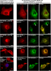
Fig. 4.. Expression analyses of disease mutations in RFP-RPGR1–19 or RFP-RPGRORF15 alone and co-expressed with wild-type YFP-RPGRIP1α1 in COS7 cells.
Disease mutations in constructs of RFP-RPGR isoforms expressed alone (1st column) were compared with identical mutant RFP-RPGR constructs co-expressed with wild-type YFP-RPGRIP1α1 for changes of their subcellular distribution pattern (2nd-4th columns) and co-localization (4th column) in COS7 cells. Expression of mutant RFP-RPGR isoforms alone do not cause changes in their subcellular distribution compared to wild-type isoforms alone, with the exception of the E230fs mutation in RFP-RPGRORF15 that in a fraction of cells leads to the formation of large vesicles of RFP-RPGRORF15 (1st column, 5th row) only when expressed alone. Co-expression of mutant RFP-RPGR1–19 or RFP-RPGRORF15 with wild-type YFP-RPGRIP1α1 neither affects the co-localization of wild-type YFP-RPGRIP1α1 with any mutant RFP-RPGR isoforms nor the subcellular localization of any of the mutant RFP-RPGR isoforms. Co-expression of either mutant RFP-RPGR isoform with wild-type YFP-RPGRIP1α1 suppresses also the formation of self-aggregates of wild-type YFP-RPGRIP1α1. Arrow depicts prominent deposits of YFP-RPGRIP1α1 in a cell expressing singly YFP-RPGRIP1α1. Inset picture shows different and dispersed intracellular distributions of RFP-RPGRORF15 with the E230fs mutation. Images shown are representative of cells examined. Scale bar, 26 µm.
Fig. 5.
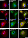
Fig. 5.. Co-expression analyses of disease mutations in YFP-RPGRIP1α1 with wild-type RFP-RPGR1–19 or RFP-RPGRORF15 in COS7 cells.
YFP-RPGRIP1α1 with disease-associated mutations (D1114G or EΔ1279) in its RID co-localizes with wild-type RFP-RPGR1–19 and RFP-RPGRORF15 isoforms and without forming intracellular deposits in COS7 cells. Images shown are representative of cells examined. Scale bar, 25 µm.
Fig. 6.
Fig. 6.. Co-expression analyses of disease mutations in YFP-RPGRIP1α1 and RFP-RPGR1–19 or RFP-RPGRORF15 in COS7 cells.
The G173R mutation in RFP-RPGR1–19 (A–C) or RFP-RPGRORF15 (D–F) co-expressed with the D1114G mutation in RID of YFP-RPGRIP1α1 cause the subcellular uncoupling (delocalization) of mutant RFP-RPGR1–19 or RFP-RPGRORF15 from mutant YFP-RPGRIP1α1 without grossly affecting the subcellular distribution pattern intrinsic to mutant RFP-RPGR1–19 or RFP-RPGRORF15 and the formation of intracellular deposits of mutant YFP-RPGRIP1α1. Pictures in (B) and (E) are enlarged views of boxed areas in (A) and (D) depicting the delocalization of mutant RFP-RPGR1–19 or RFP-RPGRORF15 from mutant YFP-RPGRIP1α1. (C) and (F) are plots of two-channel fluorescent intensities along the line scans of images (B) and (E), respectively. Scale bars, 13.3 µm; 2.6 µm for inset pictures. In fluorescent plots of (E) and (F), the distance of 56 units = 1 µm. Images shown are representative of cells examined.
Fig. 7.
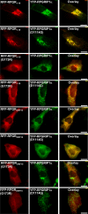
Fig. 7.. Mutations singly in RFP-RPGR1–19 or RFP-RPGRORF15 but not YFP-RPGRIP1α1 cause dissociation of YFP-RPGRIP1α1 from RFP-RPGR isoforms and the clustering of YFP-RPGRIP1α1 in a cone photoreceptor cell line (661W).
Wild-type RFP-RPGR1–19 or RFP-RPGRORF15 co-localize with wild-type YFP-RPGRIP1α1 in 661W cells. The D1114G mutation alone in YFP-RPGRIP1α1 had no effect on its co-localization with wild-type RFP-RPGR1–19 or RFP-RPGRORF15, whereas the G173R mutation in RFP-RPGR1–19 or RFP-RPGRORF15 is sufficient to delocalize wild-type YFP-RPGRIP1α1 from either mutant RFP-RPGR isoform and promote the clustering of YFP-RPGRIP1α1 throughout the cytosol. Images shown are representative of cells examined. Scale bar, 26 µm.
Fig. 8.
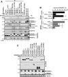
Fig. 8.. Effects of disease mutations on the physical tethering of RFP-RPGR isoforms with YFP-RPGRIP1α1 and limited proteolysis of YFP-RPGRIP1α1.
(A) Qualitative analyses of co-immunoprecipitation assays with extracts of COS7 cells (∼6×10⁶) co-transfected with wild-type or mutant RFP-RPGR isoforms and YFP-RPGRIP1α1 to examine the effects of disease mutations on the physical association of RPGR isoforms and other partners with RPGRIP1α1. Mutations singly in RPGR isoforms or RPGRIP1α1 weaken their physical association, whereas the presence of mutations in both RPGR isoforms and RPGRIP1α1 suppresses the physical association between these. The ectopically expressed wild-type and mutant RPGRIP1-RPGR complex also co-precipitate endogenous NPHP4, but not endogenous NPHP5 and SDCCAG8. Note that compared to RPGR1–19, RPGRORF15 enhances the expression levels of RPGRIP1α1.IP, immunoprecipitation; IB, immunoblot. (B) Quantitative analyses of co-immunoprecipitation assays on extracts of ∼6×10⁶ COS7 cells co-transfected with wild-type and mutant RFP-RPGR isoforms and YFP-RPGRIP1α1. Independent co-immunoprecipitation assays in triplicate (n = 3) with mutant constructs were normalized against the amount of wild-type YFP-RPGRIP1α1 co-precipitated by either wild-type RFP-RPGR1–19 (black bars) or RFP-RPGRORF15 (gray bars). Double disease mutations in RPGR1–19 or RPGRORF15 and RPGRIP1α1 cause maximal untethering of these partners, whereas the G173R mutation singly in either RPGR1–19 or RPGRORF15 typically has a stronger effect than the D1114G mutation singly in RPGRIP1α1. Data represent the mean ± S.D, n = 3; *, comparison of mutant constructs' group with wild-type; #, comparison between single and double mutant constructs; *,#, P<0.05 (Mann-Whitney test). (C) The lack of C-terminal proteolytic processing of YFP-RPGRIP1α1 upon co-expression with RFP-RPGRORF15, but not RFP-RPGR1-19, supports RPGRORF15 protects RPGRIP1α1 from proteolytic cleavage, whereas mutations in RPGRORF15 destroy such protection (middle panel). Note that expression of YFP-RPGRIP1α1 alone also suppresses its limited proteolysis because of the formation of self-aggregates of YFP-RPGRIP1α1 in cell culture. In comparison to the wild type protein, G173R in RFP-RPGRORF15, but not RFP-RPGR1–19, induce a significant upward electrophoretic mobility shift and a reduction of its expression level (upper panel). Lower panel is an immunoblot with anti-Hsc70 (loading control).
Fig. 9.
Fig. 9.. Temporal dynamics of the dispersion of pre-existing intracellular deposits of YFP-RPGRIP1α1 upon expression of RFP-RPGRORF15 in COS7 cells.
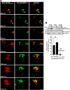
(A) Pre-existing YFP-RPGRIP1α1 deposits (arrows) were monitored by time-lapse imaging after subsequent expression of RFP-RPGRORF15. Time points represent still snapshots of a focused optical slice along the Z-axis captured from a time-lapse sequence and featuring significant events. Image capturing began 5 hours (300 min) after transfection of RFP-RPGRORF15 in COS7 cells expressing already YFP-RPGRIP1α1 for 16 hours. RFP-RPGRORF15 co-localizes with YFP-RPGRIP1α1 deposits (300–690 min). Then, aggregates of YFP-RPGRIP1α1 begin to diffuse and bursts of strong co-localization of YFP-RPGRIP1α1 with RFP-RPGRORF15 are visible during the rest of the 18.5 hour time-lapse experiment (810–1410 min). Note that an atrophic cell (arrowhead at 810, 1320 and 1350 min) with prominent YFP-RPGRIP1α1 deposits becomes phagocytized by another cell co-expressing RFP-RPGRORF15 and YFP-RPGRIP1α1 at 1350 min (e.g. arrowhead pointing to a restricted green area within the cell in yellow of the overlay image). This event leads to another strong burst of dispersed co-localization signal between YFP-RPGRIP1α1 and RFP-RPGRORF15 throughout the cell at 1410 min. All live images captured during the time-lapse imaging experiment were converted into a movie, which is presented in supplementary material Movie 1. Scale bar, 20 µm. (B) Time-course and biochemical partitioning of intracellular deposits of YFP-RPGRIP1α1 from non-cytosolic to cytosolic fractions upon expression of RFP-RPGRORF15 in COS7 cells with pre-existing aggregates of YFP-RPGRIP1α1. Non-cytosolic and cytosolic fractions of 1×10⁶ COS7 cells expressing YFP-RPGRIP1α1 for 16 hours and subsequently transfected with RFP-RPGRORF15 were analyzed at 0, 6 and 20 hours after transfection by qualitative (upper panel) and quantitative (lower panel) immunoblot analyses. The levels of YFP-RPGRIP1α1 decrease and increase in the non-cytosolic and cytosolic fractions, respectively, after 20 hr of expression of RFP-RPGRORF15. The ratio of non-cytosolic (NC) to cytosolic (C) of normalized levels of YFP-RPGRIP1α1 strongly decreases by ∼30-fold after 20 hours of expression of RFP-RPGRORF15 (lower panel). Results shown represent the mean ± S.D. (n = 4). *, comparison of 20 to 6 and 0 hours; ns (non-significant), comparison of 6 to 0 hours; P<0.05 is considered significant (Mann-Whitney test). A.U., arbitrary units.
Fig. 10.
Fig. 10.. Model of cell-context-dependent effects of XlRP3 and LCA mutations in the tethering of RPGR-RPGRIP1 complex.
In yeast cells, the lack of mammalian compensatory factors complementing the tethering of RPGR with RPGRIP1 causes the uncoupling of the RHD of either RPGR isoform (RPGR1–19 or RPGRORF15) from the RID of RPGRIP1 upon mutations affecting either domain of these proteins. In the 661W photoreceptor cell line, there is at least an accessory X factor, whose role compensates for the loss-of-function caused by the D1114G in RPGRIP1, but not for the loss-of-function of G173R mutation in the RHD of either RPGR isoform. In COS7 cells, the association between RPGR isoforms and RPGRIP1 depends on two accessory factors, X and Y, whose roles compensate for the loss of function in either RPGR isoform or RPGRIP1, but not both. In Hep2B liver cells, concomitant mutations in RPGR and RPGRIP1 do not cause the uncoupling of these, because of the expression of a third (Z) accessory and tethering factor, which compensates for functional deficits in RPGR and RPGRIP1. Hence, differences in expression of accessory and compensatory cell-type selective factors of the RPGRIP1 interactome may underlie the clinical and subcellular expression of the mutational load affecting components of the RPGRIP1 interactome and causing syndromic XlRP3, LCA or other diseases, such as nephronophthisis (NPHP4) and Senior-Løken syndrome (SLSN) affecting the retina, kidney or both. Legend: red bar, G173R substitution in RHD of RPGR isoforms causing XlRP3 disease; blue bar, D1114G substitution in RID of RPGRIP1 causing LCA disease. Mutant complex refers to mutations scenarios in RPGR, RPGRIP1 or both, in various cell types. Outcome refers to the effect of mutations in RPGR, RPGRIP1 or both, in the untethering of components of the RPGRIP1 interactome in various cell types.

References

    1. Abagyan R. A., Batalov S. (1997). Do aligned sequences share the same fold? J. Mol. Biol. 273, 355–368 10.1006/jmbi.1997.1287 - DOI - PubMed
    1. Abagyan R., Totrov M. (1994). Biased probability Monte Carlo conformational searches and electrostatic calculations for peptides and proteins. J. Mol. Biol. 235, 983–1002 10.1006/jmbi.1994.1052 - DOI - PubMed
    1. Abagyan R., Totrov M., Kuznetsov D. (1994). ICM-a new method for protein modeling and design: applications to docking and structure prediction from the distorted native conformation. J. Comput. Chem. 15, 488–506 10.1002/jcc.540150503 - DOI
    1. Abagyan R., Orry A., Raush E., Totrov M. (2010). ICM User Guide 3.7. La Jolla, CA: Molsoft LLC
    1. al-Ubaidi M. R., Font R. L., Quiambao A. B., Keener M. J., Liou G. I., Overbeek P. A., Baehr W. (1992). Bilateral retinal and brain tumors in transgenic mice expressing simian virus 40 large T antigen under control of the human interphotoreceptor retinoid-binding protein promoter. J. Cell Biol. 119, 1681–1687 10.1083/jcb.119.6.1681 - DOI - PMC - PubMed