. 2012 Mar 15;1(3):161-72.
doi: 10.1242/bio.2012043. Epub 2011 Dec 28.

Sumoylation is tumor-suppressive and confers proliferative quiescence to hematopoietic progenitors in Drosophila melanogaster larvae

Affiliations

Sumoylation is tumor-suppressive and confers proliferative quiescence to hematopoietic progenitors in Drosophila melanogaster larvae

Marta E Kalamarz et al. Biol Open. .

Abstract

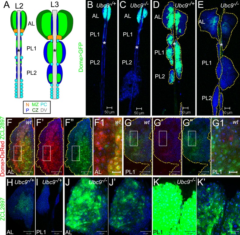
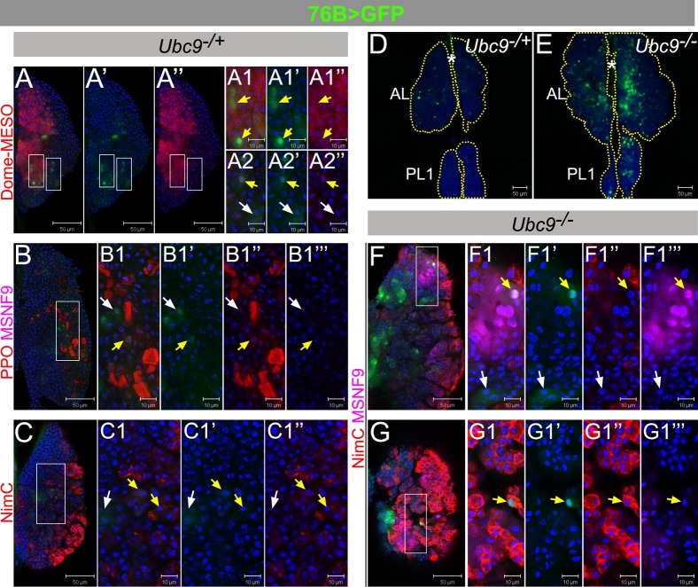
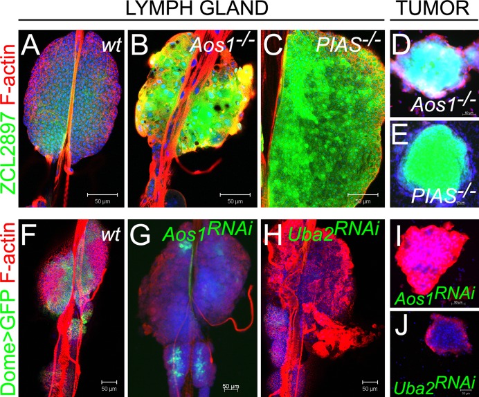
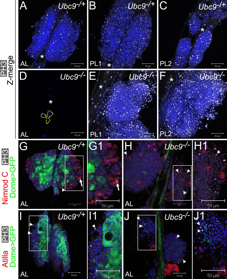
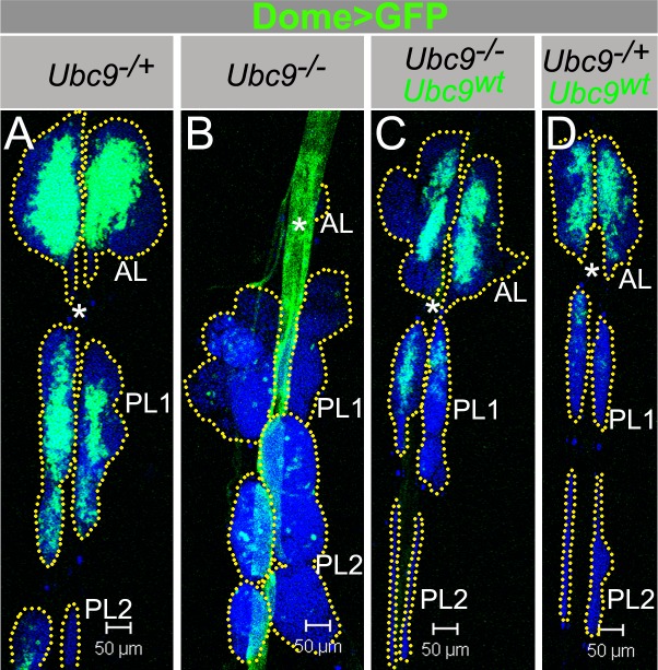
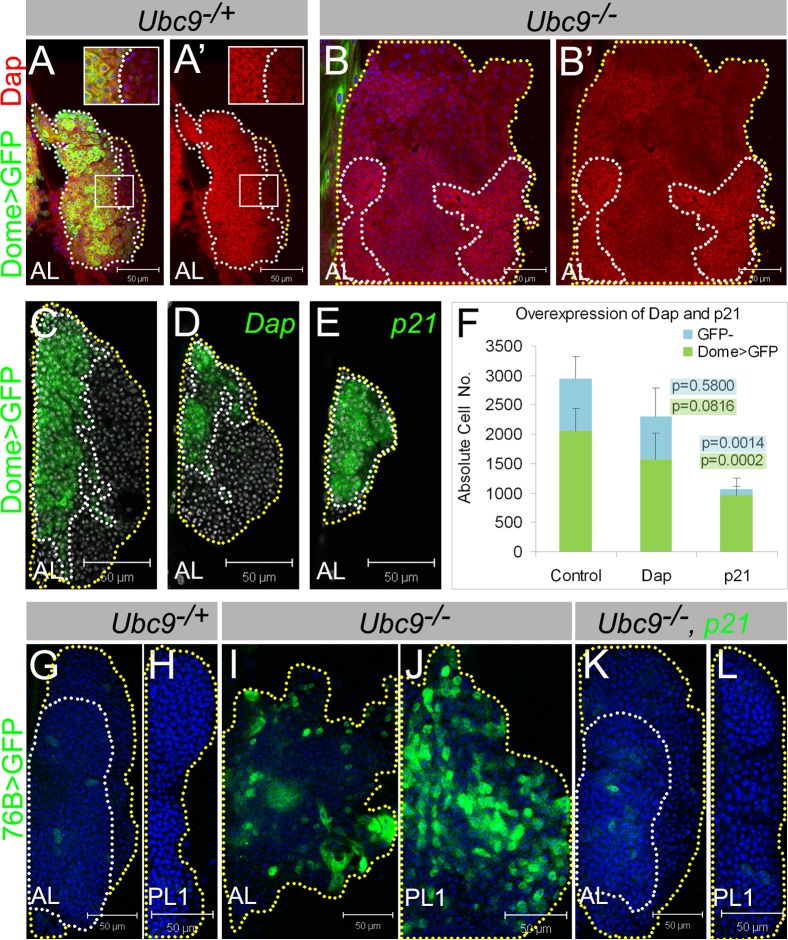
How cell-intrinsic regulation of the cell cycle and the extrinsic influence of the niche converge to provide proliferative quiescence, safeguard tissue integrity, and provide avenues to stop stem cells from giving rise to tumors is a major challenge in gene therapy and tissue engineering. We explore this question in sumoylation-deficient mutants of Drosophila. In wild type third instar larval lymph glands, a group of hematopoietic stem/progenitor cells acquires quiescence; a multicellular niche supports their undifferentiated state. However, how proliferative quiescence is instilled in this population is not understood. We show that Ubc9 protein is nuclear in this population. Loss of the SUMO-activating E1 enzyme, Aos1/Uba2, the conjugating E2 enzyme, Ubc9, or the E3 SUMO ligase, PIAS, results in a failure of progenitors to quiesce; progenitors become hyperplastic, misdifferentiate, and develop into microtumors that eventually detach from the dorsal vessel. Significantly, dysplasia and lethality of Ubc9 mutants are rescued when Ubc9(wt) is provided specifically in the progenitor populations, but not when it is provided in the niche or in the differentiated cortex. While normal progenitors express high levels of the Drosophila cyclin-dependent kinase inhibitor p21 homolog, Dacapo, the corresponding overgrown mutant population exhibits a marked reduction in Dacapo. Forced expression of either Dacapo or human p21 in progenitors shrinks this population. The selective expression of either protein in mutant progenitor cells, but not in other hematopoietic populations, limits overgrowth, blocks tumorogenesis, and restores organ integrity. We discuss an essential and complex role for sumoylation in preserving the hematopoietic progenitor states for stress response and in the context of normal development of the fly.

Keywords: Dacapo; Ubc9; dysplasia; hematopoiesis; microtumor; niche; organ integrity; p21; quiescence; stem cell; sumoylation; tumor suppressor.


Figures

Fig. 1.
Fig. 1.. Aberrant gene expression in progenitors of Ubc9 lymph glands.
Labeling: AL – anterior lobe(s), PL1 – first set of posterior lobes, PL2 – second set of posterior lobes; asterisk – dorsal vessel (DV). (A) Lymph glands in second (L2) and third (L3) larval instars. Medullary zone (MZ, light green), cortical zone (CZ, dark green); the niche (N, orange); unclassified cells (navy blue, P); pericardial cells (PC, light blue). Pairs of lobes aligned along the antero-posterior axis; PL1, PL2 consist of smaller lobes (2–3 pairs each) distinguishable at L2, but forming a continuous lobe at L3. (B–E) Dome>GFP (green) in lymph glands of 4-day L2: Ubc9−/+ (B), Ubc9−/−(C) and 6-day L3: Ubc9−/+ (D), Ubc9−/− (E). Lobes outlined in dotted marking (D,E). (F–G1) ZCL2897 (green) and Dome>DsRed (red) in wild type L3: AL (F–F”), PL (G–G”). Dome>DsRed (F',G') and ZCL2897 (F”,G”) shown separately; overlap of expressions (F,F1,G,G1). Regions of F,G (white rectangles) shown magnified in F1,G1, respectively. Yellow dotted markings outline the lobes (F–F”,G–G”). (H–K') ZCL2897 (green) expression in Ubc9−/+ (H, AL; I, PL1) and Ubc9−/− (J, AL; K, PL1) lymph glands; note: lobes in J and K are representative examples from different lymph glands. Increased ZCL2897 expression in Ubc9−/− (J,K) produced overexposure and samples were therefore re-imaged after reducing detector gain (J',K'). K–K' is a fragment of S2L. Brightness and contrast were slightly modified in panels F–G1 for clarity in merged images. Confocal sections (B–K'). Scale bars: 50 µm, except F1,G1 – 10 µm.
Fig. 2.
Fig. 2.. 76B-Gal4 characterization in control and Ubc9 lymph glands.
(A–C1″) L3 (6-day) Ubc9−/+ anterior lobes expressing 76B>GFP (green), co-labeled with Dome-MESO (red; A–A2″), or Pro-Phenol Oxidase and misshapen (PPO, red, and MSNF9, magenta, respectively; B–B1‴), or Nimrod C (NimC, red; C–C1″). (D–E) 76B>GFP (green) expression in 6-day Ubc9−/+ (D) and Ubc9−/−(E) lymph glands. (F–G1‴) L3 (6-day) Ubc9−/− anterior lobes expressing 76B>GFP (green), labeled with Nimrod C and misshapen (NimC, red and MSNF9, magenta, respectively); presented are lower (F–F1‴) and upper (G–G1‴) optical sections of two lymph glands. Split-channel images show green (′), red (″) or magenta (‴). Selected regions (white rectangles) are shown as high magnifications (panels labeled with numbers 1–2). White arrows – cells expressing singly 76B>GFP, and yellow arrows – two markers; star–DV. Confocal sections (A–G1‴). Scale bars: 50 µm (A–A″,B,C,D,E,F,G) and 10 µm (remaining panels).
Fig. 3.
Fig. 3.. Sumoylation enzymes in larval hematopoiesis.
(A–E) ZCL2897 (green) expression in wild type (A), Aos1−/− (B) and PIAS−/− (C) lymph glands and in tumors of Aos1−/− (D) and PIAS−/− (E) larvae. (F–H) Dome>GFP (green) in control lymph gland (F, without RNAi constructs). Reduction of Dome>GFP in lymph glands expressing Dome>Aos1RNAi (G) and Dome>Uba2RNAi (H). (I, J) Tumors form in animals expressing Dome>Aos1RNAi (I) and Dome>Uba2RNAi (J). Parental UAS-Aos1RNAi and UAS-Uba2RNAi classes do not produce tumors. Confocal sections (A–C,F,H,J) and fluorescent microscopy (D,E,G,I). Scale bars: 50 µm.
Fig. 4.
Fig. 4.. Overproliferation of immature cells in Ubc9 lymph gland.
(A–F) Phosphorylated histone H3 (PH3, white) in 6.5–7-day L3 Ubc9−/+ AL, PL1, PL2 (A,B,C, respectively) and Ubc9−/− AL (D; remaining cells outlined), PL1, PL2 (E,F, respectively). Star marks DV. Optical Z-sections merged (A–C,E–F). Mutant PL1 and PL2 (E,F) are partially detached from the DV and misaligned; lobe orientation (top – anterior, bottom – posterior) is reverse of the DV. (G–J1) Dome>GFP (green), PH3 (white; arrowheads) and Nimrod C (red, G–H1), or Atilla (red, I–J1) in 6-day AL in Ubc9−/+ (G,G1,I,I1) and Ubc9−/− (H,H1,J,J1) animals. PH3/Nimrod C localization in the same cell (G1, arrow). Regions indicated in G,H,I,J magnified in G1,H1,I1,J1, respectively. Confocal sections (A–J1). Scale bars: 50 µm.
Fig. 5.
Fig. 5.. Dome>Ubc9wt restores Ubc9 lymph gland size and Dome>GFP expression.
(A–D) Dome>GFP in Ubc9−/+ (A); Ubc9−/− (B); Ubc9−/−, Dome>Ubc9wt (C); Ubc9−/+, Dome>Ubc9wt (D). Asterisk (DV); dotted line outlines the lobes (A–D). Confocal sections (A–D). Scale bars: 50 µm.
Fig. 6.
Fig. 6.. Lymph gland cells respond to cell cycle inhibitors Dacapo/p21 and human p21 rescues Ubc9 tumorogenesis.
(A–B') Dacapo (red) and Dome>GFP (green) expression in L3 Ubc9−/+ (A,A') and Ubc9−/− (B,B') lymph glands. Indicated regions (A,A') are magnified in the insets. (A',B') show Dacapo staining only. (C–F) Lymph glands with Dome>GFP (C); Dome>Dap, GFP (D); Dome>p21, GFP (E) expression; nuclei labeled in white. Absolute cell numbers in Dome>GFP; Dome>Dap, GFP; Dome>p21, GFP (F; average ± SE, n≥5 animals per genotype); p values relative to control are shown on the graph (for Dome>GFP populations with green highlight, for remaining GFP cells with blue highlight). Dotted lines outline MZ (white) and CZ (yellow). (G–L) 76B>GFP (green) in Ubc9−/+ AL (G), PL1 (H); Ubc9−/− AL (I), PL1 (J); Ubc9−/−, 76B>p21, GFP AL (K), PL1 (L). Outlines: compact tissue (white line), lobe edges (yellow). Confocal sections (A–L). Brightness of images in A–B' was slightly increased for clarity without modifying the result. Scale bars: 50 µm.
Fig. 7.
Fig. 7.. Sumoylation controls proliferation of progenitor cells along with Dacapo.
Hematopoietic progenitors express high level of either one or both, ZCL2897 (green) and Dome>GFP (red). Some of these cells and cells in transition zone express 76B-Gal4 (cyan). At third instar stage, progenitors enter quiescence. Heterogeneity and absence of mature marker expression suggest similarity to mammalian transit amplifying cells (Morrison and Spradling, 2008; Shaker and Rubin, 2010) or Drosophila testis progenitors (Shivdasani and Ingham, 2003). We propose that sumoylation regulates multiple events including maintenance of high levels of Dacapo protein in these cells. In the absence of sumoylation enzymes Aos1/Uba2, Ubc9, and PIAS, these cells fail to quiesce and progress into G2/M phase (cells with purple outline) and misdifferentiate (dark green cells); dysplasia and tumorogenesis follow.

Similar articles

Cited by

References

    1. Adnane J., Jackson R. J., Nicosia S. V., Cantor A. B., Pledger W. J., Sebti S. M. (2000). Loss of p21WAF1/CIP1 accelerates Ras oncogenesis in a transgenic/knockout mammary cancer model. Oncogene 19, 5338–5347 10.1038/sj.onc.1203956 - DOI - PubMed
    1. Apionishev S., Malhotra D., Raghavachari S., Tanda S., Rasooly R. S. (2001). The Drosophila UBC9 homologue lesswright mediates the disjunction of homologues in meiosis I. Genes Cells 6, 215–224 10.1046/j.365-2443.2001.00413.x - DOI - PubMed
    1. Asha H., Nagy I., Kovacs G., Stetson D., Ando I., Dearolf C. R. (2003). Analysis of Ras-induced overproliferation in Drosophila hemocytes. Genetics 163, 203–215 - PMC - PubMed
    1. Bourbon H. M., Gonzy-Treboul G., Peronnet F., Alin M. F., Ardourel C., Benassayag C., Cribbs D., Deutsch J., Ferrer P., Haenlin M. et al. (2002). A P-insertion screen identifying novel X-linked essential genes in Drosophila. Mech. Dev. 110, 71–83 10.1016/S0925-4773(01)00566-4 - DOI - PubMed
    1. Brand A. H., Perrimon N. (1993). Targeted gene expression as a means of altering cell fates and generating dominant phenotypes. Development 118, 401–415 - PubMed

LinkOut - more resources