. 2012 Mar 15;1(3):237-46.
doi: 10.1242/bio.2012027. Epub 2012 Feb 3.

The adult retinal stem cell is a rare cell in the ciliary epithelium whose progeny can differentiate into photoreceptors

Affiliations

The adult retinal stem cell is a rare cell in the ciliary epithelium whose progeny can differentiate into photoreceptors

Brian G Ballios et al. Biol Open. .

Abstract

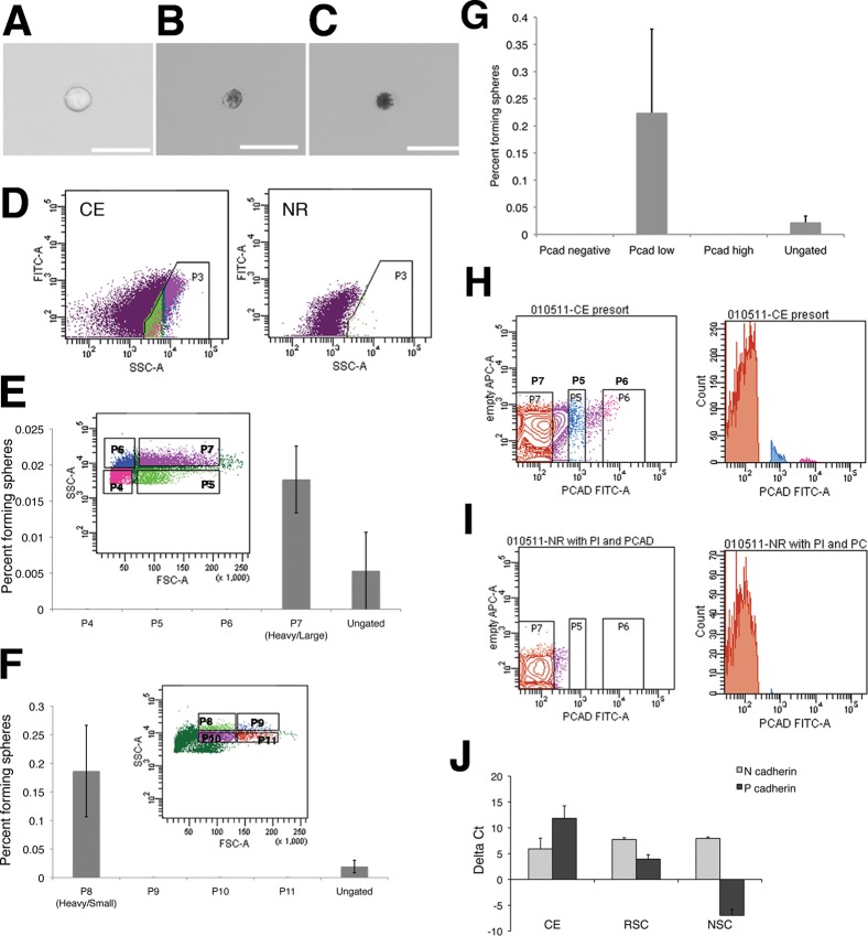
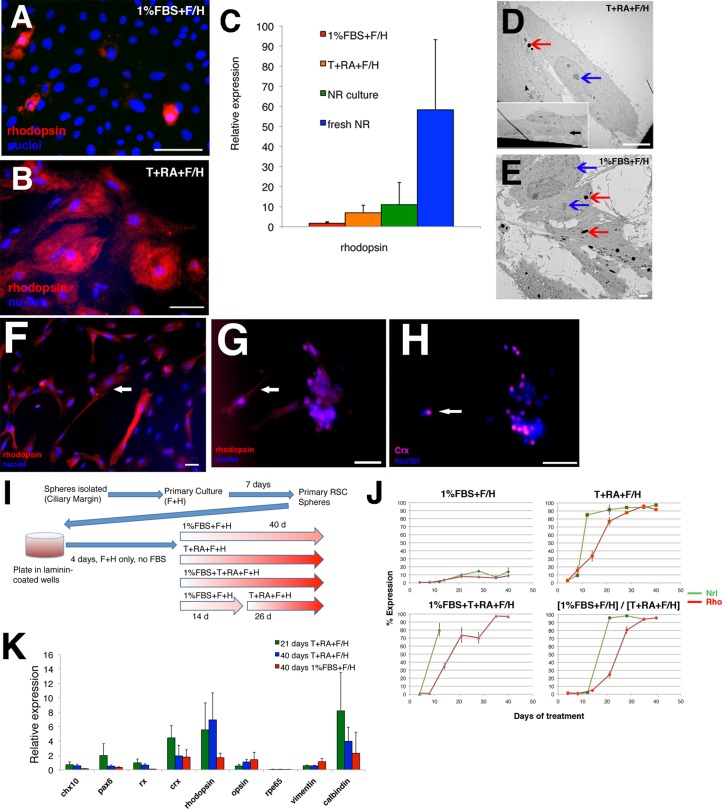
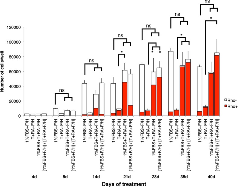
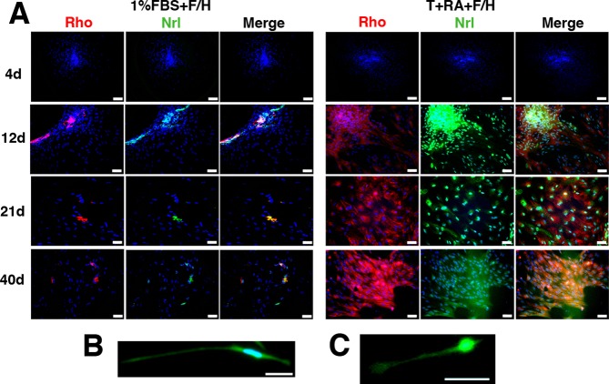
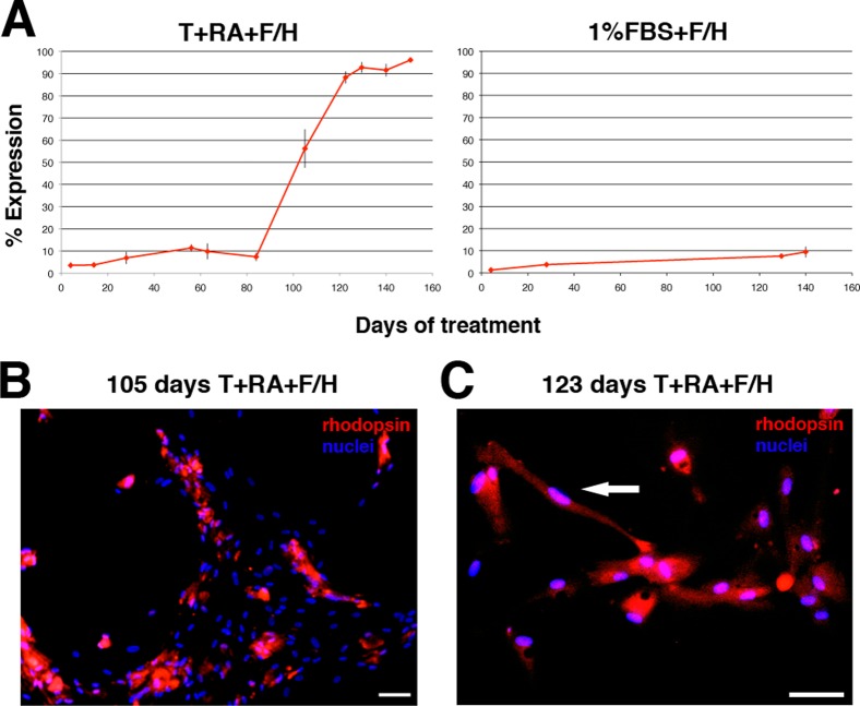
Self-renewing, multipotential retinal stem cells (RSCs) reside in the pigmented ciliary epithelium of the peripheral retina in adult mammals. RSCs can give rise to rhodopsin positive-cells, which can integrate into early postnatal retina, and represent a potentially useful option for cellular therapy. The ability to purify a stem cell population and direct the differentiation toward a particular cell lineage is a challenge facing the application of stem cells in regenerative medicine. Here we use cell sorting to prospectively enrich mouse RSCs based on size, granularity and low expression of P-cadherin and demonstrate that only rare cells with defined properties proliferate to form colonies. We show that clonally-derived mouse and human RSC progeny are multipotent and can differentiate into mature rhodopsin-positive cells with high efficiency using combinations of exogenous culture additives known to influence neural retinal development, including taurine and retinoic acid. This directed RSC differentiation follows the temporal sequence of photoreceptor differentiation in vivo, and the cells exhibit morphology, protein and gene expression consistent with primary cultures of rods in vitro. These results demonstrate that the RSC, an adult stem cell, can be enriched and directed to produce photoreceptors as a first step toward a targeted cell replacement strategy to treat retinal degenerative disease.

Keywords: photoreceptor differentiation; retina; stem cells.


Conflict of interest statement

Competing interests: None to declare.

Figures

Fig. 1.
Fig. 1.. RSCs can be isolated from ciliary epithelial (CE) cells using forward and side scatter and express the surface adhesion molecule P-cadherin.
CE dissection yields a variety of cell types: single non-pigmented cells are present (A) along with (B) lightly pigmented, and (C) heavily pigmented cells. Scale bars represent 25 µm. (D) Pigmented cells from dissociated CE make up a population with high side scatter that is not present in dissociated neural retina (NR). (E) This pigmented cell population was divided into 4 quadrants, of which only one gave rise to RSC colonies. (F) The population of large, heavily pigmented cells (P7) was subdivided into 4 new quadrants and RSCs colonies formed only from the smaller, most heavily pigmented cells. Cells were plated at clonal density (10 cells/µL) for colony forming assays. Error bars represent s.e.m., N = 3 for each.. Dissociated CE cells were immunostained for P-cadherin. Cells were divided into negative (P7), low (P5), and high (P6) P-cadherin level (G), FITC intensity along horizontal axis), which was not present in the secondary-only controls. (H) A histogram of cell counts for sorting of dissociated CE cells, while sorting dissociated NR (I) confirms that gate P7 is sorting for P-cadherin-negative cells, as NR cells do not express P-cadherin. All RSC colonies are derived from P-cadherin-low cells, N = 2. Freshly isolated CE expresses high levels of mRNA for P-cadherin, expressed in the pigmented CE and N-cadherin, expressed in the non-pigmented layer. (J) RSC colonies are derived from pigmented CE cells, but also express high levels of both cadherins as spheres are a mix of pigmented RSCs and non-pigmented progenitors. Data shown are deltaCt compared to the endogenous control rplp0 ± s.e.m., N = 2 or 3.
Fig. 2.
Fig. 2.. Exogenous factors can induce directed-differentiation of mouse RSC progeny to a rod photoreceptor fate in a pattern consistent with developmental rod production.
(A,B) Representative images of rhodopsin-positive cells (red, cytoplasmic) following 40 days of culture in (A) 1% FBS + F/H, (B) T+RA+F/H. Nuclei are shown with Hoechst staining (blue). Scale bars represent 50 µm. (C) Q-PCR analysis comparing rhodopsin expression between 1% FBS + F/H and T+RA+F/H culture conditions, compared to primary cultures of dissociated adult neural retina and undissociated (fresh) adult neural retina. Data are reported relative to expression in primary undifferentiated RSC spheres. Error bars represent s.e.m. (1% FBS+F/H, N = 4; T+RA+F/H, N = 6; NR culture, N = 2; fresh NR, N = 3). (D–E) Representative electron microscopic images of cells differentiated at 40 days in (D) T+RA+F/H or (E) 1%FBS+F/H. Cells differentiated in T+RA+F/H show a morphology lacking in ciliation and pigmentation (D and inset, black arrowhead), compared to the extensive ciliation and retention of pigment granules seen with cells differentiated in serum. Light grey intracellular regions represent lysosomes and are not pigment granules. Coloured arrowheads have been used to indicate the difference between pigment granules (red arrowheads) and cellular organelles (blue arrowheads) – which include lysosomes and nucleoli. Scale bars represent 10 µm (2 µm inset) (D) or 2 µm (E). (F) RSC-derived rods staining positive for rhodopsin (red) at 28 days differentiation in taurine/retinoic acid show a neuronal morphology (arrows) with long process extending from a central cell body (arrowheads), similar to cultured primary rod photoreceptors from adult mice for 4 days in T+RA. Scale bar represents 50 μm. (G,H) Primary adult rods are positive for (G) rhodopsin (red cytoplasmic) and (H) Crx (purple nuclear). Where these cells form aggregates when plated, the morphology looks similar to aggregates of RSC-derived rod photoreceptors (see Fig. 4). Nuclei are shown with Hoechst staining (blue). Scale bars represent 50 µm. (I) Schematic representation of the differentiation protocol developed for directed-differentiation of rod photoreceptors from primary RSCs isolated from Nrl.gfp animals. (J) Expression profiles of Nrl (green) and rhodopsin (red) over 40 days of differentiation in 1% FBS+F/H, T+RA+F/H, 1% FBS+T+RA+F/H, and the [1% FBS+F/H]/[T+RA+F/H] sequential protocol. Nrl expression precedes rhodopsin expression by approximately 1 week in T+RA, and is consistent regardless of a two week expansion in 1% FBS+F/H. Error bars represent s.e.m. (N>3). (K) Q-PCR analysis comparing genes for multiple retinal cell types between 1% FBS+F/H (40 days), T+RA+F/H (21 days), and T+RA+F/H (40 days). Data are reported relative to expression in primary undifferentiated RSC spheres. Error bars represent the s.e.m. of independent experiments (N = 4–6).
Fig. 3.
Fig. 3.. Absolute numbers of cells/well over 40 days of RSC differentiation in various culture conditions.
Red bars represent the number of rhodopsin-positive cells present in each condition at each time point. Significantly greater proliferation is observed in media supplemented with 1% FBS than taurine and retinoic acid (* P<0.05); however, the absolute number of rhodopsin-positive cells is greater at 40 days. Error bars represent s.e.m.
Fig. 4.
Fig. 4.. Nrl and rhodopsin protein expression profiles in differentiating RSCs.
(A) Representative images of differentiating cultures in 1% FBS+F/H and T+RA+F/H conditions at 4, 12, 21, and 40 days. In T+RA conditions, 12 days corresponds to high Nrl and lower rhodopsin expression, while at 21 days both Nrl and rhodopsin expression are high. By 40 days, cytoplasmic spreading results from further differentiation and the GFP is more widespread in the cultures. The images show overlap of Nrl (gfp, green) and rhodopsin (red) expression in the cell cytoplasm, including a number of Nrl+/rho+ and Nrl+/rho− cells. Nuclei are shown with Hoechst staining (blue). Scale bars represent 100 µm (4d) or 50 µm (12d, 21d, 40d). (B,C) Similar morphology is exhibited at the single cell level in low-density culture, between (B) a mature RSC-derived rhodopsin-positive cell (28 days) and (C) a cultured primary adult rod photoreceptor (3 days in vitro). The cytoplasmic morphology is visualized with ACTN.GFP (B) and Nrl.gfp (C). Scale bars represent 50 µm.
Fig. 5.
Fig. 5.. Exogenous factors can induce directed-differentiation of human RSC progeny to a rod photoreceptor fate.
(A) Expression profiles of rhodopsin (red) in human RSC progeny over 150 days of differentiation in T+RA+F/H, 1% FBS+F/H. Error bars represent s.e.m. (N = 2–3). (B,C) Representative images of rod photoreceptor progeny arising in differentiating cultures of human RSC progeny at 105 and 123 days of differentiation in T+RA+F/H. Note the mature polarized neuronal morphology characteristic of mature rods adopted by isolated RSC-derived rods (arrowhead). Nuclei are shown with Hoechst staining (blue). Scale bars represent 100 µm.

Similar articles

Cited by

References

    1. Aaku-Saraste E., Hellwig A., Huttner W. B. (1996). Loss of occludin and functional tight junctions, but not ZO-1, during neural tube closure-Remodeling of the neuroepithelium prior to neurogenesis. Dev. Biol. 180, 664–679 10.1006/dbio.1996.0336 - DOI - PubMed
    1. Adler R., Hatlee M. (1989). Plasticity and differentiation of embryonic retinal cells after terminal mitosis. Science 243, 391–393 10.1126/science.2911751 - DOI - PubMed
    1. Akimoto M., Cheng H., Zhu D., Brzezinski J. A., Khanna R., Filippova E., Oh E. C. T., Jing Y., Linares J.-L., Brooks M. et al. (2006). Targeting of GFP to newborn rods by Nrl promoter and temporal expression profiling of flow-sorted photoreceptors. Proc. Natl. Acad. Sci. USA 103, 3890–3895 10.1073/pnas.0508214103 - DOI - PMC - PubMed
    1. Altshuler D., Lo, Turco J. J., Rush J., Cepko C. (1993). Taurine promotes the differentiation of a vertebrate retinal cell type in vitro. Development 119, 1317–1328 - PubMed
    1. Baas D., Bumsted K. M., Martinez J. A., Vaccarino F. M., Wikler K. C., Barnstable C. J. (2000). The subcellular localization of Otx2 is cell-type specific and developmentally regulated in the mouse retina. Brain Res. Mol. Brain Res. 78, 26–37 10.1016/S0169-328X(00)00060-7 - DOI - PubMed