. 2012 Mar 15;1(3):275-85.
doi: 10.1242/bio.2012380. Epub 2012 Jan 19.

The Aplnr GPCR regulates myocardial progenitor development via a novel cell-non-autonomous, Gα(i/o) protein-independent pathway

Affiliations

The Aplnr GPCR regulates myocardial progenitor development via a novel cell-non-autonomous, Gα(i/o) protein-independent pathway

Sivani Paskaradevan et al. Biol Open. .

Abstract

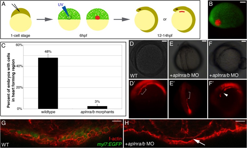
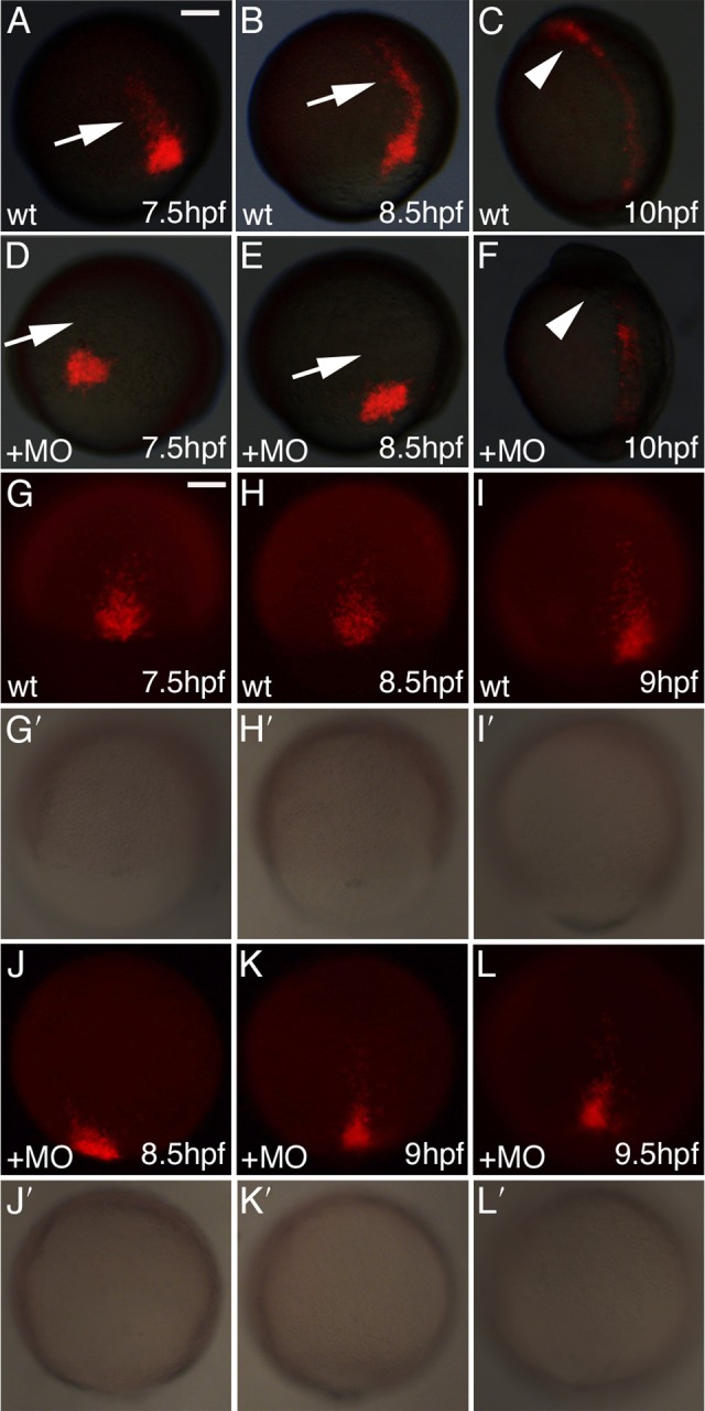
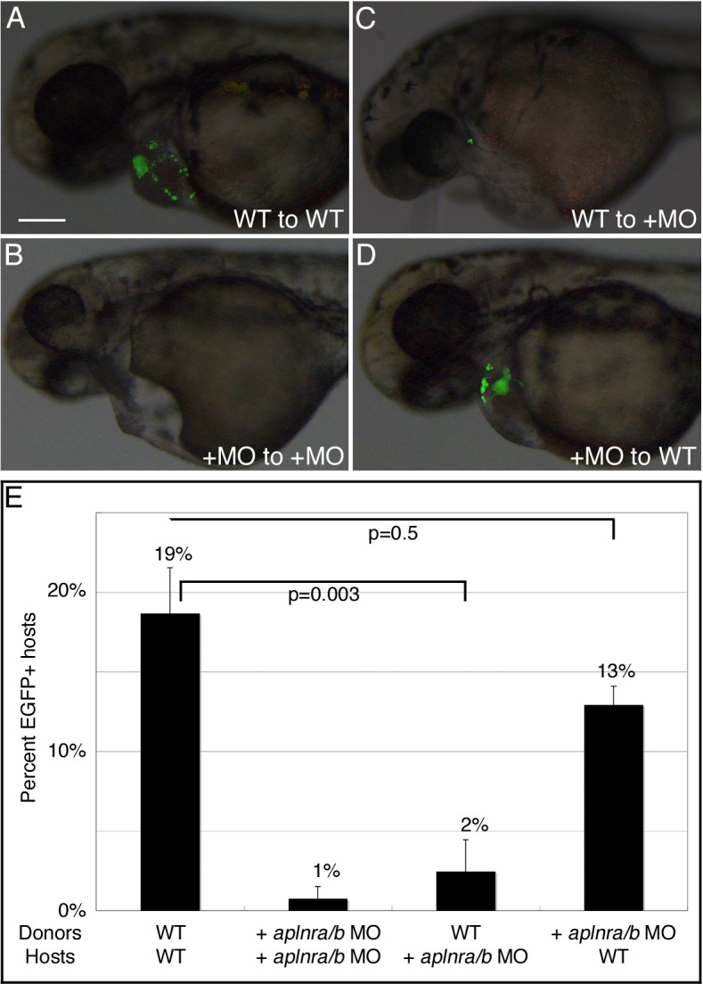
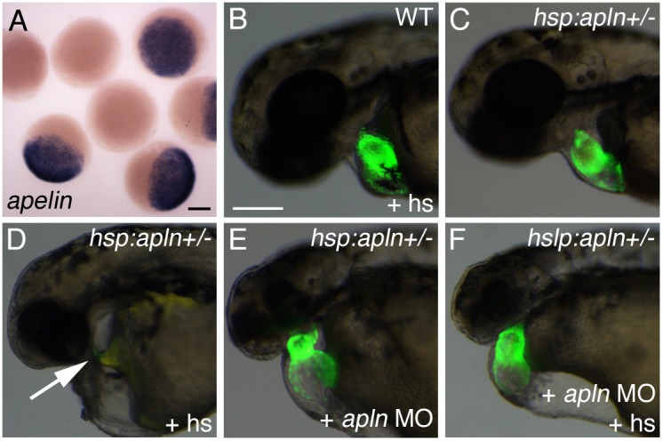
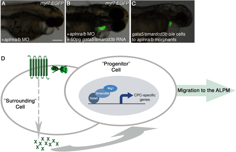
Myocardial progenitor development involves the migration of cells to the anterior lateral plate mesoderm (ALPM) where they are exposed to the necessary signals for heart development to proceed. Whether the arrival of cells to this location is sufficient, or whether earlier signaling events are required, for progenitor development is poorly understood. Here we demonstrate that in the absence of Aplnr signaling, cells fail to migrate to the heart-forming region of the ALPM. Our work uncovers a previously uncharacterized cell-non-autonomous function for Aplnr signaling in cardiac development. Furthermore, we show that both the single known Aplnr ligand, Apelin, and the canonical Gα(i/o) proteins that signal downstream of Aplnr are dispensable for Aplnr function in the context of myocardial progenitor development. This novel Aplnr signal can be substituted for by activation of Gata5/Smarcd3 in myocardial progenitors, suggesting a novel mechanism for Aplnr signaling in the establishment of a niche required for the proper migration/development of myocardial progenitor cells.

Keywords: Apelin Receptor; Gastrulation; Myocardial Progenitors; Zebrafish.


Figures

Fig. 1.
Fig. 1.. Cells from the lateral embryonic margin fail to reach the heart-forming region in aplnra/b morphants.
(A) Schematic of photoconversion method. mRNA encoding KikGR is injected at the 1-cell stage. Cells at the lateral embryonic margin (90° from dorsal, anatomically marked by the shield) are photoconverted by UV light at 6hpf. Embryos are scored at 12–14hpf for the presence of cells in the heart-forming region of the ALPM. (B) Merged fluorescent views of 6hpf embryo following photoconversion of lateral margin cells. (C) Graph of percentage of embryos with photoconverted cells that reach the heart-forming region. In wildtype embryo labeled cells are found in the lateral plate mesoderm (D′), including the heart-forming region (bracket in D′). In aplnra/b morphants labeled cells are found in the lateral plate mesoderm but fail to extend anteriorly to the heart-forming region (bracket in E′) or are excluded from the heart-forming region (arrowhead in F′) while cells are found both and anterior and posterior to this region. (D–F) are bright field images of embryos in (D′-F′). (D,F) lateral views with anterior to the left, (E) dorsal view with anterior to the top. For (C) N = 3, n = 57 for WT; N = 4, n = 104 for aplnra/b morphants; p = 0.001. (G,H) cross-sections through the ALPM of 20hpf wildtype (WT) and aplnra/b morphant myl7:EGFP embryos, respectively, stained with rhodamine-phalloidin to visualize cells. EGFP+ cardiac progenitors are present in wildtype embryos (G) while there is and absence of cells in the heart-forming region in aplnra/b morphants (arrow in H). See also supplementary material Fig. S3. Scale bars represent 100μm (B, D/D′, E/E′ and F/F′), 17μm (G) and 12μm (H).
Fig. 2.
Fig. 2.. Cells from the lateral embryonic margin display a defect in the initiation of migration.
(A–F) Embryos injected with KikGR RNA were photoconverted at shield stage to mark cells in the lateral margin. Representative images of wildtype and aplnra/b morphant embryos at 70% epiboly (7.5hpf), 80% epiboly (8.5hpf), and bud (10hpf) stages. Arrows indicate leading streak of migrating cells in wildtype embryos and their absence in morphant embryos. Arrowheads indicate cells in the heart-forming region of wildtype embryos and absence of cells in morphants. (G–L) Time-lapse image of one wildtype and one aplnra/b morphant embryo during gastrulation demonstrating a delay in the initiation of migration of morphant cells. Corresponding images of the embryos are shown in (G′-L′) for each fluorescent image. Scale bar represents 100μm (A).
Fig. 3.
Fig. 3.. Aplnr signaling functions cell-non-autonomously in the development of cardiomyocytes.
Transplantation of wildtype (WT) or aplnra/b MO-injected (+MO) myl7:EGFP donor embryos to the margin WT or aplnra/b MO-injected host embryos was carried out at 4hpf. (A–D) Representative host embryos at 2dpf following transplantation; lateral view, anterior to left. (C) wildtype cells are unable to contribute to the heart in aplnra/b morphant host embryos, whereas aplnra/b morphant cells are able to contribute to the heart in wildtype hosts (D). (E) Graph of percentage of host embryos with contribution from myl7:EGFP donor cells. N = 5, n = 236 for wildtype donors and hosts; N = 4, n = 130 for aplnra/b morphant donors and hosts; N = 4, n = 190 for wildtype donors and aplnra/b morphant hosts; N = 3, n = 190 for aplnra/b morphant donors and wildtype hosts. Views in (A–D) are lateral views, with anterior to the left. Scale bar represents 200μm (A).
Fig. 4.
Fig. 4.. Apelin is not necessary for the development of cardiomyocytes.
(A) Embryos produced from crossing hsp70:apln+/- and wildtype fish showing robust up-regulation of apelin expression by RNA in situ hybridization following heat shock in embryos carrying the hsp70:apln transgene. (B) Wildtype fish are unaffected by heat-shock (+hs). (C) Unshocked hsp70:apln+/- embryos are phenotypically wildtype. (D) Overexpression of apelin leads to a loss in cardiac tissue (arrow shows empty cardiac region). (E) Injection of apelin MO leads to a heart that is present, but dismorphic. (F) Injection of apelin MO followed by heat shock of hsp70:apln embryos results in phenocopy of the apelin morphant heart phenotype. Scale bars represent 200μm (A and B, B scale bar is for images B–F).
Fig. 5.
Fig. 5.. Aplnr activity is independent of Gαi/o proteins in cardiomyocyte development.
(A–C) Bright field images of wildtype (WT), aplnra/b morphant, and PTX overexpressing myl7:EGFP embryos at 2dpf. (A–C) Fluorescent images of embryos in (A–C), (A″–C″) are overlays of (A–C) and (A′–C′). Overexpression of PTX (C) recapitulates the aplnra/b morphant (B) cardiac phenotype. (D–F) Transplantation of myl7:EGFP donor cells from WT or PTX overexpressing cells to WT or PTX overexpressing host embryos at 4hpf was carried out. Hosts were subsequently scored at 2dpf for EGFP +′ve donor cells in the heart. (D–E) PTX overexpressing cells are able to contribute to the heart in wildtype embryos (D), and wildtype cells are able to contribute to the heart in PTX overexpressing host embryos (E), suggesting that Gαi/o proteins are not downstream of Aplnr signaling in cardiomyocyte development. (F) Graph showing percentage of host embryos with contribution from myl7:EGFP donor cells in transplants performed. N = 5, n = 236 for wildtype donors and hosts; N = 2, n = 80 for PTX O/E donors and hosts; N = 3, n = 158 for wildtype donors and PTX O/E hosts; N = 3, n = 216 for PTX O/E donors and wildtype hosts. O/E = overexpressing. (G,H) Cross sections through the ALPM of 20hpf myl7:EGFP wildtype and PTX overexpressing embryos. The ALPM is evident at the level of the heart-forming region following PTX addition (arrow in H) but cells fail to differentiate into cardiomyocytes as noted by the absence of EGFP +′ve cells. (A–E) are lateral views, with anterior to the left. Scale bars represent 200μm (A and D), 17μm (G) and 22μm (H).
Fig. 6.
Fig. 6.. Gata5/smarcd3b function downstream of Aplnr signaling to direct cells to the heart-forming region.
(A,B) Overexpression of gata5 and smarcd3b is able to rescue the aplnra/b morphant cardiac phenotype in myl7:EGFP embryos. (C) Cells from myl7:EGF embryos overexpressing gata5 and smarcd3b can contribute to the heart when transplanted into aplnra/b morphant embryos. (D) Working model for Aplnr signaling in cardiac progenitor development. Aplnr activity functions independently of Gαi/o proteins in the production of extracellular factor(s)/cue(s). These factor(s)/cue(s) act cell-non-autonomously upon neighboring cells in the lateral embryonic margin, wherein gata5/smarcd3b, as part of the cBAF complex, functions cell-autonomously, facilitating their migration to the ALPM for development into cardiac progenitors. Aplnr activity is not required in progenitor cells. o/e: overexpressing. Scale bar represents 200μm (A).

Similar articles

Cited by

References

    1. Ashley E. A., Powers J., Chen M., Kundu R., Finsterbach T., Caffarelli A., Deng A., Eichhorn J., Mahajan R., Agrawal R. et al. (2005). The endogenous peptide apelin potently improves cardiac contractility and reduces cardiac loading in vivo. Cardiovasc. Res. 65, 73–82 10.1016/j.cardiores.2004.08.018 - DOI - PMC - PubMed
    1. Auda-Boucher G., Bernard B., Fontaine-Perus J., Rouaud T., Mericksay M., Gardahaut M. F. (2000). Staging of the commitment of murine cardiac cell progenitors. Dev. Biol. 225, 214–225 10.1006/dbio.2000.9817 - DOI - PubMed
    1. Barnes G., Japp A. G., Newby D. E. (2010). Translational promise of the apelin--APJ system. Heart 96, 1011–1016 10.1136/hrt.2009.191122 - DOI - PubMed
    1. Berghmans S., Murphey R. D., Wienholds E., Neuberg D., Kutok J. L., Fletcher C. D., Morris J. P., Liu T. X., Schulte-Merker S., Kanki J. P. et al. (2005). tp53 mutant zebrafish develop malignant peripheral nerve sheath tumors. Proc. Natl. Acad. Sci. USA 102, 407–412 10.1073/pnas.0406252102 - DOI - PMC - PubMed
    1. Bondue A., Blanpain C. (2010). Mesp1: a key regulator of cardiovascular lineage commitment. Circ. Res. 107, 1414–1427 10.1161/CIRCRESAHA.110.227058 - DOI - PubMed