. 2012 May 15;1(5):488-97.
doi: 10.1242/bio.2012885. Epub 2012 Apr 11.

Drosophila Syncrip binds the gurken mRNA localisation signal and regulates localised transcripts during axis specification

Affiliations

Drosophila Syncrip binds the gurken mRNA localisation signal and regulates localised transcripts during axis specification

Suzanne M McDermott et al. Biol Open. .

Abstract

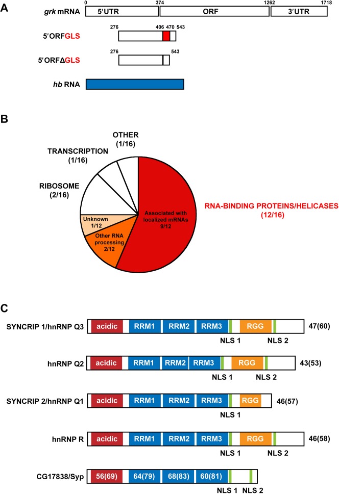
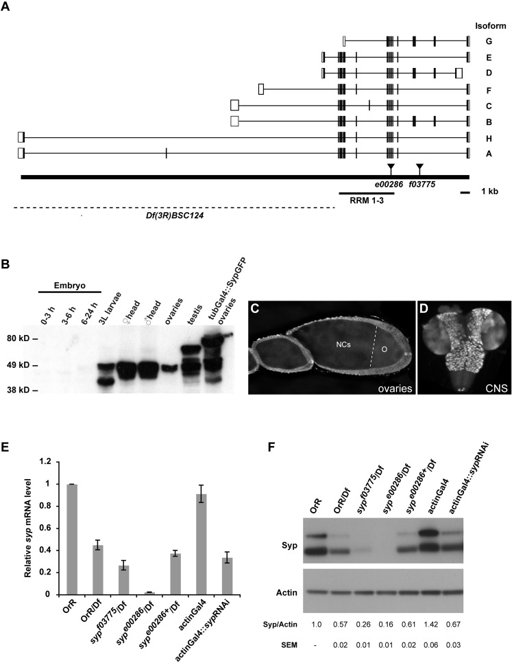
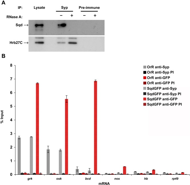
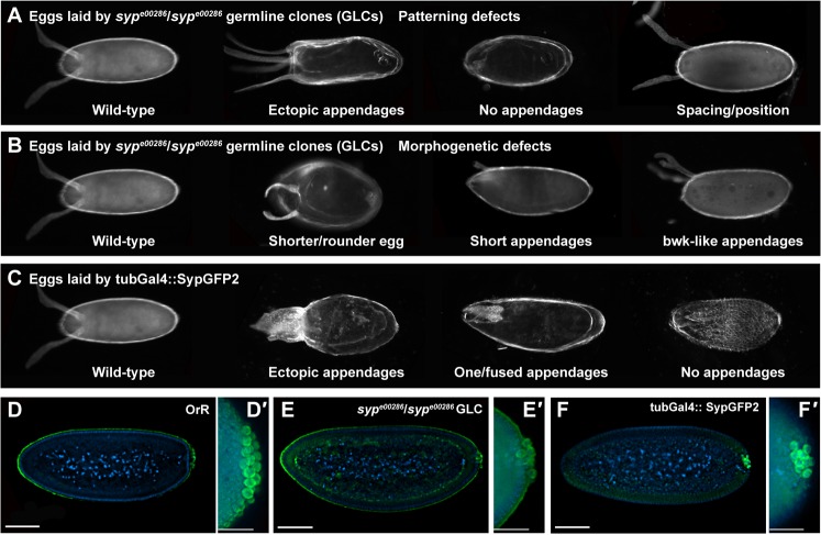
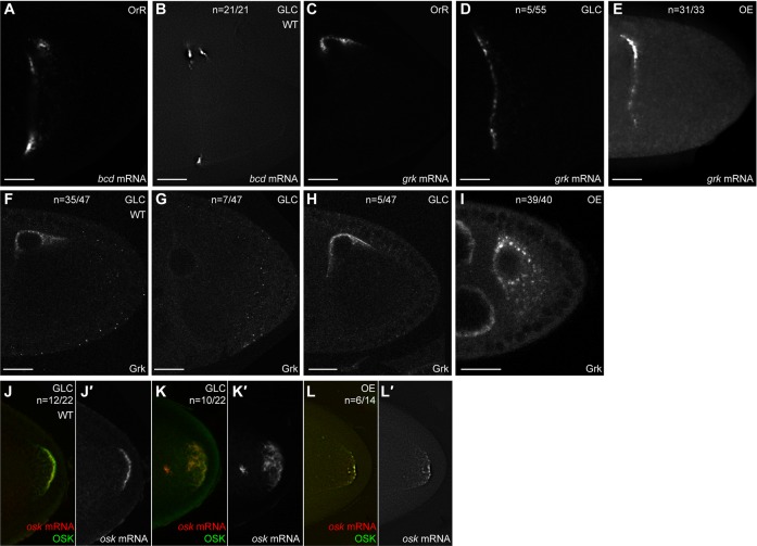
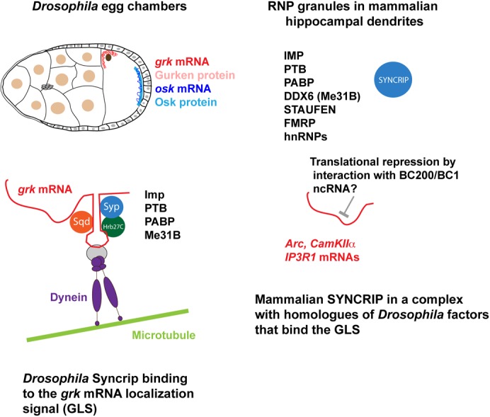
In the Drosophila oocyte, mRNA transport and localised translation play a fundamental role in axis determination and germline formation of the future embryo. gurken mRNA encodes a secreted TGF-α signal that specifies dorsal structures, and is localised to the dorso-anterior corner of the oocyte via a cis-acting 64 nucleotide gurken localisation signal. Using GRNA chromatography, we characterised the biochemical composition of the ribonucleoprotein complexes that form around the gurken mRNA localisation signal in the oocyte. We identified a number of the factors already known to be involved in gurken localisation and translational regulation, such as Squid and Imp, in addition to a number of factors with known links to mRNA localisation, such as Me31B and Exu. We also identified previously uncharacterised Drosophila proteins, including the fly homologue of mammalian SYNCRIP/hnRNPQ, a component of RNA transport granules in the dendrites of mammalian hippocampal neurons. We show that Drosophila Syncrip binds specifically to gurken and oskar, but not bicoid transcripts. The loss-of-function and overexpression phenotypes of syncrip in Drosophila egg chambers show that the protein is required for correct grk and osk mRNA localisation and translational regulation. We conclude that Drosophila Syncrip is a new factor required for localisation and translational regulation of oskar and gurken mRNA in the oocyte. We propose that Syncrip/SYNCRIP is part of a conserved complex associated with localised transcripts and required for their correct translational regulation in flies and mammals.

Keywords: Drosophila; Syncrip; localised translation; mRNA localization; oogenesis.


Conflict of interest statement

Competing interests: The authors declare that there are no competing interests.

Figures

Fig. 1.
Fig. 1.. GRNA affinity chromatography identifies the previously uncharacterized Drosophila homologue of mammalian SYNCRIP.
(A) Illustration of the RNA fragments used to prepare GRNA resins. 5′ORF RNA consists of the GLS surrounded by 211 nucleotides spanning the 3′ of the 5′UTR into the 5′ of the grk open reading frame. 5′ORFΔGLS is the same sequence, but lacks the GLS. (B) Classification of the proteins able to specifically associate with 5′ORF RNA by protein type and function. 16 proteins were found to specifically associate with 5′ORF RNA, 12 of which were RNA binding proteins or RNA helicases. Nine of these proteins, or their orthologues, have also been shown by previous studies to be associated with localised mRNAs in a variety of model organisms. (C) Schematic representation of the mouse and human SYNCRIP/hnRNP Q isoforms, hnRNP R and Drosophila CG17838 (isoform F)/Syp. Acidic, protein domain rich in acidic amino acids; RRM, RNA Recognition Motif; NLS, Nuclear Localisation Signal; RGG, arginine-glycine-glycine-rich domain. hnRNP Q2 has a deletion within RRM 2, whilst hnRNP Q1 lacks the second putative nuclear localisation signal and has a unique C-terminus. hnRNP R differs only in the identity of a number of amino acids at the very N-terminus of the protein. The N-terminal acidic domain and the three RRMs are highly conserved in Drosophila CG17838, which lacks the C-terminal RGG boxes. Percent amino acid identity and similarity shared by the Drosophila protein is indicated for each mammalian protein and for individual domains. Percent similarity is given in brackets.
Fig. 2.
Fig. 2.. Syp isoforms are expressed during oogenesis and in the larval nervous system.
(A) The genomic structure of the syp gene and associated alleles. Black bars represent coding sequence exons and white boxes represent untranslated sequence exons. Alternative splicing is predicted to produce a number of transcripts corresponding to isoforms A to H. The sype00286 allele is an insertion within an exon coding for RRM 3, whilst sypf03775 lies within a C-terminal intron. The deficiency Df(3R)BSC124 uncovers a large portion of the syp gene. (B) Western blot of extracts from a range of tissues and developmental stages, probed with guinea-pig anti-Syp antibodies. Extracts are OrR, unless otherwise stated. (C) Wild-type (OrR) egg chambers stained with guinea-pig anti-Syp antibodies. Syp is expressed both in the germline and in the somatic follicle cells. The dashed line delineates the approximate boundary between the oocyte (O) and nurse cells (NCs). (D) Wild-type (OrR) third instar larval brain stained with guinea-pig anti-Syp antibodies. (E) The levels of syp mRNA in third instar larvae of the indicated genotypes were quantified by qRT-PCR and normalized to rp49 mRNA. Error bars indicate the mean ± SEM from three independent experiments. qPCR primers were designed to amplify all syp isoforms. The syp RNAi line is 33012 from VDRC. (F) Syp protein levels in larvae of the same genotypes as in (E) were assessed by western blot analysis and quantified by densitometry using actin for normalization. The upper Syp band was used for quantification. Data are shown as means ± SEM from three independent experiments. The reduction in Syp protein levels in sype00286/Df(3R)BSC124, sypf03775/Df(3R)BSC124, and actinGal4::sypRNAi larvae shows that the anti-Syp antibody is highly specific.
Fig. 3.
Fig. 3.. Syp is a component of grk and osk RNP complexes.
(A) Western blot of immunoprecipitation from wild-type ovarian lysate with anti-Syp antibodies or pre-immune serum with (+) or without (−) RNase A treatment. An amount of lysate equal to 0.5% of that used for immunoprecipitation was also loaded. (B) RNA immunoprecipitation from wild-type (OrR) or SqdGFP ovarian lysate with anti-Syp or anti-GFP antibodies, or pre-immune sera (PI). The indicated mRNAs were detected by RT-qPCR analysis. Results are expressed as the percentage of input mRNA. Data are shown as means ± SEM from three independent experiments.
Fig. 4.
Fig. 4.. Altering the level of Syp in the germline results in grk and osk phenotypes.
(AC) The range of dorso-ventral patterning and morphogenetic phenotypes of eggs laid by females with a sype00286 mutant or SypGFP2 overexpressing germline. ‘bwk-like appendages’ refers to a dorsal appendage phenotype resembling that observed in bullwinkle mutants. See also Table 3. (DF) Cellular blastoderm embryos collected from wild-type (OrR) females and females with a sype00286 mutant or SypGFP2 overexpressing germline were stained with anti-Vasa antibody (green) to visualize pole cells. Many embryos from mutant germline and overexpressing females have a reduced number of Vasa-positive pole cells. Scale bars 40 µm. See also Table 4.
Fig. 5.
Fig. 5.. Syp modulates grk and osk mRNA localisation and translation.
(AE) In situ hybridization using a grk or bcd probe was carried out on wild-type (OrR), sype00286 germline clone (GLC) or SypGFP2 overexpressing (OE) egg chambers. WT indicates a wild-type pattern of mRNA localisation. Scale bar 20 µm. (FI) sype00286 germline clone (GLC) or SypGFP2 overexpressing (OE) egg chambers were stained with anti-Grk antibodies. WT indicates a wild-type distribution of Grk protein. Scale bar 20 µm. (JL) In situ hybridization using an osk probe was carried out on sype00286 germline clone (GLC) and SypGFP2 overexpressing (OE) egg chambers that were also stained with anti-Osk antibodies. WT indicates a wild-type distribution of osk mRNA and Osk protein.
Fig. 6.
Fig. 6.. Syncrip is a conserved regulator of mRNA localisation and translational repression in Drosophila oocytes, as well as mammalian hippocampal dendrites.

Similar articles

Cited by

References

    1. Adolph S. K., DeLotto R., Nielsen F. C., Christiansen J. (2009). Embryonic expression of Drosophila IMP in the developing CNS and PNS. Gene Expr. Patterns 9, 138–143 10.1016/j.gep.2008.12.001 - DOI - PubMed
    1. Arn E. A., Cha B. J., Theurkauf W. E., Macdonald P. M. (2003). Recognition of a bicoid mRNA localization signal by a protein complex containing Swallow, Nod, and RNA binding proteins. Dev. Cell 4, 41–51 10.1016/S1534-5807(02)00397-0 - DOI - PubMed
    1. Ashraf S. I., McLoon A. L., Sclarsic S. M., Kunes S. (2006). Synaptic protein synthesis associated with memory is regulated by the RISC pathway in Drosophila. Cell 124, 191–205 10.1016/j.cell.2005.12.017 - DOI - PubMed
    1. Bannai H., Fukatsu K., Mizutani A., Natsume T., Iemura S., Ikegami T., Inoue T., Mikoshiba K. (2004). An RNA-interacting protein, SYNCRIP (heterogeneous nuclear ribonuclear protein Q1/NSAP1) is a component of mRNA granule transported with inositol 1,4,5-trisphosphate receptor type 1 mRNA in neuronal dendrites. J. Biol. Chem. 279, 53427–53434 10.1074/jbc.M409732200 - DOI - PubMed
    1. Barbee S. A., Estes P. S., Cziko A. M., Hillebrand J., Luedeman R. A., Coller J. M., Johnson N., Howlett I. C., Geng C., Ueda R. et al. (2006). Staufen- and FMRP-containing neuronal RNPs are structurally and functionally related to somatic P bodies. Neuron 52, 997–1009 10.1016/j.neuron.2006.10.028 - DOI - PMC - PubMed