. 2012 Jul 15;1(7):640-7.
doi: 10.1242/bio.20121420. Epub 2012 May 21.

CD81 and CD9 work independently as extracellular components upon fusion of sperm and oocyte

Affiliations

CD81 and CD9 work independently as extracellular components upon fusion of sperm and oocyte

Naoko Ohnami et al. Biol Open. .

Abstract

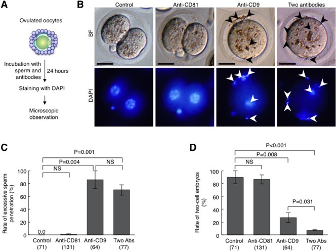
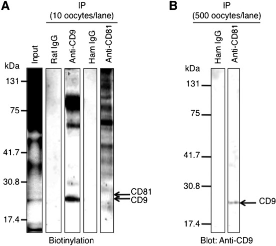
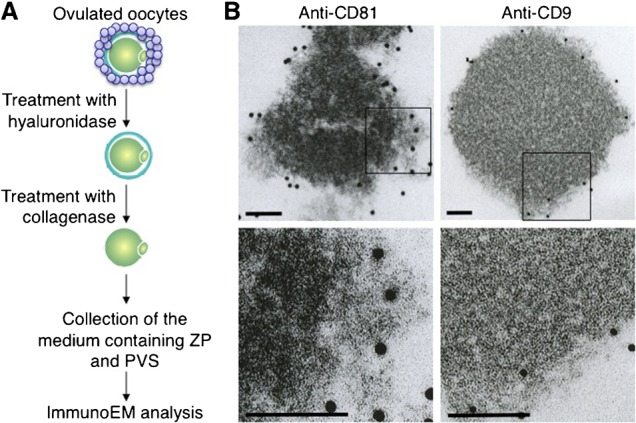
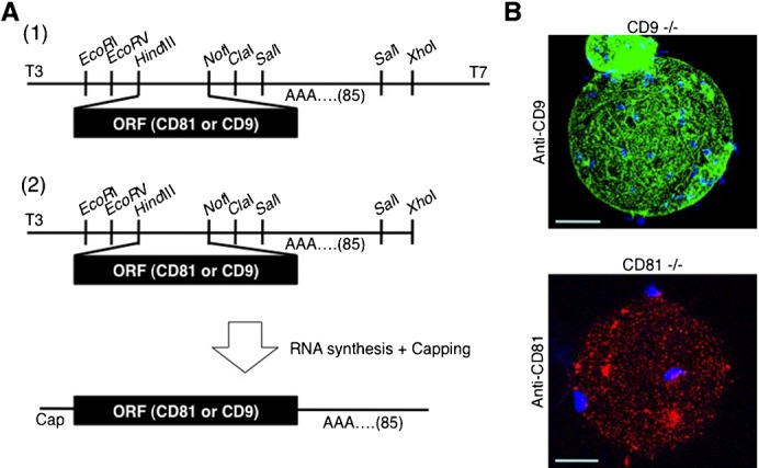
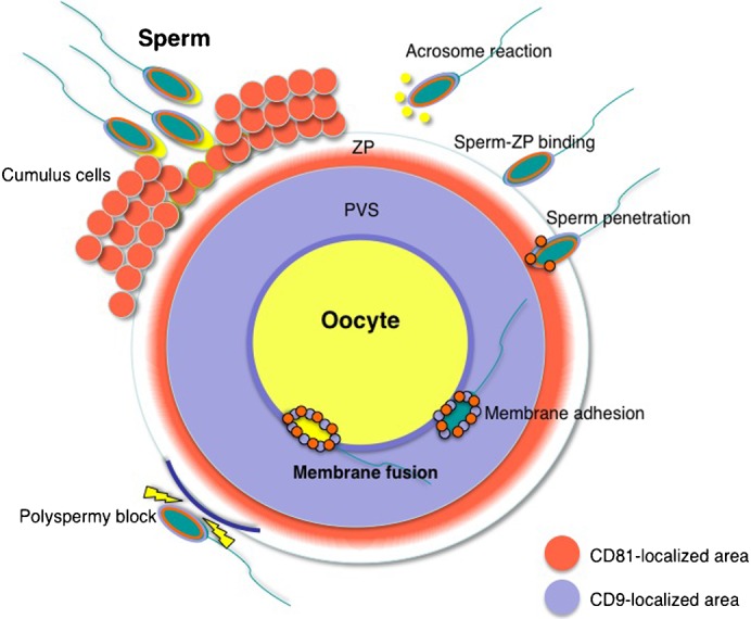
When a sperm and oocyte unite into one cell upon fertilization, membranous fusion between the sperm and oocyte occurs. In mice, Izumo1 and a tetraspanin molecule CD9 are required for sperm-oocyte fusion as one of the oocyte factors, and another tetraspanin molecule CD81 is also thought to involve in this process. Since these two tetraspanins often form a complex upon cell-cell interaction, it is probable that such a complex is also formed in sperm-oocyte interaction; however, this possibility is still under debate among researchers. Here we assessed this problem using mouse oocytes. Immunocytochemical analysis demonstrated that both CD9 and CD81 were widely distributed outside the oocyte cell membrane, but these molecules were separate, forming bilayers, confirmed by immunobiochemical analysis. Electron-microscopic analysis revealed the presence of CD9- or CD81-incorporated extracellular structures in those bilayers. Finally, microinjection of in vitro-synthesized RNA showed that CD9 reversed a fusion defect in CD81-deficient oocytes in addition to CD9-deficient oocytes, but CD81 failed in both oocytes. These results suggest that both CD9 and CD81 independently work upon sperm-oocyte fusion as extracellular components.

Keywords: CD81; CD9; Exosome; Membrane fusion.


Conflict of interest statement

Competing interests: The authors declare that there are no competing financial interests.

Figures

Fig. 1.
Fig. 1.. Inhibitory effects of anti-CD9 and anti-CD81 on fertilization.
(A) Experimental flow for testing the rate of excessive sperm penetration in perivitelline space (PVS) of an oocyte and the rate of two-cell embryos. Oocytes collected from oviducts of superovulated female mice were subjected to IVF in the presence of anti-CD9 (50 µg/ml) and/or anti-CD81 (50 µg/ml) or a preimmune Ab (50 µg/ml) for 24 hours. They were then stained with DAPI. BF, bright field. (B) Embryos 24 hours after IVF in the presence of Abs. Arrowheads marked sperm accumulated at the PVS. Scale bars: 20 µm. (C) The rate of embryos exhibiting excess sperm penetration. Parentheses  =  number of oocytes examined. NS, not significant. Values are the mean±s.e.m. (D) The rate of two-cell embryos 24 hours after IVF in the presence of Abs. Parentheses  =  number of oocytes examined. NS, not significant. Values are the mean±s.e.m.
Fig. 2.
Fig. 2.. Subcellular localization of CD9 and CD81 in oocytes.
(A) Experimental flow. Oocytes were isolated from oviducts, and cumulus cells were removed from oocytes by treatment with hyaluronidase. Oocytes were then reacted with anti-CD9 and anti-CD81 without fixation and observed under a confocal microscope. (B) Oocytes immunostained with anti-CD9 and anti-CD81. In each panel, boxes in the middle set of panels were enlarged and shown on the lower. BF, bright field; PVS, perivitelline space; ZP, zona pellucida. In each panel, scale bars: 20 µm.
Fig. 3.
Fig. 3.. Biochemical analysis for identification of CD9 and CD81 in the extracellular region of an oocyte.
(A) Experimental flow. A total of 40 oocytes were collected from oviducts of superovulated female mice, and cumulus cells were removed from oocytes by treatment with hyaluronidase. The ZP was removed from oocytes by treatment with collagenase. Extracellular components containing PVS and ZP and denuded oocytes were then subjected to immunoblotting together with epididymal sperm (1.5×10³). (B) Immunoblotting using anti-CD9 and anti-CD81.
Fig. 4.
Fig. 4.. Immunoprecipitation patterns of oocytes.
(A) Immunoprecipitation (IP) of oocyte lysates using anti-CD9 and anti-CD81. A total of 200 oocytes were collected from oviducts of superovulated female mice, and cumulus cells were removed from oocytes. Oocytes were biotinylated for 1 hour at 4°C and then lysed in 1% Brij 97-containing buffer for 3 hours at 4°C. This input lysate was next reacted with each anti-CD9 or anti-CD81 for 2 hours at 4°C, and precipitated with Sepharose beads conjugated with secondary Abs. After immunoprecipitation, the lysates corresponding to 10 oocytes were electrophoresed per lane. The preimmune rat IgG and hamster IgG (ham IgG) were concomitantly reacted with the oocyte lysates as negative controls. (B) Immunoblotting of the precipitate after reaction with anti-CD81. 500 oocytes/lane were collected from oviducts and lysed in Brij 97-containing buffer for 3 hours at 4°C. The lysates were reacted with anti-CD81 for 2 hours at 4°C and precipitated with Sepharose beads conjugated with secondary Ab. As a negative control, the oocyte lysates were precipitated with the preimmune hamster IgG (ham IgG). The precipitates corresponding to 500 oocytes were then electrophoresed per lane and immunoblotted with anti-CD9.
Fig. 5.
Fig. 5.. Electron-microscopic analysis of extracellular components containing CD9 and CD81.
(A) Experimental flow for observing CD9-containing or CD81-containing extracellular structures. Oocytes were collected from oviducts of superovulated female mice, and cumulus cells were removed from oocytes by treatment with hyaluronidase. After ZP removal by collagenase, extracellular components containing ZP and PVS were collected, reacted with anti-CD9 or anti-CD81, and then incubated with 10 nm colloidal gold particles coupled to the secondary Abs for 1 hour at room temperature. The materials conjugated with the gold particles were spun down at 3,000 rpm for 10 min at room temperature, and the precipitates were washed with TYH medium three times. The final precipitates were fixed and subjected to electron-microscopic analysis. (B) Electron-microscopic analysis of materials bound to the gold particles. In each panel, boxes in the middle set of panels were enlarged and shown below. In each panel, scale bars: 100 nm.
Fig. 6.
Fig. 6.. In vitro synthesis of RNAs encoding CD81 and CD9 and subsequent forced expression of mRNA in oocytes.
(A) Experimental flow for in vitro synthesis of RNAs encoding mouse CD81 and CD9. (1) Subcloning of CD9 and CD81 cDNAs into plasmid vectors. The ORF corresponding to each cDNA was PCR-amplified, and the amplified DNA fragments were subcloned into the Hin dIII and Not I sites in pBluescript SKII-A85, a vector containing poly(A) repeats (comprising 85 adenines) instead of polyadenylation signal. (2) RNA synthesis. The cDNA-inserted vectors were linearized by digestion with Xho I and used as templates for RNA synthesis using the mCAP RNA Capping Kit. (B) Forced expression of mRNA encoding CD9 or CD81 in oocytes. GV-stage oocytes were collected from ovaries of CD9−/− and CD81−/− female mice and subjected to RNA injection. CD9 RNA was microinjected into CD9−/− oocytes, while CD81 RNA was injected into CD81−/− oocytes. After maturing in vitro for 24 hours, these oocytes were subjected to IVF, after which they were stained with DAPI, immunostained with anti-CD9 or anti-CD81, and observed with a confocal microscope. In each panel, scale bars: 20 µm.
Fig. 7.
Fig. 7.. Increased rate of sperm-oocyte fusion after forced expression of CD9 and CD81 mRNAs in CD9-deficient and CD81-deficient oocytes.
(A) Experimental flow for evaluating the rate of sperm-oocyte fusion. ZP-free oocytes were preincubated with DAPI for 1 hour prior to IVF. The number of fused sperm per oocyte was then counted as shown in the right panel, in which a wild-type ZP-free oocyte fused with several sperm. Arrowheads, sperm fused to an oocyte; arrow, oocyte chromosomes; BF, bright field. Scale bars: 20 µm. (B) Number of sperm fused per RNA-injected CD9-deficient oocyte. Poly(A)+ RNA was in vitro synthesized as depicted in Fig. 6A, and microinjected into GV-stage oocytes. After maturing in vitro for 24 h, these oocytes were subjected to IVF. Parentheses indicate the number of oocytes examined. Values are the mean±s.e.m. (C) Number of sperm fused per RNA-injected CD81-deficient oocyte. Preparation of poly(A)+RNAs, microinjection and subsequent IVF are as described in (B). Parentheses indicate the number of oocytes examined. Values are the mean±s.e.m.
Fig. 8.
Fig. 8.. Schematic representation of the distribution of CD81 and CD9 in oocytes upon fertilization.
In fertilization, a sperm first interacts with cumulus cells. After the sperm has separated the cumulus cells from an oocyte by its own enzymatic activities, it commences an acrosomal reaction and then adheres to ZP (‘sperm-ZP binding’) (Jin et al., 2011). After sperm-ZP binding, the sperm penetrates the ZP and adheres to the oocyte cell membrane. At this time, membrane fusion occurs between the sperm and oocyte. Once a sperm has fused to the oocyte cell membrane, cortical granule exudates cause ZP modification (‘zona hardening’) to block polyspermic penetration. Upon fertilization, CD81 localizes in the inner region of the ZP, whereas CD9 localizes at the PVS. When the sperm penetrates the PVS, CD81 and CD9 molecules appear to adhere to the sperm surface via exosomes (Miyado et al., 2008; Ito et al., 2010; Kawano et al., 2011). Orange area, CD81-localized area; light blue area, CD9-localized area. ZP, zona pellucida; PVS, perivitelline space.

Similar articles

Cited by

References

    1. Barroso G., Valdespin C., Vega E., Kershenovich R., Avila R., Avendaño C., Oehninger S. (2009). Developmental sperm contributions: fertilization and beyond. Fertil. Steril. 92, 835–848 10.1016/j.fertnstert.2009.06.030 - DOI - PubMed
    1. Bobrie A., Colombo M., Raposo G., Théry C. (2011). Exosome secretion: molecular mechanisms and roles in immune responses. Traffic 12, 1659–1668 10.1111/j.1600-0854.2011.01225.x - DOI - PubMed
    1. Caplan M. J., Kamsteeg E. J., Duffield A. (2007). Tetraspan proteins: regulators of renal structure and function. Curr. Opin. Nephrol. Hypertens. 16, 353–358 10.1097/MNH.0b013e328177b1fa - DOI - PubMed
    1. Chen M. S., Tung K. S., Coonrod S. A., Takahashi Y., Bigler D., Chang A., Yamashita Y., Kincade P. W., Herr J. C., White J. M. (1999). Role of the integrin-associated protein CD9 in binding between sperm ADAM 2 and the egg integrin alpha6beta1: implications for murine fertilization. Proc. Natl. Acad. Sci. USA 96, 11830–11835 10.1073/pnas.96.21.11830 - DOI - PMC - PubMed
    1. de Gassart A., Geminard C., Fevrier B., Raposo G., Vidal M. (2003). Lipid raft-associated protein sorting in exosomes. Blood 102, 4336–4344 10.1182/blood-2003-03-0871 - DOI - PubMed