. 2012 Jul 15;1(7):677-91.
doi: 10.1242/bio.20121701. Epub 2012 May 29.

The loss of Hoxa5 function promotes Notch-dependent goblet cell metaplasia in lung airways

Affiliations

The loss of Hoxa5 function promotes Notch-dependent goblet cell metaplasia in lung airways

Olivier Boucherat et al. Biol Open. .

Abstract

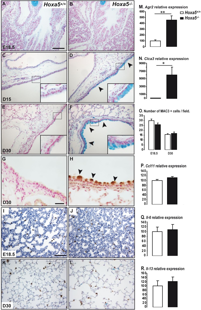
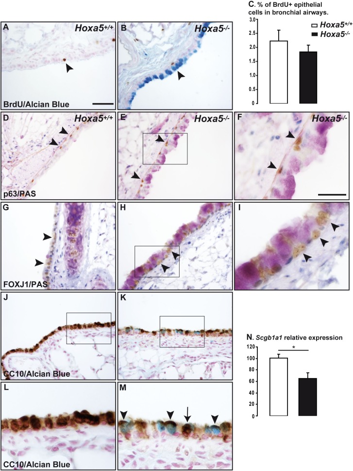
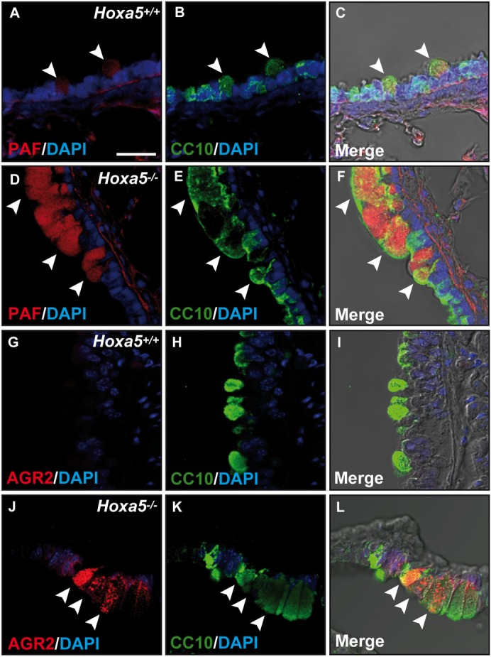
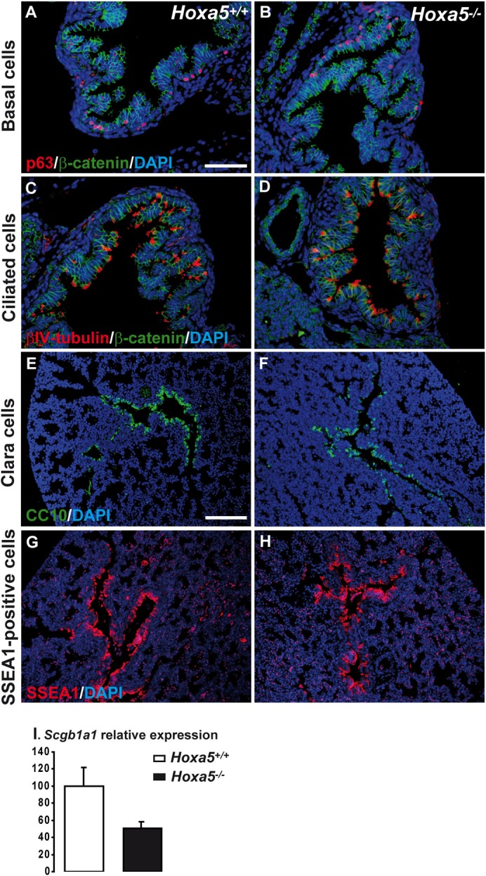
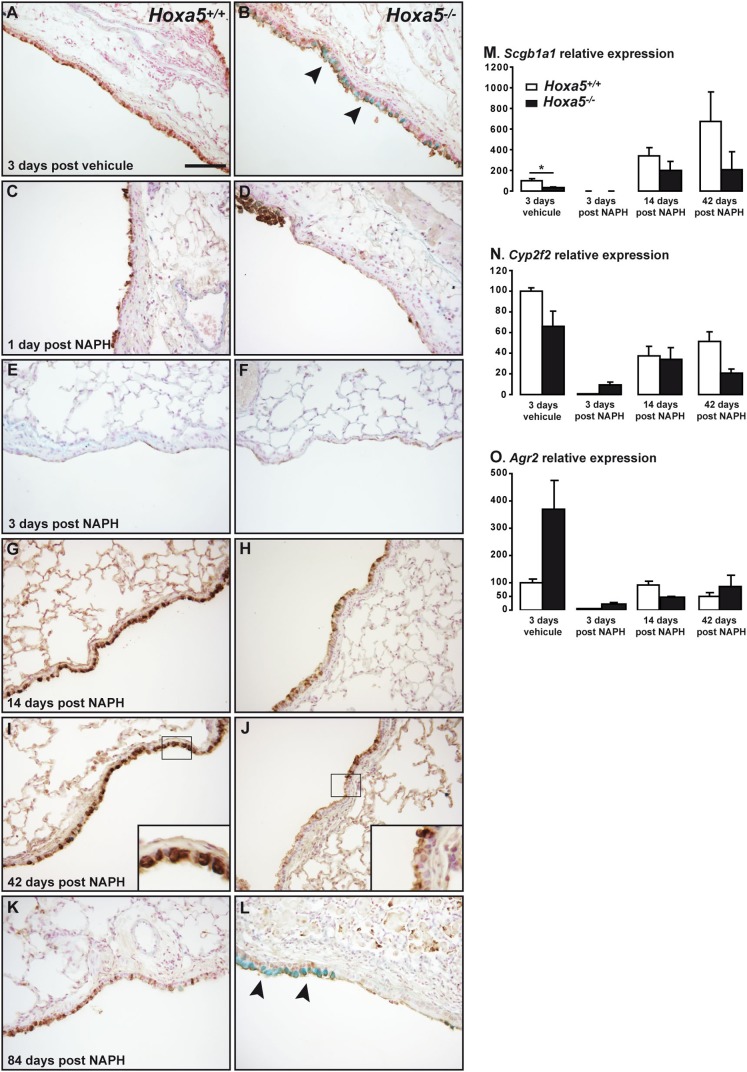
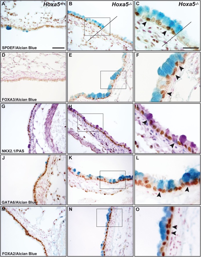
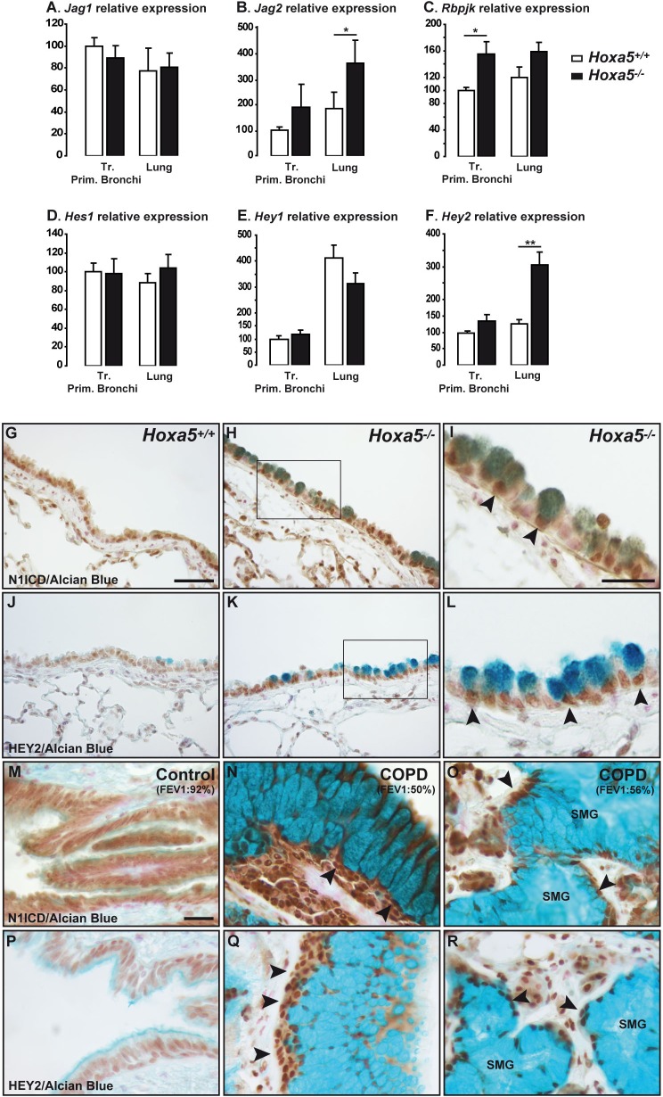
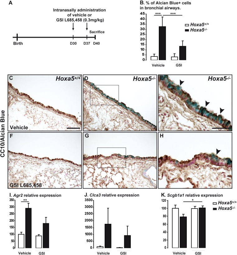
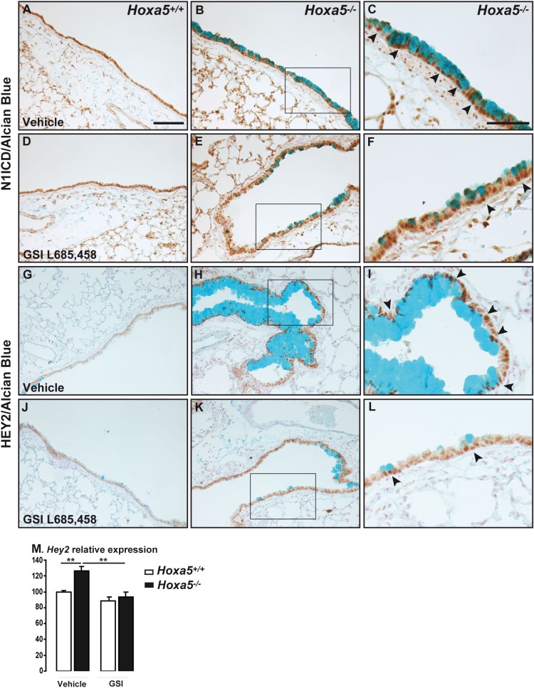
Hox genes encode transcription factors controlling complex developmental processes in various organs. Little is known, however, about how HOX proteins control cell fate. Herein, we demonstrate that the goblet cell metaplasia observed in lung airways from Hoxa5(-/-) mice originates from the transdifferentiation of Clara cells. Reduced CC10 expression in Hoxa5(-/-) embryos indicates that altered cell specification occurs prior to birth. The loss of Hoxa5 function does not preclude airway repair after naphthalene exposure, but the regenerated epithelium presents goblet cell metaplasia and less CC10-positive cells, demonstrating the essential role of Hoxa5 for correct differentiation. Goblet cell metaplasia in Hoxa5(-/-) mice is a FOXA2-independent process. However, it is associated with increased Notch signaling activity. Consistent with these findings, expression levels of activated NOTCH1 and the effector gene HEY2 are enhanced in patients with chronic obstructive pulmonary disease. In vivo administration of a γ-secretase inhibitor attenuates goblet cell metaplasia in Hoxa5(-/-) mice, highlighting the contribution of Notch signaling to the phenotype and suggesting a potential therapeutic strategy to inhibit goblet cell differentiation and mucus overproduction in airway diseases. In summary, the loss of Hoxa5 function in lung mesenchyme impacts on epithelial cell fate by modulating Notch signaling.

Keywords: Goblet cells; Hox genes; Notch pathway.


Conflict of interest statement

Competing interests: The authors declare that there are no competing interests.

Figures

Fig. 1.
Fig. 1.. Goblet cell hyperplasia in Hoxa5−/− mice.
(A–F) Detection of goblet cells by AB staining in lungs from wt and Hoxa5−/− E18.5 embryos, and D15 and D30 mice. Goblet cells were scarce in wt specimens (A,C,E), but numerous in bronchi from D15 and D30 Hoxa5−/− mice (arrowheads; D,F). MUC5AC-positive cells were solely detected in Hoxa5−/− specimens (arrowheads; G,H). (I–L) Detection of macrophages by MAC3 immunostaining in lungs from wt and Hoxa5−/− E18.5 embryos and D30 mice. No major difference was observed. Values are expressed as mean ± SD (O; n = 5–7 animals/group). (M,N) qPCR analysis for Agr2 and Clca3 expression in lungs from wt and Hoxa5−/− D30 mice revealed respectively a 4.5- and 60-fold increase in mutants. (P–R) qPCR analysis for Ccl11, Il-6 and Il-13 expression in lungs from wt and Hoxa5−/− D30 mice did not show difference. Values are expressed as mean ± SEM (n = 5–8 animals/group). *p<0.05; **p<0.01. Scale bars, 100 µm (A–F, I–L), 25 µm (G,H). (C–F) Insets correspond to a 7.5× magnification of the boxed areas.
Fig. 2.
Fig. 2.. Characterization of the respiratory epithelium of Hoxa5−/− mice.
(A–C) BrdU-labeled airway cells (arrowheads) from wt and Hoxa5−/− D30 mice. BrdU labeling index was similar for both genotypes. Values are expressed as mean ± SD (n = 4–5 animals/group). (D–I) Correct specification of basal and ciliated cells as shown respectively by immunostaining with p63 (arrowheads; D–F) and FOXJ1 (arrowheads; G–I) in lungs from wt and Hoxa5−/− D30 mice. (J–M) Decreased CC10-staining in Hoxa5−/− D30 mice. AB-stained goblet cells displayed weak but positive CC10 immunolabeling in mutants (arrowheads, the arrow points to a Clara cell; M). (N) qPCR analysis of Scgb1a1 expression in lungs from wt and Hoxa5−/− D60 mice revealed decreased expression in mutants. Values are expressed as mean ± SEM (n = 6–8 animals/group). *p<0.05. Scale bars, 50 µm (A,B,D,E,G,H,J,K), 25 µm (F,I,L,M).
Fig. 3.
Fig. 3.. Clara to goblet cell transdifferentiation in Hoxa5−/− airway epithelium.
(A–L) Proximal airway sections from wt and Hoxa5−/− D60 mice. Clara cells were detected by CC10 immunolabeling (green) and goblet cells by PAF staining or AGR2 immunolabeling (red). Nuclei were counterstained with DAPI. Co-localization of CC10 and PAF or AGR2 within Hoxa5−/− airway epithelial cells is shown by the yellow color (arrowheads; C,F,I,L). Scale bar, 25 µm.
Fig. 4.
Fig. 4.. Characterization of airway epithelium in Hoxa5−/− embryos.
Correct specification of basal (A,B) and ciliated cells (C,D), shown respectively by immunostaining with p63 (red) and βIV-tubulin (red) in lungs from wt and Hoxa5−/− E18.5 embryos. (E,F) CC10 immunolabeling (green) showed a reduced number of Clara cells in Hoxa5−/− specimens. (G,H) SSEA1 immunostaining (red) was similar for both genotypes. (I) qPCR analysis for Scgb1a1 expression in lungs from E18.5 wt and Hoxa5−/− embryos indicated decreased expression in mutants. Values are expressed as mean ± SEM (n = 7 animals/group). Scale bars, 50 µm (A–D), 100 µm (E–H).
Fig. 5.
Fig. 5.. Impact of naphthalene-induced injury in Hoxa5−/− mice.
CC10 IHC combined to AB staining was performed on lungs from vehicle-treated (A,B) and naphthalene-treated wt and Hoxa5−/− mice (C–L). At days 1 and 3 after naphthalene injection, Clara and goblet cells were shed into the airway lumen in wt and Hoxa5−/− mice. On days 14, 42, and 84 after injury, CC10 expression was restored in wt mice, while CC10 staining was less intense in Hoxa5−/− mice. Goblet cells were detected in Hoxa5−/− specimens 84 days after injury (arrowheads; L). (M–O) qPCR analysis for Scgb1a1, Cyp2f2, and Agr2 expression in lungs from wt and Hoxa5−/− mice treated or not with naphthalene. Values are expressed as mean ± SEM (n = 3–4 animals/group). *p<0.05. Scale bar, 100 µm. (I,J) Insets correspond to a 7.5× magnification of the boxed areas.
Fig. 6.
Fig. 6.. Expression of transcriptional regulators of airway epithelial cell differentiation in Hoxa5−/− mice.
Expression of SPDEF (A–C), FOXA3 (D–F), NKX2.1 (G–I), GATA6 (J–L), and FOXA2 (M–O) was assessed in bronchial tissues from wt and Hoxa5−/− D30 mice. SPDEF and FOXA3 immunostaining was markedly increased at sites of goblet cell metaplasia. No NKX2.1 expression was detected in goblet cells from both genotypes. GATA6 and FOXA2 were expressed along the airway epithelium with no difference between wt and Hoxa5−/− lungs. Arrowheads indicate goblet cells. Scale bars, 50 µm (A,B,D,E,G,H,J,K,M,N), 25 µm (C,F,I,L,O).
Fig. 7.
Fig. 7.. Increased Notch signaling in airways from Hoxa5−/− mice and COPD patients.
(A–F) qPCR analysis for Jag1, Jag2, Rbpjk, Hes1, Hey1, and Hey2 expression in trachea/primary bronchi and lungs from wt and Hoxa5−/− D30 mice. Values are expressed as mean ± SEM (n = 7–8 animals/group). *p<0.05, **p<0.01. (G–L) Immunostaining for N1ICD (G–I) and HEY2 (J–L) was performed in lungs from wt and Hoxa5−/− D30 mice. A salt-and-pepper N1ICD staining was observed in airway epithelium of controls. Goblet cells in Hoxa5−/− lungs displayed a strong N1ICD staining. HEY2 staining was weak along the bronchial epithelium of controls, but intense in goblet cells lining the airways of Hoxa5−/− mice. (M–R) Expression of N1ICD (M–O) and HEY2 (P–R) in lungs from controls and COPD patients. A strong N1ICD and HEY2 expression was detected in areas of goblet cell metaplasia along the airway epithelium and in submucosal glands (SMG) from COPD patients. Arrowheads indicate positive cells. FEV1: forced expiratory volume in 1 second. Scale bars, 50 µm (G,H,J,K), 25 µm (I,L–R).
Fig. 8.
Fig. 8.. Attenuated goblet cell metaplasia in Hoxa5−/− mice after GSI treatment.
(A) Diagram of the experimental protocol. (B) Hoxa5−/− mice displayed less goblet cells after GSI treatment when compared to vehicule-treated mutants. Percentage of goblet cells was calculated as the number of AB-positive bronchial epithelial cells relative to the total number of bronchial epithelial cells. Values are expressed as mean ± SD (n = 5–6 animals/group). (C–H) CC10 IHC combined to AB staining in wt and Hoxa5−/− mice after vehicule (C–E) or GSI (F–H) treatments. Arrowheads indicate goblet cells. (I–K) qPCR analysis for Agr2, Clca3, and Scgb1a1 expression in lungs from wt and Hoxa5−/− mice treated with vehicule or GSI. Values are expressed as mean ± SEM (n = 5-6 animals/group). *p<0.05, **p<0.01, ***p<0.001. Scale bars, 100 µm (C,D,F,G), 50 µm (E,H).
Fig. 9.
Fig. 9.. Decreased Notch signaling activity in Hoxa5−/− mice after GSI treatment.
Immunostaining for N1ICD (A–F) and HEY2 (G–L) was performed in lungs from wt and Hoxa5−/− mice treated with vehicule (A–C,G–I) or GSI (D–F,J–L). Reduced N1ICD and HEY2 staining was observed along the epithelium of GSI-treated Hoxa5−/− mice. Arrowheads indicate goblet cells. (M) qPCR analysis of Hey2 expression in lungs from wt and Hoxa5−/− mice treated with vehicule or GSI. Values are expressed as mean ± SEM (n = 5 animals/group). **p<0.01. Scale bars, 100 µm (A,B,D,E,G,H,J,K), 50 µm (C,F,I,L).

Similar articles

Cited by

References

    1. Alescio T., Cassini A. (1962). Induction in vitro of tracheal buds by pulmonary mesenchyme grafted on tracheal epithelium. J. Exp. Zool. 150, 83–94 10.1002/jez.1401500202 - DOI - PubMed
    1. Aubin J., Lemieux M., Tremblay M., Bérard J., Jeannotte L. (1997). Early postnatal lethality in Hoxa-5 mutant mice is attributable to respiratory tract defects. Dev. Biol. 192, 432–445 10.1006/dbio.1997.8746 - DOI - PubMed
    1. Aubin J., Déry U., Lemieux M., Chailler P., Jeannotte L. (2002). Stomach regional specification requires Hoxa5-driven mesenchymal-epithelial signaling. Development 129, 4075–4087. - PubMed
    1. Bogue C. W., Gross I., Vasavada H., Dynia D. W., Wilson C. M., Jacobs H. C. (1994). Identification of Hox genes in newborn lung and effects of gestational age and retinoic acid on their expression. Am. J. Physiol. 266, L448–L454. - PubMed
    1. Boucherat O., Franco-Montoya M. L., Thibault C., Incitti R., Chailley-Heu B., Delacourt C., Bourbon J. R. (2007). Gene expression profiling in lung fibroblasts reveals new players in alveolarization. Physiol. Genomics 32, 128–141 10.1152/physiolgenomics.00108.2007 - DOI - PubMed