The overexpression of nuclear envelope protein Lap2β induces endoplasmic reticulum reorganisation via membrane stacking
- PMID: 23213473
- PMCID: PMC3507222
- DOI: 10.1242/bio.20121537
The overexpression of nuclear envelope protein Lap2β induces endoplasmic reticulum reorganisation via membrane stacking
Abstract
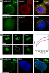
Some nuclear envelope proteins are localised to both the nuclear envelope and the endoplasmic reticulum; therefore, it seems plausible that even small amounts of these proteins can influence the organisation of the endoplasmic reticulum. A simple method to study the possible effects of nuclear envelope proteins on endoplasmic reticulum organisation is to analyze nuclear envelope protein overexpression. Here, we demonstrate that Lap2β overexpression can induce the formation of cytoplasmic vesicular structures derived from endoplasmic reticulum membranes. Correlative light and electron microscopy demonstrated that these vesicular structures were composed of a series of closely apposed membranes that were frequently arranged in a circular fashion. Although stacked endoplasmic reticulum cisternae were highly ordered, Lap2β could readily diffuse into and out of these structures into the surrounding reticulum. It appears that low-affinity interactions between cytoplasmic domains of Lap2β can reorganise reticular endoplasmic reticulum into stacked cisternae. Although the effect of one protein may be insignificant at low concentrations, the cumulative effect of many non-specialised proteins may be significant.
Keywords: Endoplasmic reticulum; Lap2β; Nuclear envelope; Overexpression.
Conflict of interest statement
Figures


Similar articles
-
The presence of intracytoplasmic confronting cisternae of the endoplasmic reticulum in osteosarcoma cells in interphase.Acta Pathol Jpn. 1986 Feb;36(2):261-7. doi: 10.1111/j.1440-1827.1986.tb01478.x. Acta Pathol Jpn. 1986. PMID: 3458351
-
Role of the nuclear envelope in synthesis, processing, and transport of membrane glycoproteins.J Biol Chem. 1985 May 10;260(9):5641-7. J Biol Chem. 1985. PMID: 2985606
-
Self-organization of cellular structures induced by the overexpression of nuclear envelope proteins: a correlative light and electron microscopy study.J Electron Microsc (Tokyo). 2011;60(1):57-71. doi: 10.1093/jmicro/dfq067. Epub 2010 Oct 6. J Electron Microsc (Tokyo). 2011. PMID: 20926432
-
Microtubules as key coordinators of nuclear envelope and endoplasmic reticulum dynamics during mitosis.Bioessays. 2014 Jul;36(7):665-71. doi: 10.1002/bies.201400022. Epub 2014 May 21. Bioessays. 2014. PMID: 24848719 Review.
-
Sorting out the nuclear envelope from the endoplasmic reticulum.Nat Rev Mol Cell Biol. 2004 Jan;5(1):65-9. doi: 10.1038/nrm1263. Nat Rev Mol Cell Biol. 2004. PMID: 14663490 Review.
Cited by
-
The N-terminal region of Jaw1 has a role to inhibit the formation of organized smooth endoplasmic reticulum as an intrinsically disordered region.Sci Rep. 2021 Jan 12;11(1):753. doi: 10.1038/s41598-020-80258-5. Sci Rep. 2021. PMID: 33436890 Free PMC article.
-
Birbeck granule-like "organized smooth endoplasmic reticulum" resulting from the expression of a cytoplasmic YFP-tagged langerin.PLoS One. 2013;8(4):e60813. doi: 10.1371/journal.pone.0060813. Epub 2013 Apr 5. PLoS One. 2013. PMID: 23577166 Free PMC article.
-
Transmembrane and coiled-coil domain family 1 is a novel protein of the endoplasmic reticulum.PLoS One. 2014 Jan 14;9(1):e85206. doi: 10.1371/journal.pone.0085206. eCollection 2014. PLoS One. 2014. PMID: 24454821 Free PMC article.
-
IntEResting structures: formation and applications of organized smooth endoplasmic reticulum in plant cells.Plant Physiol. 2021 Apr 2;185(3):550-561. doi: 10.1104/pp.20.00719. Plant Physiol. 2021. PMID: 33822222 Free PMC article.
-
Carboxyl-terminal Tail-mediated Homodimerizations of Sphingomyelin Synthases Are Responsible for Efficient Export from the Endoplasmic Reticulum.J Biol Chem. 2017 Jan 20;292(3):1122-1141. doi: 10.1074/jbc.M116.746602. Epub 2016 Dec 7. J Biol Chem. 2017. PMID: 27927984 Free PMC article.
References
LinkOut - more resources
Full Text Sources

