. 2012 Sep 15;1(9):863-73.
doi: 10.1242/bio.20121834. Epub 2012 Jul 11.

MRE11 facilitates the removal of human topoisomerase II complexes from genomic DNA

Affiliations

MRE11 facilitates the removal of human topoisomerase II complexes from genomic DNA

Ka Cheong Lee et al. Biol Open. .

Abstract

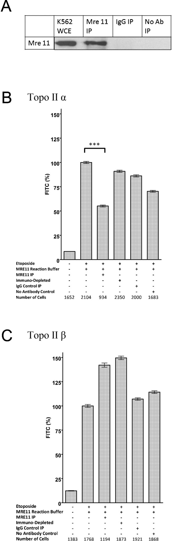
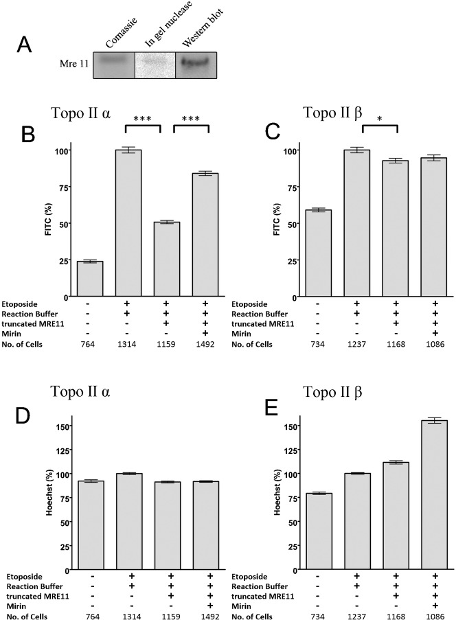
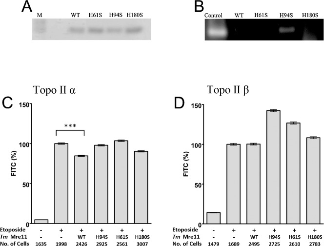
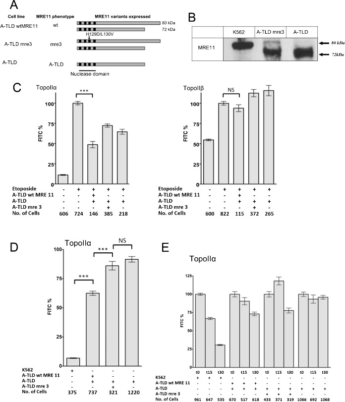
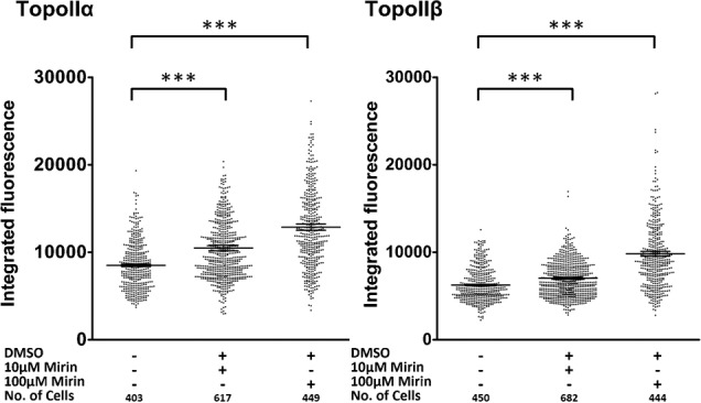
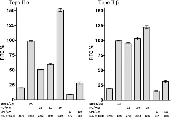
Topoisomerase II creates a double-strand break intermediate with topoisomerase covalently coupled to the DNA via a 5'-phosphotyrosyl bond. These intermediate complexes can become cytotoxic protein-DNA adducts and DSB repair at these lesions requires removal of topoisomerase II. To analyse removal of topoisomerase II from genomic DNA we adapted the trapped in agarose DNA immunostaining assay. Recombinant MRE11 from 2 sources removed topoisomerase IIα from genomic DNA in vitro, as did MRE11 immunoprecipitates isolated from A-TLD or K562 cells. Basal topoisomerase II complex levels were very high in A-TLD cells lacking full-length wild type MRE11, suggesting that MRE11 facilitates the processing of topoisomerase complexes that arise as part of normal cellular metabolism. In K562 cells inhibition of MRE11, PARP or replication increased topoisomerase IIα and β complex levels formed in the absence of an anti-topoisomerase II drug.

Keywords: A-TLD; DSB repair; MRE11; Protein-DNA adducts; Topoisomerase II.


Conflict of interest statement

Competing interests: The authors have no competing interests to declare.

Figures

Fig. 1.
Fig. 1.. Use of TARDIS to assay activities capable of removing topoisomerase II covalent DNA complexes from genomic DNA.
(A) Scheme of assay method. The TARDIS assay allows quantitative assessment of topoisomerase II-DNA covalent complexes that are formed in vivo on genomic DNA (Willmore et al., 1998; Cowell et al., 2011a). Cells treated with topoisomerase poison (in this case 100 µM etoposide) are embedded in agarose and spread onto a microscope slide. After salt/detergent extraction of soluble components, covalently bound topoisomerase II remaining in the embedded genomic DNA is detected by immunofluorescence. To assay for components capable of removing topoisomerase II covalent DNA complexes, slides are incubated with extracts or purified proteins, before antibody probing and immunofluorescence (see Materials and Methods). (B,C) Validation of assay. K562 cells were either untreated (first column) or treated with etoposide (100 µM, 2 hr, columns 2–6). After extraction slides were either untreated or treated as shown with mung bean nuclease (MB) or proteinase K (PK) in the relevant buffer or with buffer alone, prior to immunofluorescence. Mean values of integrated fluorescence per nucleus were determined and the mean of all the cells analysed is shown ± standard error of the mean. All treatments were normalised to the mean values obtained for cells treated with 100 µM etoposide alone. Left graph, Hoechst fluorescence for DNA content; right graph, FITC fluorescence for remaining topoisomerase II. ***  =  p value <0.0001.
Fig. 2.
Fig. 2.. Immunopurified MRE11 removes topoisomerase IIα covalent complexes from genomic DNA.
(A) Western blot probing K562 whole cell extract, K562 MRE11 immunoprecipitate and IgG and no antibody control immunoprecipitates for MRE11 (B,C) K562 cells were treated with 100 µM etoposide for two hours prior to embedding in agarose on microscope slides. The slides were incubated with various immunoprecipitates, the fluorescence levels for the topoisomerase II covalent complexes remaining after the incubations were measured, the fluorescence value for each nucleus was determined and normalised to the mean of the positive control incubated in buffer (100%). These are shown as percentage of FITC signal remaining on a slide after incubation with MRE11 IP  =  eluted components from MRE11 IP; Depl MRE11 IP  =  immunodepleted MRE 11 IP eluate, Ig cont IP  =  IgG control IP, No Ab cont IP  =  control IP without antibody. ***  =  p value <0.0001.
Fig. 3.
Fig. 3.. Purified recombinant MRE11 removes topoisomerase IIα covalent complexes from genomic DNA.
(A). Coommassie, in-gel nuclease and Western analysis of recombinant MRE11. (B–E) K562 cells were treated with 100 µM etoposide for two hours. Slides bearing agarose-embedded cells (1–2×10⁶ cells in 100 µl 1% agarose in PBS spread across the slide surface) were incubated with MRE11 buffer or 1 µg MRE11 in MRE11 buffer in the presence or absence of the MRE11 nuclease inhibitor mirin and quantitative immunofluorescence was carried out for topoisomerase IIα or -β. (B,C) The mean FITC fluorescence for each nucleus was normalised to the 100 µM etoposide positive control, and the mean±SEM are shown. ***  =  p value, 0.0001, *  =  p value <0.05. (D,E) The mean hoechst fluorescence for each nucleus was normalised to the positive control, and the mean±SEM are shown.
Fig. 4.
Fig. 4.. Purified recombinant MRE11 from Thermotoga maritima removes topoisomerase IIα covalent complexes from genomic DNA.
Purified recombinant MRE11 from Thermotoga maritima (A) SDS-PAGE of TmMre11, TmMre11H94S, TmMre11H61S or TmMre11H180S proteins. (B) Endonuclease assays were performed with single-stranded bacteriophage φX174 DNA as a substrate with TmMre11, TmMre11H94S, TmMre11H61S or TmMre11H180S. K562 cells were treated with 100 µM etoposide for two hours prior to being embedded in agarose on microscope slides. Slides bearing agarose-embedded cells (1–2×10⁶ cells in 100 µl 1% agarose in PBS spread across the slide surface) were incubated with MRE11 buffer (positive control) or 1 µg TmMre11, TmMre11H94S, TmMre11H61S or TmMre11H180S. Quantitative immunofluorescence was carried out for topoisomerase IIα (C) or -β (D). The mean fluorescence for each nucleus was normalised to the positive control, and the mean±SEM are shown. ***  =  p value, 0.0001.
Fig. 5.
Fig. 5.. Functional MRE11 is required to maintain low topoisomerase II covalent complex levels.
(A) Schematic of the MRE11 variants expressed in the respective A-TLD cell lines. (B) Western blot showing the full length MRE11 in K562 whole cell extracts, full length and 72 kDa MRE11 in A-TLDmre-3 whole cell extract and only the truncated 72 KDa MRE11 in A-TLD whole cell extracts. (C) K562 cells were treated with 100 µM etoposide or vehicle alone. Cells were prepared for TARDIS analysis as described in Fig. 1 and slides were treated with IP eluates from cells expressing the MRE11 variants shown (columns 3–5 of each graph). Topoisomerase II immunofluorescence was measured and mean values for each nucleus were normalised to the mean signal obtained with 100 µM etoposide with no IP treatment, the mean of the cell means is shown ±SEM. (D,E) Topoisomerase IIα complex levels were determined by TARDIS assay in A-TLD (s) cells. The levels of complexes in the cells were determined in the absence and presence of etoposide. The mean level of complexes as determined by FITC fluorescence, with 100 µM etoposide was set at 100% for each cell line. (D). The untreated control levels are shown for each of the 3 A-TLD cell lines. For comparison the levels in untreated K562 cells are also shown as a percentage of the signal obtained in cells treated with 100 µM etoposide. (E) Change in topoisomerase IIα complex levels in cells, indicative of the rate of removal of the complexes are shown for K562, A-TLDwtMRE11, A-TLDmre-3 and A-TLD cells following removal from medium containing etoposide. ***  =  p value, 0.0001.
Fig. 6.
Fig. 6.. The MRE11 nuclease inhibitor mirin increases topoisomerase II DNA covalent complexes.
K562 cells were exposed to mirin for 24 hours at the concentrations shown and topoisomerase II complexes were quantified by TARDIS. Mann-Whitney significance values are shown. ***  =  p value, 0.0001.
Fig. 7.
Fig. 7.. Hydroxyurea exposure leads to elevated topoisomerase II DNA covalent complex levels.
K562 cells were treated with etoposide for 2 hours (Etop), hydroxyurea for 18 hours (HU) or camptothecin for 18 hours (CPT). Topoisomerase II levels were measured by TARDIS. Mean fluorescence signals were normalised to the mean level obtained with 100 µM etoposide.

References

    1. Adachi N., Suzuki H., Iiizumi S., Koyama H. (2003). Hypersensitivity of nonhomologous DNA end-joining mutants to VP-16 and ICRF-193: implications for the repair of topoisomerase II-mediated DNA damage. J. Biol. Chem. 278, 35897–35902 10.1074/jbc.M306500200 - DOI - PubMed
    1. Adachi N., Iiizumi S., So S., Koyama H. (2004). Genetic evidence for involvement of two distinct nonhomologous end-joining pathways in repair of topoisomerase II-mediated DNA damage. Biochem. Biophys. Res. Commun. 318, 856–861 10.1016/j.bbrc.2004.04.099 - DOI - PubMed
    1. Alchanati I., Teicher C., Cohen G., Shemesh V., Barr H. M., Nakache P., Ben-Avraham D., Idelevich A., Angel I., Livnah N. et al. (2009). The E3 ubiquitin-ligase Bmi1/Ring1A controls the proteasomal degradation of Top2α cleavage complex - a potentially new drug target. PLoS ONE 4, e8104 10.1371/journal.pone.0008104 - DOI - PMC - PubMed
    1. Arthur L. M., Gustausson K., Hopfner K. P., Carson C. T., Stracker T. H., Karcher A., Felton D., Weitzman M. D., Tainer J., Carney J. P. (2004). Structural and functional analysis of Mre11-3. Nucleic Acids Res. 32, 1886–1893 10.1093/nar/gkh343 - DOI - PMC - PubMed
    1. Ayene I. S., Ford L. P., Koch C. J. (2005). Ku protein targeting by Ku70 small interfering RNA enhances human cancer cell response to topoisomerase II inhibitor and γ radiation. Mol. Cancer Ther. 4, 529–536 10.1158/1535-7163.MCT-04-0130 - DOI - PubMed