. 2012 Sep 15;1(9):874-83.
doi: 10.1242/bio.20121982. Epub 2012 Jul 9.

Defects in GPI biosynthesis perturb Cripto signaling during forebrain development in two new mouse models of holoprosencephaly

Affiliations

Defects in GPI biosynthesis perturb Cripto signaling during forebrain development in two new mouse models of holoprosencephaly

David M McKean et al. Biol Open. .

Abstract

Holoprosencephaly is the most common forebrain defect in humans. We describe two novel mouse mutants that display a holoprosencephaly-like phenotype. Both mutations disrupt genes in the glycerophosphatidyl inositol (GPI) biosynthesis pathway: gonzo disrupts Pign and beaker disrupts Pgap1. GPI anchors normally target and anchor a diverse group of proteins to lipid raft domains. Mechanistically we show that GPI anchored proteins are mislocalized in GPI biosynthesis mutants. Disruption of the GPI-anchored protein Cripto (mouse) and TDGF1 (human ortholog) have been shown to result in holoprosencephaly, leading to our hypothesis that Cripto is the key GPI anchored protein whose altered function results in an HPE-like phenotype. Cripto is an obligate Nodal co-factor involved in TGFβ signaling, and we show that TGFβ signaling is reduced both in vitro and in vivo. This work demonstrates the importance of the GPI anchor in normal forebrain development and suggests that GPI biosynthesis genes should be screened for association with human holoprosencephaly.

Keywords: Cripto; GPI; Holoprosencephaly (HPE); Nodal; Pgap1; Pign; TGFβ; forebrain; glycerophosphatidyl inositol.


Conflict of interest statement

Competing interests: The authors have no competing interests to declare.

Figures

Fig. 1.
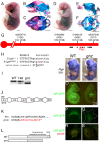
Fig. 1.. Mutation of the glycerophosphatidyl inositol biosynthesis enzyme Pign leads to anterior truncations in Gonzo mutant embryos.
Wildtype (A–C) and gnz (D–F) mutant E18.5 embryos with lateral (B,E) and top (C,F) views of skull stained for bone (red) and cartilage (blue). Exoccipital (eo), supraoccipital (so), basioccipital (bo), tectum synoticum (tso) and interparietal (ip) elements are indicated. Critical 5.14 Mb region of chromosome 1 that the gnz mutation mapped to by meiotic recombination (G). Recombinants are shown in parentheses divided by number of recombination opportunities. SNP and SSLP markers and known or predicted genes (horizontal bars) are indicated. There is a T to A transversion in the splice donor of Pign's intron 23 (H) that causes skipping of exons 23 and 24 (J) as shown by PCR amplification of wildtype (WT), heterozygote (Het) and gnz mutant cDNA (I). Mis-splicing of exon 22 to 25 results in a frame shift and premature stop codon within 13 amino acids from the mutation (K) leading to truncation and partial loss of the catalytic domain and KKXX ER-retention motif (L). Pign mRNA detected by whole mount in situ hybridization in E14.5 wildtype (M) and Pigngnz/gnz mutant (N) embryos, showing ubiquitous and stable expression in gnz mutants. GFP fluorescence was detected in E13.5 Pign+/+; TgGPI-GFP (O -left) but was greatly reduced in Pigngnz/gnz; TgGPI-GFP (O -right) embryos and MEFs (P–S; P and R are Pign+/+; TgGPI-GFP MEFs and Q and S are Pigngnz/gnz; TgGPI-GFP MEFs). Panels R and S were immunostained for the Golgi marker βCOP (red). See also supplementary material Fig. S1 for mutant phenotypes. Ratios in lower right of panels N,O and subsequent figures indicate number of similar phenotypes observed out of number of mutant embryos analyzed. Scale bars (A–F) = 2 mm.
Fig. 2.
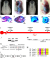
Fig. 2.. Mutation of the glycerophosphatidyl inositol deacylase Pgap1 results in anterior truncations in Bkr mutant embryos.
Wildtype (A) and bkr mutant (C) E11.5 embryos reveal HPE in bkr mutants. Wildtype (B,E,F) and bkr mutant (D,G,H) E18.5 embryos with lateral (E,G) and top (F,H) views of skull stained for bone and cartilage. Abbreviations as in Fig. 1. (I) 23.8 Mb region of chromosome 1 that the bkr mutation mapped to by meiotic recombination. The Pgap1 gene (red horizontal bar) is shown but not the other 144 genes in this interval. There is a T to C transition in the splice donor of intron 19 of Pgap1 (K) that causes skipping of exon 19 (L) as shown by PCR of wildtype (WT), heterozygote (het) and bkr mutant cDNA (J) and sequencing. Mis-splicing of exon 18 to 20 results in an in-frame deletion of 13 amino acids that are conserved (identical amino acids highlighted in yellow, conserved in pink) among human (HS), chick (GG) and zebrafish (DR) Pgap1 orthologs (N). These deleted amino acids lie outside of domains of known function (M). Scale bars (A–H) = 2 mm.
Fig. 3.
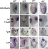
Fig. 3.. Forebrain patterning markers are misexpressed in both Gnz and Bkr mutants.
Gnz mutant embryos (B,F,J,N) and wildtype littermates (A,E,I,M) and bkr mutant embryos (D,H,L,P) and their wildtype littermates (C,G,K,O) were analyzed by whole mount RNA in situ hybridization for Brachyury (A–D), Six3 and Krox20 (E–H), Fgf8 (I–L) and Shh (M–P). Embryos were at E6.5 (A,B), E7.5 (C,D), E8.5 (E–H,M,N) and E9.5 (I–L,O,P). Black and white asterisks in E denote Krox20 and Six3 expression, respectively. Abbreviations: is = isthmus; ba = branchial arches; tb = tailbud. Arrows and arrowheads denote presence or absence, respectively of Fgf8 expression in the ANR (I–L). Black bars mark prechordal plate (M,O).
Fig. 4.
Fig. 4.. Forebrain organizing centers are mislocalized in both Gnz and Bkr mutants.
E7.5 wildtype (A) and gnz mutant (B) littermates and E7.75 wildtype (C) and bkr mutant (D) littermates were analyzed by whole mount RNA in situ hybridization for Otx2. Gnz mutant embryos (F,J) and wildtype littermates (E,I) and bkr mutant (H,L) and wildtype littermates (G,K), all containing Hex-GFP transgene were analyzed by GFP fluorescence. Hex-GFP fluorescence marked the ADE in E7.5 embryos (E–H) and DVE/AVE in E6.5 embryos (I–L).
Fig. 5.
Fig. 5.. Cripto/Nodal signaling is defective in MEFs derived from Gnz and Bkr embryos.
Wildtype and Pigngnz/gnz MEFs (A) or wildtype and Pgap1bkr/bkr MEFs (B) were transfected with HA-Cripto-pcDNA3 expression vector (Cripto) or pcDNA3 vector (Empty), serum starved, then treated with 250 ng/ml recombinant Nodal protein (+) or vehicle (−) for 1 hour. Whole cell extract was separated by SDS-PAGE, and phosphorylated (P-Smad2) and total Smad2 (T-Smad2) and Cripto proteins detected by western blot. (C) Quantification of at least three independent Cripto/Nodal signaling experiments. * represents statistical significance (P value<0.05) by Student's T-test. (D) Endogenous Cripto protein was immunoprecipitated from serum-free conditioned medium overlying Pign+/+, Pigngnz/gnz, Pgap1+/+ and Pgap1bkr/bkr MEFs, separated by SDS-PAGE and detected by western blot.

References

    1. Allen B. L., Tenzen T., McMahon A. P. (2007). The Hedgehog-binding proteins Gas1 and Cdo cooperate to positively regulate Shh signaling during mouse development. Genes Dev. 21, 1244–1257 10.1101/gad.1543607 - DOI - PMC - PubMed
    1. Andersson O., Reissmann E., Jörnvall H., Ibáñez C. F. (2006). Synergistic interaction between Gdf1 and Nodal during anterior axis development. Dev. Biol. 293, 370–381 10.1016/j.ydbio.2006.02.002 - DOI - PubMed
    1. Chazaud C., Rossant J. (2006). Disruption of early proximodistal patterning and AVE formation in Apc mutants. Development 133, 3379–3387 10.1242/dev.02523 - DOI - PubMed
    1. Chu J., Shen M. M. (2010). Functional redundancy of EGF-CFC genes in epiblast and extraembryonic patterning during early mouse embryogenesis. Dev. Biol. 342, 63–73 10.1016/j.ydbio.2010.03.009 - DOI - PMC - PubMed
    1. Chu J., Ding J., Jeays-Ward K., Price S. M., Placzek M., Shen M. M. (2005). Non-cell-autonomous role for Cripto in axial midline formation during vertebrate embryogenesis. Development 132, 5539–5551 10.1242/dev.02157 - DOI - PubMed