. 2012:2012:180259.
doi: 10.1155/2012/180259. Epub 2012 Nov 19.

Evaluation of resection margins in breast conservation therapy: the pathology perspective-past, present, and future

Affiliations

Evaluation of resection margins in breast conservation therapy: the pathology perspective-past, present, and future

Rajyasree Emmadi et al. Int J Surg Oncol. 2012.

Abstract

Tumor surgical resection margin status is important for any malignant lesion. When this occurs in conjunction with efforts to preserve or conserve the afflicted organ, these margins become extremely important. With the demonstration of no difference in overall survival between mastectomy versus lumpectomy and radiation for breast carcinoma, there is a definite trend toward smaller resections combined with radiation, constituting "breast-conserving therapy." Tumor-free margins are therefore key to the success of this treatment protocol. We discuss the various aspects of margin status in this setting, from a pathology perspective, incorporating the past and current practices with a brief glimpse of emerging future techniques.


Figures
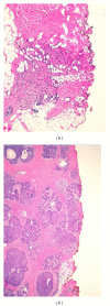
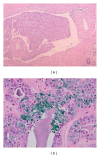
Figure 1
Figure 1
(a)   Section of an invasive carcinoma that extends to and is transected in the surgical margin. (b) Section on extensive ductal carcinoma in situ focally transected in a surgical margin. (H and E).
Figure 2
Figure 2
(a)  Lymphatic invasion in a margin is not considered a “positive” margin. However, such disease present in a margin indicates the patient has high risk of both residual and systemic disease. (b) Image of lobular carcinoma in situ (LCIS) in an inked margin; however, the surgical margin is not defined as being positive for carcinoma.
Figure 3
Figure 3
Section of ductal carcinoma in situ close to a margin but not surgically transected. There is some agreement that a clearance less than 2 mm is inadequate and places a patient at high risk for residual disease. (H and E).
Figure 4
Figure 4
On left, gross inspection in theory: A clearly defined mass measurable from the margins. On right: Reality, an ill-defined mass with indistinct borders and irregular specimen edges.
Figure 5
Figure 5
Specimen radiograph of a wire localization excission taken without compression. The cluster of abnormal calcifications is present but is not at the edge of the specimen. Often a second image is taken after rotating the specimen 90 degrees.
Figure 6
Figure 6
Serially sectioned excision specimen and its Faxitron X-ray. The X-ray image shows a stellate mass in the sixth section from the left with fingers extending very close to the surgical margins. (Image courtesy Dr. A. Sahin, MD Anderson Medical Center).
Figure 7
Figure 7
Method of obtaining touch imprints and/or smears. A slide is pressed against the surface of an excision specimen or the surface is scraped and smeared on a slide. The slide is then stained and examined for malignant cells. The microscopic image at the right shows enlarged irregular nuclei, consistent with carcinoma.
Figure 8
Figure 8
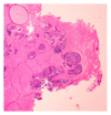
Frozen section slide showing thermal artifact, which obliterates microscopic details that a pathologist needs to diagnose carcinoma. The area bottom center on the edge of the tissue is ductal carcinoma in situ that has been transected in a margin.
Figure 9
Figure 9
Method of obtaining a (pathologic) shave margin from a specimen. A thin piece is taken from the surface of a specimen and either frozen or processed for microscopy. The slide and microscopy show tumor present in the tissue. This would be considered a “positive” margin.
Figure 10
Figure 10
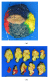
(a) Different colored inks placed on the surface of a specimen maintain the orientation during sectioning and processing. (b) Serial sectioning of inked specimen showing the different inks on the edges of the slices.
Figure 11
Figure 11
Close up of a perpendicular section of margins with tumor. The tumor is distant from the margin at the top of the image, but very close to a margin at the bottom right of the image.

Similar articles

Cited by

References

    1. Fisher B, Anderson S, Bryant J, et al. Twenty-year follow-up of a randomized trial comparing total mastectomy, lumpectomy, and lumpectomy plus irradiation for the treatment of invasive breast cancer. New England Journal of Medicine. 2002;347(16):1233–1241. - PubMed
    1. Veronesi U, Cascinelli N, Mariani L, et al. Twenty-year follow-up of a randomized study comparing breast-conserving surgery with radical mastectomy for early breast cancer. New England Journal of Medicine. 2002;347(16):1227–1232. - PubMed
    1. Van Dongen JA, Voogd AC, Fentiman IS, et al. Long-term results of a randomized trial comparing breast-conserving therapy with mastectomy: European organization for research and treatment of cancer 10801 trial. Journal of the National Cancer Institute. 2000;92(14):1143–1150. - PubMed
    1. Poortmans PM, Collette L, Horiot JC, et al. Impact of the boost dose of 10 Gy versus 26 Gy in patients with early stage breast cancer after a microscopically incomplete lumpectomy: 10-year results of the randomised EORTC boost trial. Radiotherapy and Oncology. 2009;90(1):80–85. - PubMed
    1. Schnitt SJ, Connolly JL, Khettry U. Pathologic findings on re-excision of the primary site in breast cancer patients considered for treatment by primary radiation therapy. Cancer. 1987;59(4):675–681. - PubMed

LinkOut - more resources