

Characterization of Kaposi's Sarcoma-Associated Herpesvirus-Related Lymphomas by DNA Microarray Analysis
- PMID: 23213546
- PMCID: PMC3504204
- DOI: 10.4061/2011/726964
Characterization of Kaposi's Sarcoma-Associated Herpesvirus-Related Lymphomas by DNA Microarray Analysis
Abstract
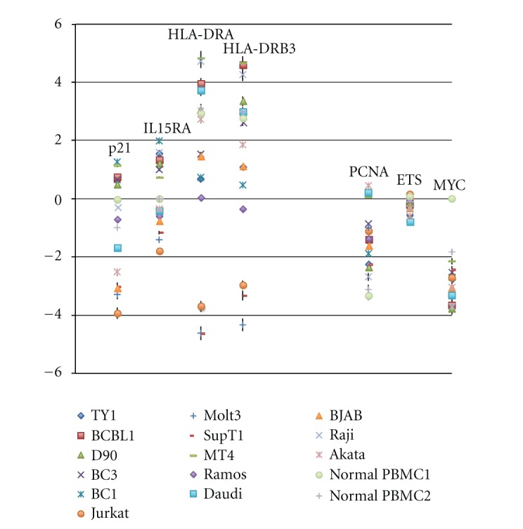
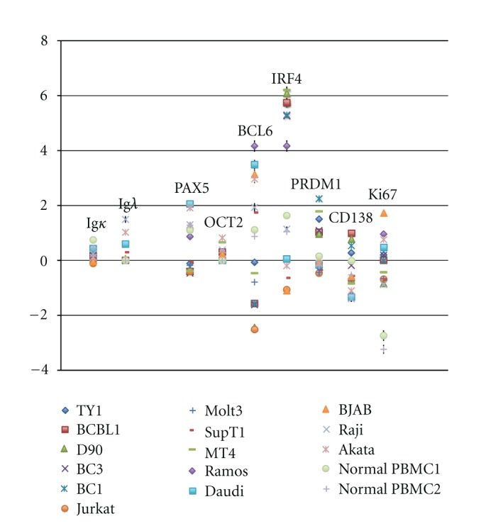
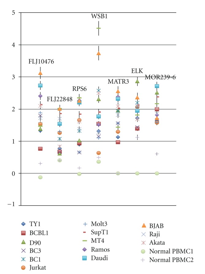
Among herpesviruses, γ-herpesviruses are supposed to have typical oncogenic activities. Two human γ-herpesviruses, Epstein-Barr virus (EBV) and Kaposi's sarcoma-associated herpesvirus (KSHV), are putative etiologic agents for Burkitt lymphoma, nasopharyngeal carcinoma, and some cases of gastric cancers, and Kaposi's sarcoma, multicentric Castleman's disease, and primary effusion lymphoma (PEL) especially in AIDS setting for the latter case, respectively. Since such two viruses mentioned above are highly species specific, it has been quite difficult to prove their oncogenic activities in animal models. Nevertheless, the viral oncogenesis is epidemiologically and/or in vitro experimentally evident. This time, we investigated gene expression profiles of KSHV-oriented lymphoma cell lines, EBV-oriented lymphoma cell lines, and T-cell leukemia cell lines. Both KSHV and EBV cause a B-cell-originated lymphoma, but the gene expression profiles were typically classified. Furthermore, KSHV could govern gene expression profiles, although PELs are usually coinfected with KSHV and EBV.
Figures
Similar articles
-
Kaposi's sarcoma-associated herpesvirus/human herpesvirus type 8-positive solid lymphomas: a tissue-based variant of primary effusion lymphoma.J Mol Diagn. 2005 Feb;7(1):17-27. doi: 10.1016/S1525-1578(10)60004-9. J Mol Diagn. 2005. PMID: 15681470 Free PMC article.
-
Mutual inhibition between Kaposi's sarcoma-associated herpesvirus and Epstein-Barr virus lytic replication initiators in dually-infected primary effusion lymphoma.PLoS One. 2008 Feb 6;3(2):e1569. doi: 10.1371/journal.pone.0001569. PLoS One. 2008. PMID: 18253508 Free PMC article.
-
Distinct patterns of viral antigen expression in Epstein-Barr virus and Kaposi's sarcoma-associated herpesvirus coinfected body-cavity-based lymphoma cell lines: potential switches in latent gene expression due to coinfection.Virology. 1999 Sep 15;262(1):18-30. doi: 10.1006/viro.1999.9876. Virology. 1999. PMID: 10489337
-
Kaposi's sarcoma-associated herpesvirus: a lymphotropic human herpesvirus associated with Kaposi's sarcoma, primary effusion lymphoma, and multicentric Castleman's disease.Semin Diagn Pathol. 1997 Feb;14(1):54-66. Semin Diagn Pathol. 1997. PMID: 9044510 Review.
-
Gamma-herpesvirus neoplasia: a growing role for COX-2.Microsc Res Tech. 2005 Nov;68(3-4):120-9. doi: 10.1002/jemt.20226. Microsc Res Tech. 2005. PMID: 16276519 Review.
Cited by
-
KSHV-Mediated Angiogenesis in Tumor Progression.Viruses. 2016 Jul 20;8(7):198. doi: 10.3390/v8070198. Viruses. 2016. PMID: 27447661 Free PMC article. Review.
-
Chromatinization of the KSHV Genome During the KSHV Life Cycle.Cancers (Basel). 2015 Jan 14;7(1):112-42. doi: 10.3390/cancers7010112. Cancers (Basel). 2015. PMID: 25594667 Free PMC article. Review.
-
Co-infections and Pathogenesis of KSHV-Associated Malignancies.Front Microbiol. 2016 Feb 15;7:151. doi: 10.3389/fmicb.2016.00151. eCollection 2016. Front Microbiol. 2016. PMID: 26913028 Free PMC article. Review.
-
Next-Generation Sequencing Analysis Reveals Differential Expression Profiles of MiRNA-mRNA Target Pairs in KSHV-Infected Cells.PLoS One. 2015 May 5;10(5):e0126439. doi: 10.1371/journal.pone.0126439. eCollection 2015. PLoS One. 2015. PMID: 25942495 Free PMC article.
References
-
- Howly PM, Lowy DR. Papillomaviruses. Philadelphia, Pa, USA: Lippincott Williams & Wilkins; 2007.
-
- Kremsdorf D, Soussan P, Paterlini-Brechot P, Brechot C. Hepatitis B virus-related hepatocellular carcinoma: paradigms for viral-related human carcinogenesis. Oncogene. 2006;25(27):3823–3833. - PubMed
-
- Levrero M. Viral hepatitis and liver cancer: the case of hepatitis C. Oncogene. 2006;25(27):3834–3847. - PubMed
-
- Matsuoka M, Jeang KT. Human T-cell leukaemia virus type 1 (HTLV-1) infectivity and cellular transformation. Nature Reviews Cancer. 2007;7(4):270–280. - PubMed
-
- Young LS, Rickinson AB. Epstein-Barr virus: 40 years on. Nature Reviews Cancer. 2004;4(10):757–768. - PubMed
LinkOut - more resources
Full Text Sources
