Review
. 2012:2012:797295.
doi: 10.1155/2012/797295. Epub 2012 Nov 19.

Activity-dependent callosal axon projections in neonatal mouse cerebral cortex

Affiliations
Review

Activity-dependent callosal axon projections in neonatal mouse cerebral cortex

Yoshiaki Tagawa et al. Neural Plast. 2012.

Abstract

Callosal axon projections are among the major long-range axonal projections in the mammalian brain. They are formed during the prenatal and early postnatal periods in the mouse, and their development relies on both activity-independent and -dependent mechanisms. In this paper, we review recent findings about the roles of neuronal activity in callosal axon projections. In addition to the well-documented role of sensory-driven neuronal activity, recent studies using in utero electroporation demonstrated an essential role of spontaneous neuronal activity generated in neonatal cortical circuits. Both presynaptic and postsynaptic neuronal activities are critically involved in the axon development. Studies have begun to reveal intracellular signaling pathway which works downstream of neuronal activity. We also review several distinct patterns of neuronal activity observed in the developing cerebral cortex, which might play roles in activity-dependent circuit construction. Such neuronal activity during the neonatal period can be disrupted by genetic factors, such as mutations in ion channels. It has been speculated that abnormal activity caused by such factors may affect activity-dependent circuit construction, leading to some developmental disorders. We discuss a possibility that genetic mutation in ion channels may impair callosal axon projections through an activity-dependent mechanism.


Figures
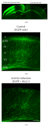
Figure 1
Figure 1
Visualization of callosal axon projections and an effect of activity reduction. (a) An in utero electroporation-mediated gene transfer method [38] was used to unilaterally express EGFP in layer 2/3 cortical neurons (electroporated side). EGFP-labeled callosal axons extend through the corpus callosum, and project densely to a narrowly restricted region in the contralateral cortex (arrow). Scale bar, 500 μm. (b) EGFP-labeled control callosal axons show lamina specific projection pattern. (c) Reduction of neuronal activity in callosal projection neurons disturbs their axonal projections. Adapted from [38]. Scale bar, 200 μm.
Figure 2
Figure 2
Effects of postsynaptic and presynaptic activity reduction on callosal axon projections. (a) The morphology of single callosal axons in the normal condition at P15. ((b) and (c)) postsynaptic (b) and presynaptic (c) activity reduction impede growth and branching of callosal axons. Scale bar, 200 μm. (d) An illustration showing the development of callosal axons in the mouse. Callosal axons reach the target innervation area around P5, arrive in the target cortical layers at P7, start to branch at P9, and elaborate their arbors afterwards. The effect of presynaptic activity reduction is apparent before axons reach the target cortical layers, but that of postsynaptic activity reduction is observed after their arrival in the target layers. Adapted from [41].
Figure 3
Figure 3
Effects of expression of several ion channels on callosal axon projections. (a) Expression of a dominant-negative KCNQ2 K+ channel [94] does not influence callosal axon development. (b) A disease-causing, dominant-negative Kir2.1 mutant does not impair callosal axon projections. (c) Expression of a gain-of-function Kir2.1 mutant in callosal projection neurons impedes their axonal projections. Scale bar, 200 μm. Adapted from [38].

Similar articles

Cited by

References

    1. Hubel D, Wiesel T. Cortical and callosal connections concerned with the vertical meridian of visual fields in the cat. Journal of Neurophysiology. 1967;30(6):1561–1573. - PubMed
    1. Shatz CJ. Anatomy of interhemispheric connections in the visual system of Boston Siamese and ordinary cats. Journal of Comparative Neurology. 1977;173(3):497–518. - PubMed
    1. van Essen D, Newsome W, Bixby J. The pattern of interhemispheric connections and its relationship to extrastriate visual areas in the macaque monkey. Journal of Neuroscience. 1982;2(3):265–283. - PMC - PubMed
    1. Blakemore C, Diao Y, Pu M, Wang Y, Xiao Y. Possible functions of the interhemispheric connexions between visual cortical areas in the cat. Journal of Physiology. 1983;337:331–349. - PMC - PubMed
    1. Olavarria J, Montero VM. Relation of callosal and striate-extrastriate cortical connections in the rat: morphological definition of extrastriate visual areas. Experimental Brain Research. 1984;54(2):240–252. - PubMed

Publication types