

Primary nonfunction of renal allograft secondary to acute oxalate nephropathy
- PMID: 23213607
- PMCID: PMC3504227
- DOI: 10.1155/2011/876906
Primary nonfunction of renal allograft secondary to acute oxalate nephropathy
Abstract
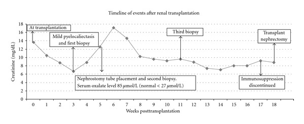
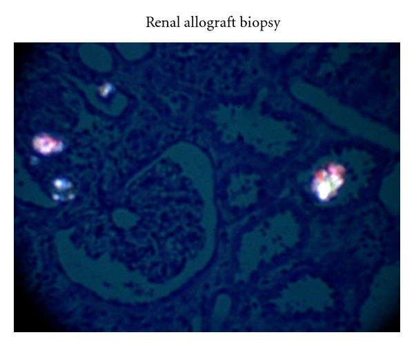
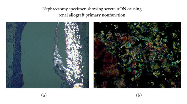
Primary nonfunction (PNF) accounts for 0.6 to 8% of renal allograft failure, and the focus on causes of PNF has changed from rejection to other causes. Calcium oxalate (CaOx) deposition is common in early allograft biopsies, and it contributes in moderate intensity to higher incidence of acute tubular necrosis and poor graft survival. A-49-year old male with ESRD secondary to polycystic kidney disease underwent extended criteria donor kidney transplantation. Posttransplant, patient developed delayed graft function (DGF), and the biopsy showed moderately intense CaOx deposition that persisted on subsequent biopsies for 16 weeks, eventually resulting in PNF. The serum oxalate level was 3 times more than normal at 85 μmol/L (normal <27 μmol/L). Allograft nephrectomy showed massive aggregates of CaOx crystal deposition in renal collecting system. In conclusion, acute oxalate nephropathy should be considered in the differential diagnosis of DGF since optimal management could change the outcome of the allograft.
Figures
References
-
- El-Zoghby ZM, Stegall MD, Lager DJ, et al. Identifying specific causes of kidney allograft loss. American Journal of Transplantation. 2009;9(3):527–535. - PubMed
-
- Stevens RB, Skorupa JY, Rigley TH, et al. Increased primary non-function in transplanted deceased-donor kidneys flushed with histidine-tryptophan-ketoglutarate solution. American Journal of Transplantation. 2009;9(5):1055–1062. - PubMed
-
- Groenewoud AF, Thorogood J, The HTK Study Group Current status of the Eurotransplant randomized multicenter study comparing kidney graft preservation with histidine-tryptophan-ketogluterate, University of Wisconsin, and Euro-Collins solutions. Transplantation Proceedings. 1993;25(1):1582–1585. - PubMed
-
- Woo YM, Jardine AG, Clark AF, et al. Early graft function and patient survival following cadaveric renal transplantation. Kidney International. 1999;55(2):692–699. - PubMed
-
- Pinheiro SH, Saraiva Câmara NO, Osaki KS, Ribeiro De Moura LA, Pacheco-Silva A. Early presence of calcium oxalate deposition in kidney graft biopsies is associated with poor long-term graft survival. American Journal of Transplantation. 2005;5(2):323–329. - PubMed
Publication types
LinkOut - more resources
Full Text Sources
