. 2013 Jan 23;135(3):1015-25.
doi: 10.1021/ja308119q. Epub 2013 Jan 11.

On the formation and properties of interstrand DNA-DNA cross-links forged by reaction of an abasic site with the opposing guanine residue of 5'-CAp sequences in duplex DNA

Affiliations

On the formation and properties of interstrand DNA-DNA cross-links forged by reaction of an abasic site with the opposing guanine residue of 5'-CAp sequences in duplex DNA

Kevin M Johnson et al. J Am Chem Soc. .

Abstract

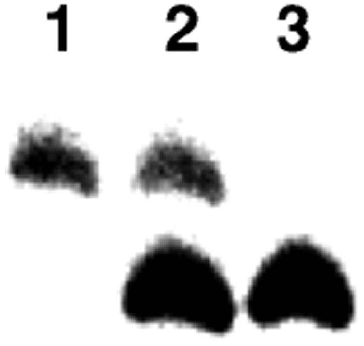
We recently reported that the aldehyde residue of an abasic (Ap) site in duplex DNA can generate an interstrand cross-link via reaction with a guanine residue on the opposing strand. This finding is intriguing because the highly deleterious nature of interstrand cross-links suggests that even small amounts of Ap-derived cross-links could make a significant contribution to the biological consequences stemming from the generation of Ap sites in cellular DNA. Incubation of 21-bp duplexes containing a central 5'-CAp sequence under conditions of reductive amination (NaCNBH(3), pH 5.2) generated much higher yields of cross-linked DNA than reported previously. At pH 7, in the absence of reducing agents, these Ap-containing duplexes also produced cross-linked duplexes that were readily detected on denaturing polyacrylamide gels. Cross-link formation was not highly sensitive to reaction conditions, and the cross-link, once formed, was stable to a variety of workup conditions. Results of multiple experiments including MALDI-TOF mass spectrometry, gel mobility, methoxyamine capping of the Ap aldehyde, inosine-for-guanine replacement, hydroxyl radical footprinting, and LC-MS/MS were consistent with a cross-linking mechanism involving reversible reaction of the Ap aldehyde residue with the N(2)-amino group of the opposing guanine residue in 5'-CAp sequences to generate hemiaminal, imine, or cyclic hemiaminal cross-links (7-10) that were irreversibly converted under conditions of reductive amination (NaCNBH(3)/pH 5.2) to a stable amine linkage. Further support for the importance of the exocyclic N(2)-amino group in this reaction was provided by an experiment showing that installation of a 2-aminopurine-thymine base pair at the cross-linking site produced high yields (15-30%) of a cross-linked duplex at neutral pH, in the absence of NaCNBH(3).


Figures

Figure 1
Figure 1
Molecular model showing the juxtaposition of the N2-amino group of the guanine residue and the abasic site at a 5′-CAp cross-linking site in B-DNA. The image was constructed using Pymol and is based on pdb entry 1dcv.
Figure 2
Figure 2
Interstrand cross-link formation in duplexes A-D under conditions of reductive amination. Duplex A, lanes 1-5, B, lanes 6-10, C, lanes 11-15, and D, lanes 16-20. The uracil-containing precursor 2′-deoxyoligonucleotides appear in lanes 1, 6, 11, and 16. The abasic-site-containing duplexes without incubation appear in lanes 2, 7, 12, and 17. The abasic-site-containing duplexes cleaved by treatment with piperidine (1 M, 95 °C, for 25 min) appear in lanes 3, 8, 13, and 18). The cross-linking reactions involving incubation of the abasic-site-containing duplex in sodium acetate buffer (750 mM, pH 5.2) and NaCNBH3 (250 mM) at 37 °C appear in lanes 4, 9, 14, and 19. The abasic-site-containing duplexes in sodium acetate buffer (750 mM, pH 5.2), NaCNBH3 (250 mM), and CH3ONH2 hydrochloride (2 mM) at 37 °C appear in lanes 5, 10, 15, and 20. The 32P-labeled 2′-deoxyoligonucleotides were resolved on a sequencing gel and the radioactivity in each band quantitatively measured by phosphorimager analysis.
Figure 3
Figure 3
Time course for the formation of cross-linked duplex under conditions of reductive amination. Duplex A was incubated in sodium acetate buffer (750 mM, pH 5.2) and NaCNBH3 (250 mM) at 37 °C and at 0, 1, 2, 4, 6, 8, 12, and 23 h aliquots were removed from the reaction and frozen prior to sequencing gel analysis (lanes 5-12). The lower bands correspond to the full length labeled 2′-deoxyoligonucleotides and the upper band cross-linked DNA. Lane 1 is the 32P-labeled uracil-containing precursor 2′-deoxyoligonucleotide, lane 2 is the 32P-labeled abasic-site-containing duplex without incubation, and lane 3 is the 32P-labeled abasic-site-containing duplexes cleaved by treatment with piperidine (1 M, 95 °C, for 25 min). Lane 4 is the abasic-site-containing duplex incubated in pH 7 buffer for 24 h at 37 °C. The 32P-labeled 2′-deoxyoligonucleotides were resolved on a sequencing gel and the radioactivity in each band quantitatively measured by phosphorimager analysis. Figure 1 shows a full image of the gel. This and subsequent gel images display only the region of the gel where full length and cross-linked DNA appear.
Figure 4
Figure 4
Cross-link formation was abrogated by replacement of the opposing guanine residue in the 5′-CAp sequence with an inosine residue. The lower bands correspond to the 32P-labeled full length labeled 2′-deoxyoligonucleotides and the upper band cross-linked DNA. Lane 1 is the abasic-site-containing 2′-deoxyoligonucleotide duplex without incubation. Lane 2 is the abasic-site-containing 2′-deoxyoligonucleotide duplex A cleaved by treatment with piperidine (1 M, 95 °C, 25 min). Lane 3 is duplex A incubated in sodium acetate buffer (750 mM, pH 5.2) and NaCNBH3 (250 mM) at 37 °C. Lane 4 is duplex E incubated in sodium acetate buffer (750 mM, pH 5.2) and NaCNBH3 (250 mM) at 37 °C. The 32P-labeled 2′-deoxyoligonucleotides were resolved on a sequencing gel and the radioactivity in each band quantitatively measured by phosphorimager analysis. The image shows the region of the gel where full-length oligonucleotide and cross-link migrate.
Figure 5
Figure 5
Detection of native (unreduced) dG-Ap cross-link duplex A and evidence that replacement of the opposing guanine residue in the 5′-CAp sequence of duplex A with inosine in duplex E abrogates cross-link formation. The lower bands correspond to the full-length 32P-labeled 2′-deoxyoligonucleotides and the upper band cross-linked DNA. Lane 1 is the uracil-containing precursor of 2′-deoxyoligonucleotide duplex A. Lane 2 is the abasic-site-containing 2′-deoxyoligonucleotide duplex A without incubation. Lane 3 is the abasic-site-containing 2′-deoxyoligonucleotide duplex A cleaved by treatment with piperidine (1 M, 95 °C, 25 min). Lane 4 is duplex A incubated in with CH3ONH2 (2 mM) in HEPES buffer (50 mM, pH 7) containing NaCl (100 mM) at 37 °C. Lane 5 is duplex A incubated in HEPES buffer (50 mM, pH 7) containing NaCl (100 mM) at 37 °C. Lane 6 is the abasic-site-containing 2′-deoxyoligonucleotide duplex E without incubation. Lane 7 is the abasic-site-containing 2′-deoxyoligonucleotide duplex E cleaved by treatment with piperidine (1 M, 95 °C, 25 min). Lane 8 is duplex E incubated in with CH3ONH2 (2 mM) in HEPES buffer (50 mM, pH 7) containing NaCl (100 mM) at 37 °C. Lane 9 is duplex E incubated in HEPES buffer (50 mM, pH 7) containing NaCl (100 mM) at 37 °C. The 32P-labeled 2′-deoxyoligonucleotides were resolved on a sequencing gel and the radioactivity in each band quantitatively measured by phosphorimager analysis. The image shows the region of the gel where full-length oligonucleotide and cross-link migrate.
Figure 6
Figure 6
The reduced and unreduced dG-Ap cross-linked duplexes can be resolved by gel electrophoresis. Lane 1 is the native (unreduced) cross-link generated by incubation of duplex A in HEPES buffer (50 mM, pH 7) containing NaCl (100 mM) at 37 °C. Lane 3 is the reduced cross-link generated by incubation of duplex A in sodium acetate buffer (750 mM, pH 5.2) and NaCNBH3 (250 mM) at 37 °C. Lane 2 is a mixture of the reduced and native cross-links. The image shows the region of the gel where the cross-link migrates.
Figure 7
Figure 7
The 2-aminopurine-thymine base pair places an exocyclic amino group in the same location as a typical G-C base pair.
Figure 8
Figure 8
Evidence for cross-link formation in 2-aminopurine-containing 2′-oligodeoxyribonucleotide duplexes G and H. The lower band corresponds to full-length labeled 2′-deoxyoligonucleotide and the upper band cross-linked DNA. The lower bands correspond to the full length labeled 2′-deoxyoligonucleotides and the upper band cross-linked DNA. Lane 1 is the uracil-containing precursor of 2′-deoxyoligonucleotide duplex G. Lane 2 is the abasic-site-containing 2′-deoxyoligonucleotide duplex G without incubation. Lane 3 is the abasic-site-containing 2′-deoxyoligonucleotide duplex G cleaved by treatment with piperidine (1 M, 95 °C, 25 min). Lane 4 is duplex G incubated with CH3ONH2 (2 mM) in HEPES buffer (50 mM, pH 7) containing NaCl (100 mM) at 37 °C. Lane 5 is duplex G incubated in HEPES buffer (50 mM, pH 7) containing NaCl (100 mM) at 37 °C. Lane 6 is the abasic-site-containing 2′-deoxyoligonucleotide duplex H without incubation. Lane 7 is the abasic-site-containing 2′-deoxyoligonucleotide duplex H cleaved by treatment with piperidine (1 M, 95 °C, 25 min). Lane 8 is duplex H incubated in with CH3ONH2 (2 mM) in HEPES buffer (50 mM, pH 7) containing NaCl (100 mM) at 37 °C. Lane 9 is duplex H incubated in HEPES buffer (50 mM, pH 7) containing NaCl (100 mM) at 37 °C. The 32P-labeled 2′-deoxyoligonucleotides were resolved on a sequencing gel and the radioactivity in each band quantitatively measured by phosphorimager analysis. The image shows the region of the gel where full-length oligonucleotide and cross-link migrate.
Figure 9
Figure 9
LC-ESI-MS and MS/MS for the analysis of the nuclease P1 digestion mixture of duplex J with a dG-Ap cross-link. Shown in (a) is the selected-ion chromatogram (SIC) for monitoring the loss of a 2′-deoxyadenosine-5′-phosphate from the [M - 2H]2− ion of the tetramer shown (i.e., the m/z 583.5 837 transition). Inset gives the higher resolution “ultra-zoom” ESI-MS for the [M - 2H]2− ion of the tetramer. Displayed in (b) is the tandem mass spectrum for the [M - 2H]2− ion of the tetramer.
Figure 10
Figure 10
LC-ESI-MS/MS/MS for the analysis of the 4-enzyme digestion mixture of duplex J with a dG-Ap cross-link. Shown in (a) is the selected-ion chromatogram (SIC) for monitoring the m/z 384→268→240 transition (proposed structures for fragment ions in this fragmentation pathway are shown on the right). Depicted in (b) is MS/MS/MS arising from the fragmentation of the ion of m/z 268 observed in MS/MS from the cleavage of the [M + H]+ ion of the completely digested crosslink remnant.
Scheme 1
Scheme 1
Scheme 2
Scheme 2
Scheme 3
Scheme 3

Similar articles

Cited by

References

    1. Gates KS, Nooner T, Dutta S. Chem Res Toxicol. 2004;17:839–856. - PubMed
    1. Gates KS. Chem Res Toxicol. 2009;22:1747–1760. - PMC - PubMed
    1. Amosova O, Coulter R, Fresco JR. Proc Nat Acad Sci USA. 2006;103:4392–4397. - PMC - PubMed
    1. Lindahl T, Nyberg B. Biochemistry. 1972;11:3610–3618. - PubMed
    1. Nakamura J, Swenberg JA. Cancer Res. 1999;59:2522–2526. - PubMed

Publication types