. 2013 Apr;168(8):1946-60.
doi: 10.1111/bph.12085.

Protein phosphatase 1 coordinates CFTR-dependent airway epithelial HCO3- secretion by reciprocal regulation of apical and basolateral membrane Cl(-)-HCO3- exchangers

Affiliations

Protein phosphatase 1 coordinates CFTR-dependent airway epithelial HCO3- secretion by reciprocal regulation of apical and basolateral membrane Cl(-)-HCO3- exchangers

James P Garnett et al. Br J Pharmacol. 2013 Apr.

Abstract

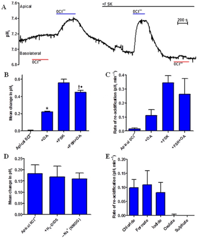
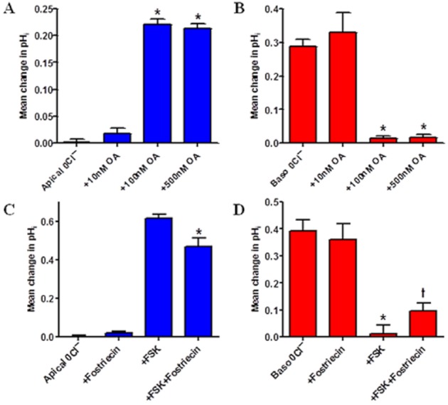
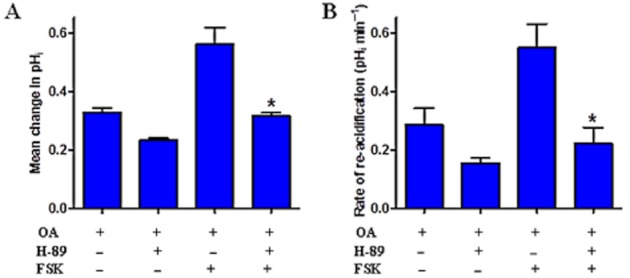
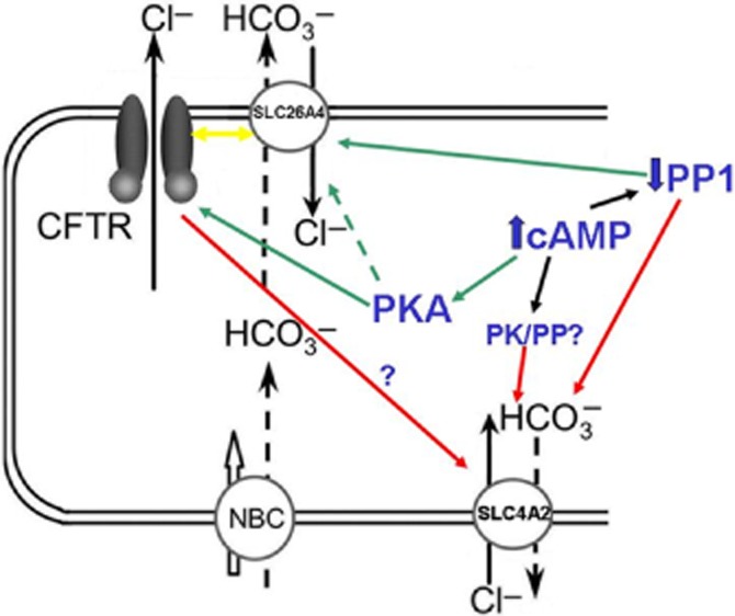
Background and purpose: Our recent studies on human airway serous-like Calu-3 cells showed that cAMP agonists stimulated a HCO3(-) rich secretion containing up to 80 mM HCO3(-). This alkaline secretion relied on a coordinated switch in the activity of distinct Cl(-)-HCO3(-) anion exchangers (AE) located at different regions of the cell. At the apical membrane, cAMP agonists activated the electroneutral AE pendrin (SLC26A4), together with cystic fibrosis transmembrane conductance regulator (CFTR), while at the basolateral membrane the agonists inhibited AE2 (SLC4A2). However, the underlying mechanism(s) that orchestrates this cAMP-dependent switch in AE activity has not been elucidated.

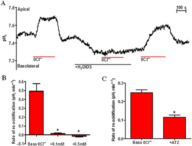
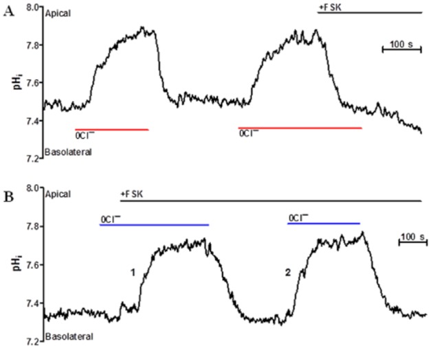
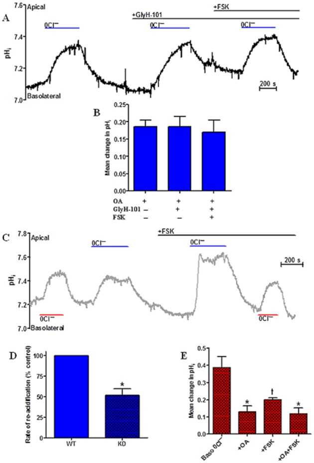
Experimental approach: Apical and basolateral Cl(-)-HCO3(-) exchange was assessed by measuring Cl(-)-dependent changes in intracellular pH (pH(i)).

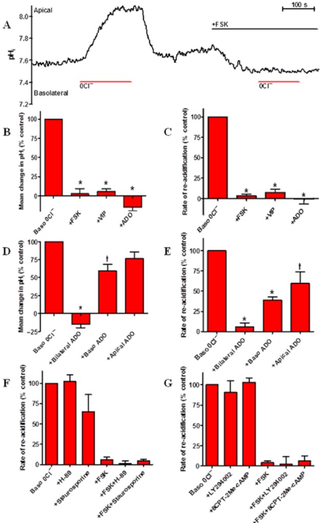
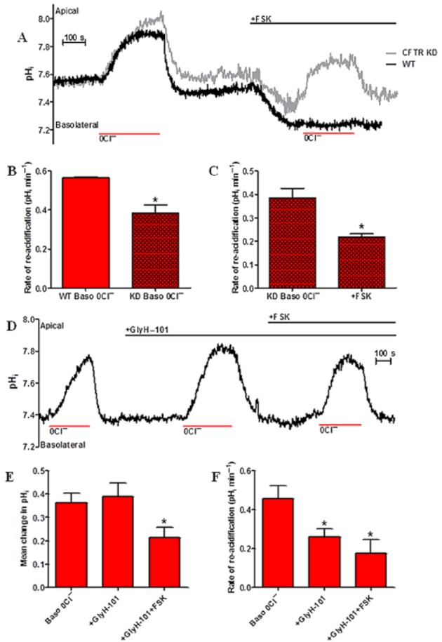
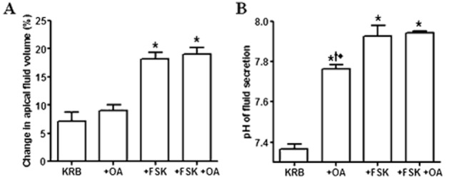
Key results: We show that protein phosphatase 1 (PP1), together with CFTR, play central roles in this reciprocal regulation of AE activity. Activation of pendrin by cAMP agonists, but not inhibition of the basolateral exchanger, was protein kinase A-dependent. Knocking down CFTR expression, or blocking its activity with GlyH-101, led to incomplete inhibition of the basolateral AE by cAMP, supporting a role for CFTR in this process. Addition of the PP1/2A inhibitor, okadaic acid, but not the PP2A specific inhibitor fostreicin, mimicked the effect of cAMP stimulation. Furthermore, okadaic acid-treated Calu-3 monolayers produced a more alkaline fluid than untreated cells, which was comparable with that produced by cAMP stimulation.

Conclusions and implications: These results identify PP1 as a novel regulator of AE activity which, in concert with CFTR, coordinates events at both apical and basolateral membranes, crucial for efficient HCO3(-) secretion from Calu-3 cells.


Figures

Figure 1
Figure 1
Pharmacological profile of basolateral Cl-dependent changes in pHi in Calu-3 cells. A: Representative trace illustrating the effect of basolateral H2-DIDS (500 μM; indicated by black bar below trace) on changes in pHi following the removal of basolateral Cl (indicated by red bars below trace). B: The effect of basolateral H2-DIDS (100 and 500 μM) on the mean rate of re-acidification in pHi upon re-addition of basolateral Cl (n = 4; paired observations. *P < 0.05 compared with Baso 0Cl). C: The effect of the carbonic anhydrase inhibitor acetazolamide (ATZ; 100 μM) on the mean rate of re-acidification in pHi upon re-addition of Cl (n = 5; *P < 0.001 compared with Baso 0Cl). Baso0Cl denotes the removal of Cl (replaced with gluconate) from the basolateral solution.
Figure 2
Figure 2
Inhibition of basolateral Cl-HCO3 exchanger by cAMP in Calu-3 cells. A: Representative trace illustrating the effect of apical forskolin (FSK) addition (5 μM; indicated by black bar above trace) on changes in pHi following the removal of basolateral Cl (indicated by red bars below trace). B: The effect of apical FSK (5 μM), basolateral VIP (150 nM) and bilateral ADO (10 μM) on the percentage mean change in pHi following the removal of basolateral Cl. pHi responses in the presence of FSK, VIP and ADO compared with control basolateral 0Cl responses. Agonists ran in parallel in separate experiments (n = 4; *P < 0.001 compared with Baso 0Cl). C: The effect of apical forskolin, basolateral VIP and bilateral ADO on the percentage mean rate of re-acidification following re-addition of basolateral Cl (n = 4; *P < 0.001 compared with Baso 0Cl). D: The effect of bilateral, apical only and basolateral only ADO (10 μM) addition on the percentage mean change in pHi produced by basolateral Cl removal. pHi responses in the presence of bilateral ADO, basolateral ADO and apical ADO compared with control baso 0Cl responses (n = 3; each condition ran in parallel in separate experiments. *P < 0.001 compared with Baso 0Cl. P < 0.01 compared with Baso 0Cl). E: The effect of bilateral, basolateral only and apical only ADO (10 μM) addition on the percentage rate of re-acidification upon basolateral Cl re-addition (n = 3; *P < 0.001 compared with Baso 0Cl. P < 0.05 compared with Baso 0Cl). F: The effect of the PKA inhibitor H-89 (50 μM) and of the protein kinase inhibitor staurosporine (1 μM) on the percentage rate of re-acidification upon basolateral Cl re-addition in non-stimulated and forskolin-stimulated cells. pHi responses in the presence of H-89 and staurosporine compared with control baso 0Cl responses (± FSK) (n = 4; H-89 and staurosporine experiments ran in parallel). G: The effect, of the PI3 kinase inhibitor LY294002 (20 μM) and Epac agonist, 8CPT-2Me-cAMP (50 μM) on the percentage mean rate of re-acidification upon basolateral Cl re-addition in non-stimulated and forskolin-stimulated cells. pHi responses in the presence of LY294002 and 8CPT-2Me-cAMP (± FSK) compared with control baso 0Cl (± FSK) responses (n = 4; Control, LY294002 and 8CPT-2Me-cAMP ran in parallel).
Figure 3
Figure 3
Forskolin-induced changes in Calu-3 cell pHi in the absence of either basolateral or apical Cl. Time course studies for examining the effect of forskolin (5 μM) on pHi when applied to cells exposed to (A) basolateral zero Cl and (B) apical zero Cl conditions. Panel B shows a comparison between the effect of forskolin (5 μM) added during apical zero Cl (response 1) or added prior to apical Cl removal (response 2). Note: bars above and below traces indicate changes to the apical and basolateral solutions, respectively.
Figure 4
Figure 4
CFTR-dependent inhibition of the basolateral Cl-HCO3 exchanger in normal and CFTR KD Calu-3 cells. A: Representative traces comparing the effects of forskolin (5 μM) on changes in pHi following the removal of basolateral Cl in normal (WT) and CFTR KD Calu-3 cells. B: Comparison of the mean rate of re-acidification upon re-addition of basolateral Cl, between non-stimulated WT and CFTR KD Calu-3 cells ran in parallel (n = 4; *P < 0.05 compared with Baso 0Cl). C: The effect of forskolin (FSK; 5 μM) on the mean rate of re-acidification upon re-addition of Cl, in CFTR KD Calu-3 cells (n = 4; paired observations. *P < 0.05 compared with Baso 0Cl). D: Representative trace illustrating the effect of apical GlyH-101 (10 μM) on basolateral Cl-dependent changes in pHi in Calu-3 cells. E: The effect of the CFTR inhibitor GlyH-101 (10 μM) on mean alkalinization of pHi following removal of basolateral Cl, in non-stimulated and forskolin-stimulated Calu-3 cells (n = 4; paired observations. *P < 0.05 compared with Baso 0Cl). F: The effect of the CFTR inhibitor GlyH-101 (10 μM) on the mean rate of re-acidification upon re-addition of Cl, in non-stimulated and forskolin-stimulated Calu-3 cells (n = 4; *P < 0.05 compared with Baso 0Cl).
Figure 5
Figure 5
OA-dependent ‘switch’ in Calu-3 Cl-HCO3 exchange activity. A: Representative pHi trace illustrating the effect of pretreating Calu-3 cells with OA (100 nM) on the pHi response to basolateral and apical Cl removal, in the absence and presence of forskolin (FSK). B: The effect of OA (100 nM) on the mean change in pHi following the removal of apical Cl in unstimulated and forskolin-stimulated (5 μM) Calu-3 cells (n = 5; OA-treated and untreated Calu-3 cell experiments were run in parallel. *P < 0.001 compared with apical 0Cl. P < 0.05 compared with +FSK. P < 0.001 compared with +OA). C: The effect of OA on the mean rate of re-acidification in pHi upon the re-addition of apical Cl in unstimulated and forskolin-stimulated WT Calu-3 cells (n = 5). D: The effects of apical H2-DIDS (500 μM) and apical Na+ removal (replaced with NMDG) on the mean alkalinization in pHi following the removal of apical Cl in OA-treated (100 nM) Calu-3 cells (n = 3). E: Anion selectivity of the apical AE in OA-treated Calu-3 cells. The rate of recovery of pHi by the introduction of monovalent (iodide, formate and chloride) and divalent (oxalate and sulphate) anions in zero Cl conditions, in okadaic acid (100 nM) treated Calu-3 cells (n = 4; paired observations).
Figure 6
Figure 6
Okadaic acid dose–response and fostriecin experiments reflect PP1-dependence of OA-mediated ‘switch’ in Calu-3 AE activity. A: Dose–response effect of okadaic acid (0, 10, 100 and 500 nM) on the mean change in pHi following the removal of apical Cl in unstimulated Calu-3 cells (n = 4; OA treatments were carried out in separate experiments that were run in parallel. *P < 0.001 compared with no OA and 10 nM OA). B: Dose–response effect of OA on mean change in pHi following the removal of basolateral Cl in non-stimulated Calu-3 cells (n = 3; *P < 0.01 compared with no OA and 10 nM OA). C: The effect of fostriecin (100 nM) on the mean change in pHi following the removal of apical Cl in unstimulated and forskolin (FSK)-stimulated Calu-3 cells (n = 4; fostriecin treated and untreated Calu-3 cell experiments were run in parallel. *P < 0.01 compared with apical 0Cl + FSK). D: The effect of fostriecin (100 nM) on the mean change in pHi following the removal of basolateral Cl in non-stimulated and forskolin-stimulated Calu-3 cells (n = 4; *P < 0.001 compared with Baso 0Cl and +Fostriecin. P < 0.01 compared with Baso 0Cl and +Fostriecin).
Figure 7
Figure 7
PKA-dependence of OA-activated apical Cl-HCO3 exchange in Calu-3 cells. A: The effect of the PKA-inhibitor H-89 on the mean alkalinization in pHi following the removal of apical Cl in unstimulated and forskolin-stimulated (5 μM) cells treated with okadaic acid (100 nM) (n = 4; *P < 0.001 compared with +OA + forskolin (FSK); paired observations). B: The effect of the PKA-inhibitor H-89 on the mean rate of re-acidification following the re-addition of apical Cl, in unstimulated and forskolin-stimulated cells treated with okadaic acid (n = 4; *P < 0.01 compared with +OA + FSK).
Figure 8
Figure 8
CFTR-dependence of OA-mediated ‘switch’ in normal and CFTR KD Calu-3 AE activity. A: Representative trace illustrating the effect of the CFTR inhibitor GlyH-101 (10 μM) present in the apical perfusate, on the change in pHi following the removal of apical Cl in unstimulated and forskolin-stimulated (5 μM) WT Calu-3 cells treated with OA (100 nM). B: The effect of apical GlyH-101 on the mean alkalinization in pHi following the removal of apical Cl, in unstimulated and forskolin-stimulated WT Calu-3 cells treated with OA (n = 4; paired observations). C: Representative trace illustrating changes in pHi following the removal of apical Cl in unstimulated and forskolin-stimulated (5 μM) CFTR KD Calu-3 cells treated with OA (100 nM). D: The mean percentage rate of re-acidification following the re-addition of apical Cl in OA-treated, forskolin-stimulated CFTR KD cells, compared with the same response in WT Calu-3 cells (n = 4; OA-treated and untreated CFTR KD Calu-3 cell experiments were run in parallel. *P < 0.01 compared with WT). E: The effect of OA on the mean change in pHi following the removal of basolateral Cl in non-stimulated and forskolin-stimulated CFTR KD Calu-3 cells (n = 4; *P < 0.01 compared with Baso 0Cl. P < 0.05 compared with Baso 0Cl).
Figure 9
Figure 9
OA increases the pH but not the volume of fluid secreted by Calu-3 cells. Apical fluid volume (% change in volume; (A) and pH (B) in untreated and OA-treated Calu-3 cells under basal and forskolin (FSK)-stimulated conditions measured after 24 h (n = 4; fluid volume and pH measurements represent paired observations from the same transwells. (*P < 0.0001 compared with KRB. P < 0.05 compared with +FSK. P < 0.01 compared with OA + FSK).
Figure 10
Figure 10
Model to explain the coordinated regulation of apical and basolateral Cl-HCO3 exchangers in Calu-3 cells. Summary of the potential mechanisms involved in the reciprocal regulation (‘switch’) of the apical Cl-HCO3 exchangers SLC26A4 (pendrin) and SLC4A2 (AE2) as determined by PK and PP inhibitor studies. Note this model assumes that the location of the two types of exchangers as well as the ability to secrete Cl and HCO3 originate from the same cell type, even though it is well documented that Calu-3 cells are not homogenous and contain both CFTR expressing (secretory) and goblet-like mucin granule containing cells in ∼60–40% ratio (Kreda et al., 2007). Green arrows indicate stimulatory regulation – cAMP elevation activates PKA, which in turn increases CFTR activity as well as pendrin, through an unknown mechanism, but which could involve interaction between the R domain of CFTR and the STAS domain of pendrin (Ko et al., 2004). As such it is not clear whether PKA directly activates pendrin or indirectly activates it via CFTR (dashed green arrow). PP1 inhibition stimulates apical Cl-HCO3 exchange independently of CFTR. Yellow arrow indicates the potential inhibitory effect of CFTR on pendrin in the absence of cAMP stimulation. Red arrows indicate inhibitory regulation – cAMP elevation inhibits the basolateral Cl-HCO3 exchanger, SLC4A2, indirectly through stimulation of CFTR, or directly by an unknown PKA-independent mechanism (black arrows), such as inhibition of PP1. Inhibiting PP1 by OA mimics the effect of cAMP elevation, but whether this is through the same mechanism is not known.

Similar articles

Cited by

References

    1. Al-Bazzaz FJ, Hafez N, Tyagi S, Gailey CA, Toofanfard M, Alrefai WA, et al. Detection of Cl-HCO3- and Na+-H+ exchangers in human airways epithelium. JOP. 2001;2:285–290. - PubMed
    1. Ballard ST, Inglis SK. Liquid secretion properties of airway submucosal glands. J Physiol. 2004;556:1–10. - PMC - PubMed
    1. Ballard ST, Spadafora D. Fluid secretion by submucosal glands of the tracheobronchial airways. Respir Physiol Neurobiol. 2007;159:271–277. - PMC - PubMed
    1. Bertrand CA, Zhang R, Pilewski JM, Frizzell RA. SLC26A9 is a constitutively active, CFTR-regulated anion conductance in human bronchial epithelia. J Gen Physiol. 2009;133:421–438. - PMC - PubMed
    1. Bizhanova A, Chew TL, Khuon S, Kopp P. Analysis of cellular localization and function of carboxy-terminal mutants of pendrin. Cell Physiol Biochem. 2011;28:423–434. - PMC - PubMed

Publication types

MeSH terms

LinkOut - more resources