. 2013 Feb 5;33(2):e00021.
doi: 10.1042/BSR20120097.

Dihydro-orotate dehydrogenase is physically associated with the respiratory complex and its loss leads to mitochondrial dysfunction

Affiliations

Dihydro-orotate dehydrogenase is physically associated with the respiratory complex and its loss leads to mitochondrial dysfunction

JingXian Fang et al. Biosci Rep. .

Abstract

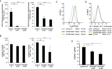
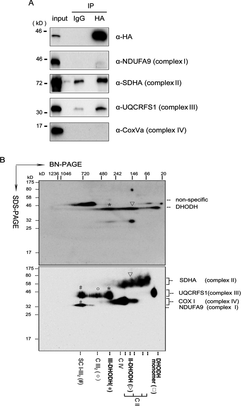
Some mutations of the DHODH (dihydro-orotate dehydrogenase) gene lead to postaxial acrofacial dysostosis or Miller syndrome. Only DHODH is localized at mitochondria among enzymes of the de novo pyrimidine biosynthesis pathway. Since the pyrimidine biosynthesis pathway is coupled to the mitochondrial RC (respiratory chain) via DHODH, impairment of DHODH should affect the RC function. To investigate this, we used siRNA (small interfering RNA)-mediated knockdown and observed that DHODH knockdown induced cell growth retardation because of G₂/M cell-cycle arrest, whereas pyrimidine deficiency usually causes G₁/S arrest. Inconsistent with this, the cell retardation was not rescued by exogenous uridine, which should bypass the DHODH reaction for pyrimidine synthesis. DHODH depletion partially inhibited the RC complex III, decreased the mitochondrial membrane potential, and increased the generation of ROS (reactive oxygen species). We observed that DHODH physically interacts with respiratory complexes II and III by IP (immunoprecipitation) and BN (blue native)/SDS/PAGE analysis. Considering that pyrimidine deficiency alone does not induce craniofacial dysmorphism, the DHODH mutations may contribute to the Miller syndrome in part through somehow altered mitochondrial function.


Figures

Figure 1
Figure 1. Effects of siRNA-mediated knockdown of DHODH
(A) HeLa cells were transfected with control siRNA or two independent DHODH siRNAs, #1 and #2, using Oligofectamine™. At 3 days after the transfection, the cells were lysed and immunoblotted with antibodies against DHODH, NDUFA9 (complex I), SDHA (complex II), complex III subunit core I, complex V, p53 and β-actin. (B) Depletion of DHODH causes growth retardation. The proliferation rate of siRNA-transfected HeLa cells is shown. HeLa cells were transfected with control or two independent DHODH siRNAs on day 0. At the indicated time, the cells were harvested and counted using a cell counter. Left-hand panel, lines represent control siRNA, DHODH siRNA #1 and DHODH siRNA #2 respectively. Uridine (1 mM) was added on day 0 and day 2 to DHODH-depleted HeLa cells. The cell number was counted at the indicated time after seeding. Right-panel panel, proliferation rate of LFN-treated HeLa cells. HeLa cells were treated with 1 mM LFN on day 0. Uridine was added on day 0 and day 2.
Figure 2
Figure 2. Cell-cycle analysis after DHODH depletion
(A, B) At 72 h after DHODH siRNA transfection, cells were fixed and stained with propidium iodide. Then, the DNA content was measured by flow cytometry. Three separate experiments were carried out, all of which exhibited similar trends. The results of one representative experiment are shown. The cell-cycle phase distribution in each condition is shown as the percentage of cells present in the G1-, S- or G2/M-phases.
Figure 3
Figure 3. Depletion of DHODH causes mitochondrial dysfunction
(A) Reduced mitochondrial respiratory activities in DHODH siRNA-treated HeLa cells. DHO-ubiquinone oxidoreductase activity and DHO-cytochrome c oxidoreductase activity were measured using cell lysates as described in the Experimental section. Reduced enzymatic activities of mitochondrial DHO-dependent oxidoreductase were observed in siRNA-treated cells. The results represent the means±S.D. of three independent experiments. **P<0.01 against controls. (B) Reduced complex III activity after DHODH siRNA-mediated depletion in HeLa cells. The activity of each complex (II and III) was measured using cell lysates as described in the Experimental section. Reduced enzymatic activity of mitochondrial complex III was observed in siRNA-treated cells. *P<0.05 against controls. (C) Increased ROS production by DHODH siRNA-mediated knockdown. HeLa cells were transfected with control (green) or the two independent DHODH siRNAs #1 and #2 (red). After 72 h, the cells were treated with 10 μM DCFH-DA for 30 min and were subjected to FACS for quantitative estimation of ROS. (D) Decreased mitochondrial membrane potential in DHODH-depleted HeLa cells. HeLa cells were stained with the fluorescent dye JC-1 and then analysed by flow cytometry at 488 and 590 nm. The fluorescence ratio of JC-1 dimer (red) to JC-1 monomer (green) is shown. Dissipation of the membrane potential with CCCP was used as a control. (E) Reduced mtDNA copy number after DHODH depletion. The amount of mtDNA per cell was estimated based on the ratio of mtDNA/nuclear DNA [ND2/HPRT (hypoxanthine-guanine phosphoribosyltransferase)] by real-time PCR. HeLa cells were treated with control or DHODH siRNA for 72 h. The value in control siRNA-treated cells was set at 100% for each gene. *P<0.05.
Figure 4
Figure 4. DHODH is associated with complexes II and III
(A) DHODH associates with respiratory complexes II and III. DHODH–HA-transfected HeLa cells were treated with DOX for 48 h. The mitochondrial fraction was purified on a Percoll density gradient and lysed with TNE buffer. After cross-linking, immunoprecipitates obtained with anti-HA and mouse IgG antibodies were separated by SDS/PAGE and immunoblotted with anti-HA, NDUFA9 (complex I), SDHA (complex II), UQCRFS1 (complex III) and COXVa (complex IV) antibodies. IP, immunoprecipitate; α, antibody. (B) BN/SDS/PAGE analysis. Isolated mitochondria before and after induction by DOX were solubilized by n-D-maltoside and protein complexes were first resolved by BN-PAGE (polyacrylamide concentration gradient, 3–12%) and resolved in a second dimension by SDS/PAGE. The transferred proteins were immunoblotted with the indicated antibodies. SC I–III2, supercomplex formed from complexes I and III2; CIII2, dimeric complex III; CIV, complex IV; II, succinate dehydrogenase complex II; III2–DHODH, association with complex III and DHODH; II–DHODH, association with complex II and DHODH. DHODH is shown as monomers. The molecular masses (in kDa) of the molecular mass standards are shown.

References

    1. McBride H. M., Neuspiel M., Wasiak S. Mitochondria: more than just a powerhouse. Curr. Biol. 2006;16:R551–R560. - PubMed
    1. DiMauro S., Schon E. A. Mitochondrial disorders in the nervous system. Annu. Rev. Neurosci. 2008;31:91–123. - PubMed
    1. Frenzel M., Rommelspacher H., Sugawa M. D., Dencher N. A. Ageing alters the supramolecular architecture of OxPhos complexes in rat brain cortex. Exp. Gerontol. 2010;45:563–572. - PubMed
    1. Wallace D. C. Mitochondrial DNA mutations in disease and aging. Environ. Mol. Mutagen. 2010;51:440–450. - PubMed
    1. Tuppen H. A., Blakely E. L., Turnbull D. M., Taylor R. W. Mitochondrial DNA mutations and human disease. Biochim. Biophys. Acta. 2010;1797:113–128. - PubMed

Publication types

MeSH terms

Supplementary concepts