Review
. 2013:75:23-47.
doi: 10.1146/annurev-physiol-030212-183802. Epub 2012 Dec 3.

The adventitia: essential regulator of vascular wall structure and function

Affiliations
Review

The adventitia: essential regulator of vascular wall structure and function

Kurt R Stenmark et al. Annu Rev Physiol. 2013.

Abstract

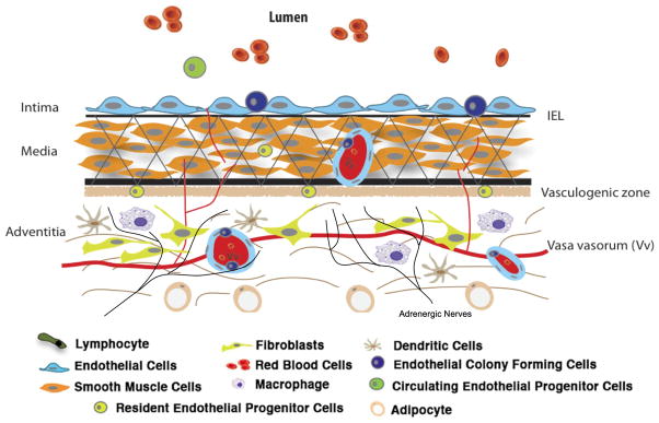
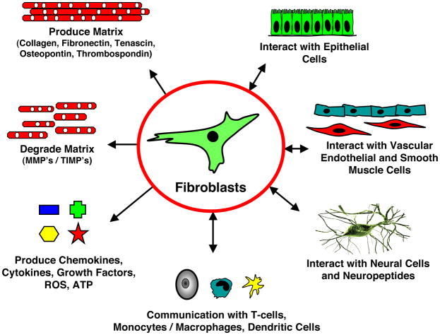
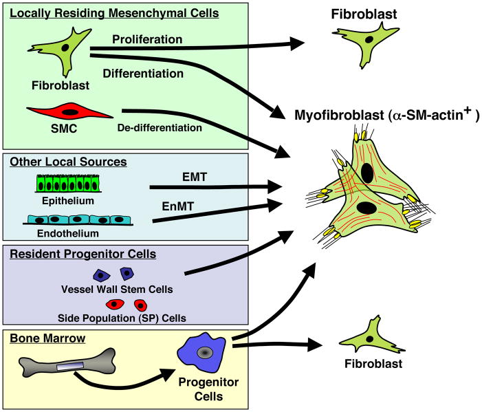
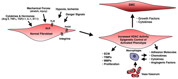
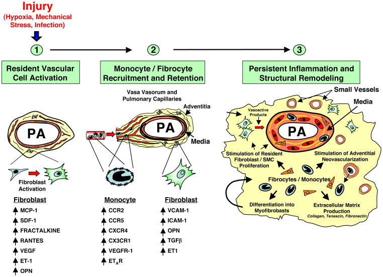
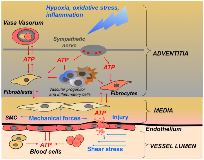
The vascular adventitia acts as a biological processing center for the retrieval, integration, storage, and release of key regulators of vessel wall function. It is the most complex compartment of the vessel wall and is composed of a variety of cells, including fibroblasts, immunomodulatory cells (dendritic cells and macrophages), progenitor cells, vasa vasorum endothelial cells and pericytes, and adrenergic nerves. In response to vascular stress or injury, resident adventitial cells are often the first to be activated and reprogrammed to influence the tone and structure of the vessel wall; to initiate and perpetuate chronic vascular inflammation; and to stimulate expansion of the vasa vasorum, which can act as a conduit for continued inflammatory and progenitor cell delivery to the vessel wall. This review presents the current evidence demonstrating that the adventitia acts as a key regulator of vascular wall function and structure from the outside in.


Figures

Figure 1
Figure 1. Complex cellular composition of the vascular adventitia
Unlike the normal intima and media, which are composed of endothelial and smooth muscle cells respectively, the normal adventitia is comprised of a wide variety of cell types, including fibroblasts, resident progenitor cells, immunomodulatory cells (dendritic cells, macrophages, T-lymphocytes), vasa vasorum endothelial cells and adrenergic nerves.
Figure 2
Figure 2. Fibroblasts play a central role in the control of vascular function
Fibroblasts produce and organize elements of the extracellular matrix (ECM), and also degrade structural elements of the ECM; they secrete a complex mixture of growth factors, cytokines, chemokines; they communicate with neural cells, with cells of hematopoietic origin (dendritic cells, macrophages, T-lymphocytes), with SMC, endothelial and epithelial cells; importantly, this communication is reciprocal (5). (Adapted from: Sorrell et al., “Fibroblasts: a diverse population at the center of it all.”).
Figure 3
Figure 3
Numerous cell types give rise to cells expressing a myofibroblast phenotype.
Figure 4
Figure 4. The constitutively activated “imprinted” phenotype of pulmonary artery AF is due, at least in part, to increased HDAC activity
This fibroblast maintains a capacity to affect the phenotype of other adjacent cells including SMC, macrophages, and vasa vasorum endothelial cells.
Figure 5
Figure 5. Essential role of the adventitial fibroblast in initiating and perpetuating vascular inflammation and, consequently, vascular remodeling
In response to hormonal, infectious, or environmental (hypoxia, hemodynamic stress, etc.) stimuli, the fibroblast is activated and secretes chemokines, cytokines and matricellular proteins involved in the recruitment of monocytes, lymphocytes and progenitor cells. With time, fibroblasts upregulate adhesion molecule expression, which promotes retention of leukocytes and progenitor cells within the adventitia. Some of the newly recruited cells can differentiate into fibroblasts and myofibroblasts, which perpetuate the cycle, thus leading to persistent inflammation and structural vascular remodeling. (PA = pulmonary artery).
Figure 6
Figure 6. Angiogenic expansion of the vasa vasorum in the pulmonary artery adventitia of calves with severe hypoxia-induced pulmonary hypertension
(A–E). Histopathology of large (A, C) and small (B, D) pulmonary arteries. Both histological, H&E (A, B) and immunofluorescent, PECAM-1/CD31 (C, D) staining’s demonstrate marked expansion of the vasa vasorum capillary network in adventitial, perivascular regions. Quantitative morphometric analyses demonstrated that the volume density (Vv) of vasa vasorum is significantly greater in neonatal calves with severe hypoxia-induced pulmonary hypertension compared with normoxic controls (E). (F, G), Angiogenic responses in the adventitia of a human patient with pulmonary fibrosis and associated pulmonary hypertension. Medium-sized pulmonary artery stained with CD31, demonstrating evidence for capillary network expansion in the perivascular area (medial/adventitial region, arrow) (F). CD31 immunohistochemical evidence of capillary proliferation (arrow) (G). Bars, 500 μm in A and C, 100 μm in B, D, F, and 25 μm in G.
Figure 7
Figure 7. Sources of extracellular ATP in the pulmonary artery vascular wall
ATP release and signaling is an integral part of hypoxia-induced response in vascular wall. ATP can be released as a result of combined action of hypoxia, inflammation, fluid shear stress (changes in blood flow), injury, mechanical forces (SMC contractility), and sympathetic neurotransmission (release from perivascular nerve together with noradrenalin and neuropeptides). Importantly, vasa vasorum endothelial cell have been identified as an abundant source of extracellular ATP in pulmonary artery adventitia (118). Collectively, within adventitial microenvironment, extracellular ATP acts in a synergistic manner with multiple pro-inflammatory and growth-promoting factors in hypoxic conditions. (SMC = smooth muscle cells; blood cells = erythrocytes, platelets, monocytes).

References

    1. Sartore S, Chiavegato A, Faggin E, Franch R, Puato M, Ausoni S, Pauletto P. Contribution of adventitial fibroblasts to neointima formation and vascular remodeling: From innocent bystander to active participant. Circ Res. 2001;89:1111–1121. - PubMed
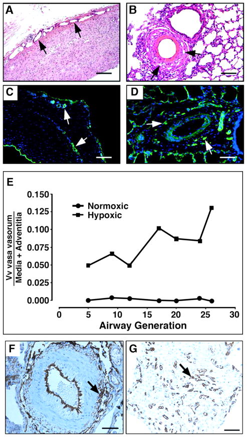
    1. Stenmark KR, Davie N, Frid M, Gerasimovskaya E, Das M. Role of the adventitia in pulmonary vascular remodeling. Physiology (Bethesda, Md. 2006;21:134–145. - PubMed
    1. Silver FH, Horvath I, Foran DJ. Viscoelasticity of the vessel wall: The role of collagen and elastic fibers. Crit Rev Biomed Eng. 2001;29:279–301. - PubMed
    1. Postlethwaite AE, Shigemitsu H, Kanangat S. Cellular origins of fibroblasts: Possible implications for organ fibrosis in systemic sclerosis. Current opinion in rheumatology. 2004;16:733–738. - PubMed
    1. Sorrell JM, Caplan AI. Fibroblasts-a diverse population at the center of it all. International review of cell and molecular biology. 2009;276:161–214. - PubMed

Publication types

LinkOut - more resources