

Adult primary retroperitoneal cavernous hemangioma: a case report
- PMID: 23216883
- PMCID: PMC3539936
- DOI: 10.1186/1477-7819-10-261
Adult primary retroperitoneal cavernous hemangioma: a case report
Abstract
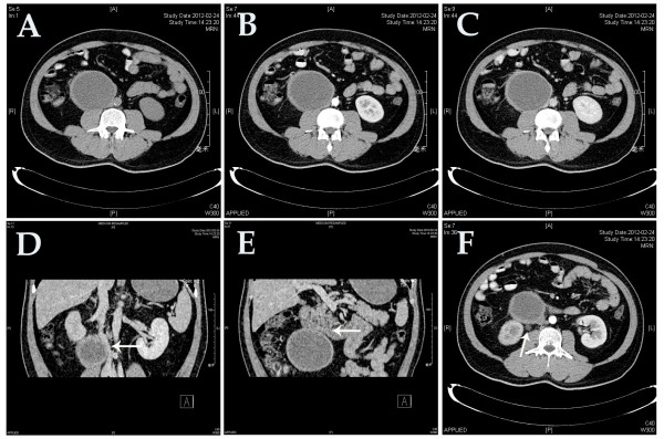
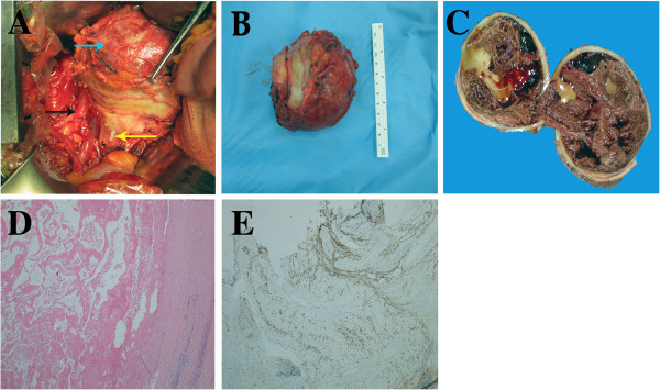
Primary retroperitoneal cavernous hemangioma (PRCH) in an adult is extremely rare. We report on the diagnosis and treatment of a patient with PRCH with subtle clinical features and atypical findings on imaging scans. A 38-year-old man was admitted to hospital with a 5-day history of epigastralgia after alcohol drinking. Using various imaging methods, we found a giant cyst-like retroperitoneal mass compressing the surrounding organs. Surgical resection of the tumor was performed, and the mass was found to be a cavernous hemangioma measuring 90 × 80 × 60 mm, with a thick fibrotic wall and extensive intracystic hemorrhage. Physicians should be aware that PRCH may mimic a cystic neoplasm, and that a large tumor size probably indicates intracystic hemorrhage. Surgical resection is a curative approach for PRCH.
Figures
References
-
- Geenen RWF, den Bakker MA, Bangma CH, Hussain SM, Krestin GR. Sonography CT MRI of giant cavernous hemangioma of the kidney: correlation with pathologic findings. Am J Roentgenol. 2004;182:411–414. - PubMed
-
- Weidenfeld J, Zakai BB, Faermann R, Barshack I, Aviel-Ronen S. Hemangioma of pancreas: a rare tumor of adulthood. Isr Med Assoc J. 2011;13:512–514. - PubMed
-
- England RJ, Woodley H, Cullinane C, McClean P, Walker J, Stringer MD. Pediatric pancreatic hemangioma: a case report and literature review. JOP. 2006;7:496–501. - PubMed
Publication types
MeSH terms
LinkOut - more resources
Full Text Sources
