. 2012 Dec 5:7:206.
doi: 10.1186/1748-717X-7-206.

Radiation resistance due to high expression of miR-21 and G2/M checkpoint arrest in breast cancer cells

Affiliations

Radiation resistance due to high expression of miR-21 and G2/M checkpoint arrest in breast cancer cells

Nataša Anastasov et al. Radiat Oncol. .

Abstract

Background: There is evidence that the extent of the G2/M arrest following irradiation is correlated with tumour cell survival and hence therapeutic success. We studied the regulation of cellular response to radiation treatment by miR-21-mediated modulation of cell cycle progression in breast cancer cells and analysed miR-21 expression in breast cancer tissue samples with long-term follow up.

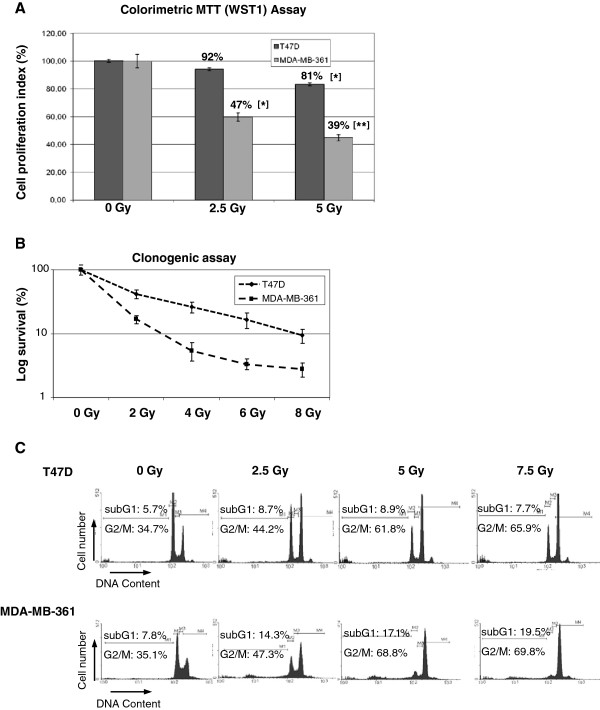
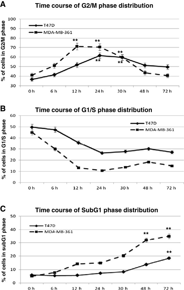
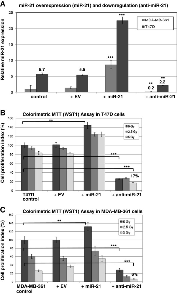
Methods: The miR-21 expression levels were quantified (qRT-PCR) in a panel of 86 cases of invasive breast carcinomas in relation to metastasis free survival. The cellular radiosensitivity of human breast cancer cells after irradiation was determined comparing two cell lines (T47D and MDA-MB-361) by cell proliferation and colony forming assays. The influence of miR-21 overexpression or downregulation on cell cycle progression and G2/M checkpoint arrest after irradiation was assessed by flow cytometric analysis.

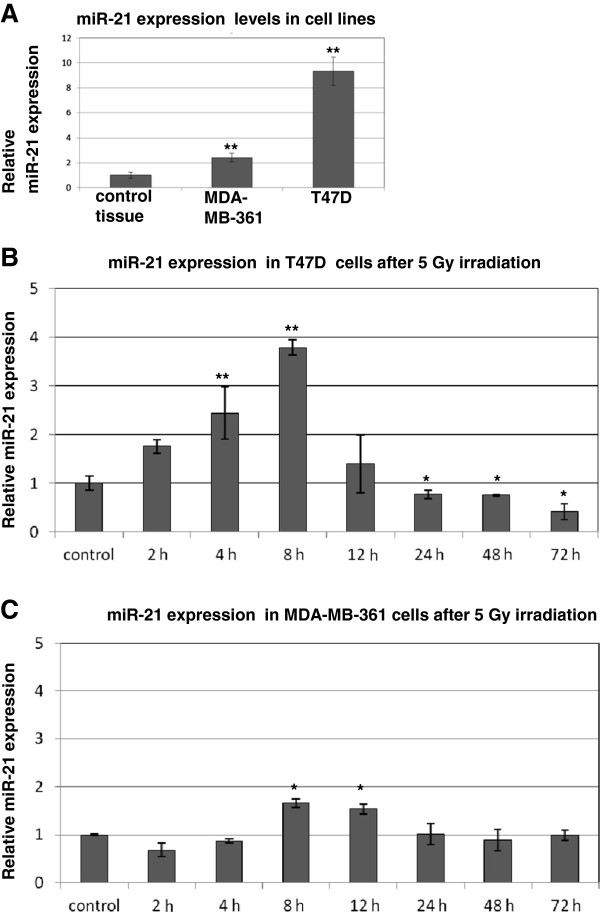
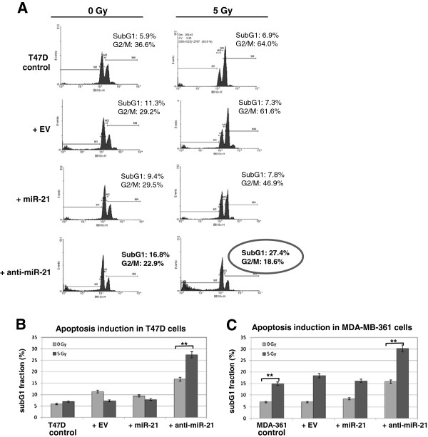
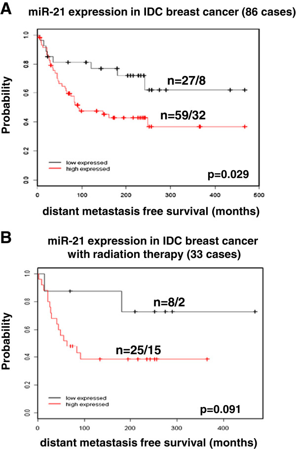
Results: The expression of miR-21 was transiently increased 8 hours after irradiation in the radioresistant T47D cells and significantly changed with lower extent in radiosensitive MDA-MB-361 cells. Anti-miR-21 treated breast cancer cells failed to exhibit the DNA damage-G2 checkpoint increase after irradiation. Apoptotic activity was significantly enhanced from 7% to 27% in T47D cells and from 18% to 30% in MDA-MB-361 cells 24 hours after 5 Gy irradiation. Additionally, we characterized expression of miR-21 in invasive breast carcinomas. In comparison to non-cancerous adjacent breast tissue, tumours samples had increased miR-21 expression that inversely correlated with the distant metastases-free survival of patients (p = 0.029).

Conclusions: Our data indicate that miR-21 expression in breast cancer cells contributes to radiation resistance by compromising cell cycle progression. These data point to the potential of combining radiotherapy with an anti-miR-21 as a potent G2/M check point inhibitor in overcoming radiation resistance of tumours.


Figures

Figure 1
Figure 1
Breast cancer cell survival and cell cycle characterisation after irradiation. (A) Growth characteristics of T47D and MDA-MB-361 breast cancer cells were determined by MTT (WST1) assay 72 hours after irradiation. Data represent the means ± SD (n=4). *p <0.05, **p <0.01 by ANOVA one-way analysis of variance. (B) clonogenic survival of breast cancer cells 10 days after irradiation. (C) Cell cycle progression in breast cancer cells was evaluated by PI staining and flow cytometry 24 hours after irradiation at the indicated doses.
Figure 2
Figure 2
Cell cycle time kinetics in breast cancer cells after 5 Gy irradiation. Cell cycle distributions were analyzed by FACS and changes in cellular fractions of G2/M (A), G1/S (B) and subG1 (C) after indicated time points are presented. Data represent the means ± SD (n=2). *p <0.05, **p <0.01 by ANOVA one-way analysis of variance.
Figure 3
Figure 3
Time kinetics of miR-21 expression in breast cancer cells after 5 Gy irradiation. (A) Relative miR-21 expression in breast cancer cell lines compared to adjacent control mammary tissue. (B) Relative miR-21 expression in T47D and (C) MDA-MB-361 breast cancer cells after 5Gy irradiation and indicated time points in relation to the expression of sham irradiated controls. Data represent the means ± SD (n=4). *p <0.05, **p <0.01 by ANOVA.
Figure 4
Figure 4
miR-21 downregulation regulates the cellular response to radiation. (A) T47D cells were infected with empty lentivirus (+ EV), with miR-21 overexpressing LV (+ miR-21) or inhibitory miR-21 LV (+ anti-mir-21) and analysed for miRNA expression in MDA-MB-361 cells (light gray boxes) and T47D cells (dark gray boxes) 72 hours after infection. MDA-MB-361 control sample was used as reference for miR-21 expression (set as 1). (B) Cell proliferation assay (WST1) in T47D cells with miR-21 overexpression or downregulation and corresponding radiation treatment after 72 hours in control cells (dark gray boxes), or after 2.5 Gy (gray boxes) and 5 Gy (light gray boxes) 72 hours after irradiation. (C) Cell proliferation assay (WST1) in MDA-MB-361 cells with miR-21 overexpression or downregulation and corresponding radiation treatment after 72 hours. Data represent the means ± SD (n=3). *p <0.05, **p <0.01, ***p <0.001 by ANOVA.
Figure 5
Figure 5
miR-21 downregulation abrogates G2/M check point accumulation after irradiation. (A) T47D cells were infected with empty lentivirus (+ EV), with miR-21 overexpressing LV (+ miR-21) or inhibitory miR-21 LV (+ anti-mir-21) and analysed for cell cycle changes 24 hours after 5 Gy irradiation. One representative FACS analysis is shown from three independent experiments. (B) Statistical analysis of subG1 cellular fraction in T47D infected cells (as in part A) and (C) in MDA-MB-361 cells (control cells - light gray boxes) or after 5 Gy irradiation (dark gray boxes). Data represent the means ± SD (n=3). *p <0.05, **p <0.01 by ANOVA.
Figure 6
Figure 6
miR-21 expression in breast cancer. (A) Kaplan Meier analyses for distant metastasis-free survival of patients. The patients were grouped into those with low (<1.8-fold) and high (>1.8-fold) miR-21 expression in tumour tissues, with a significantly better prognosis for patients with low miR-21 expression (p = 0.029). (B) Expression of miR-21 in radiotherapy treated patients group. Although this analysis did not reach significance (p = 0.091) a clear trend is obvious for better prognosis of patients with low miR-21 expression.

References

    1. Bartel DP. MicroRNAs: target recognition and regulatory functions. Cell. 2009;136(2):215–233. doi: 10.1016/j.cell.2009.01.002. - DOI - PMC - PubMed
    1. Calin GA, Croce CM. MicroRNA signatures in human cancers. Nat Rev Cancer. 2006;6(11):857–866. doi: 10.1038/nrc1997. - DOI - PubMed
    1. Ventura A, Jacks T. MicroRNAs and cancer: short RNAs go a long way. Cell. 2009;136(4):586–591. doi: 10.1016/j.cell.2009.02.005. - DOI - PMC - PubMed
    1. Volinia S, Calin, Liu GA, Ambs CG, Cimmino S, Petrocca A, Visone F, Iorio R, Roldo M, Ferracin C. et al.A microRNA expression signature of human solid tumors defines cancer gene targets. Proc Natl Acad Sci USA. 2006;103(7):2257–2261. doi: 10.1073/pnas.0510565103. - DOI - PMC - PubMed
    1. Lu J, Getz, Miska G, Alvarez-Saavedra EA, Lamb E, Peck J, Sweet-Cordero D, Ebert A, Mak BL, Ferrando RH. et al.MicroRNA expression profiles classify human cancers. Nature. 2005;435(7043):834–838. doi: 10.1038/nature03702. - DOI - PubMed

Publication types

MeSH terms