

Mucinous adenocarcinoma of the intestinal type arising from mature cystic teratoma of the ovary: a rare case report and review of the literature
- PMID: 23216975
- PMCID: PMC3524777
- DOI: 10.1186/1757-2215-5-41
Mucinous adenocarcinoma of the intestinal type arising from mature cystic teratoma of the ovary: a rare case report and review of the literature
Abstract
Background: Mature cystic teratomas (MCTs) are the most common germ cell tumors of the ovary. Malignant tranformation occurs in 1-2% of these neoplasms. Although most of the malignancies arising from MCTs are squamous cell carcinomas, adenocarcinoma of the gastrointestinal type is extremery rare. We herein present a case of adenocarcinoma of the intestinal type arising from a MCT.
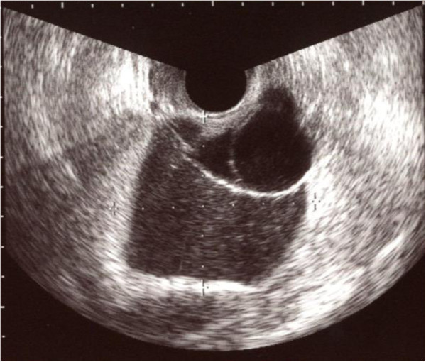
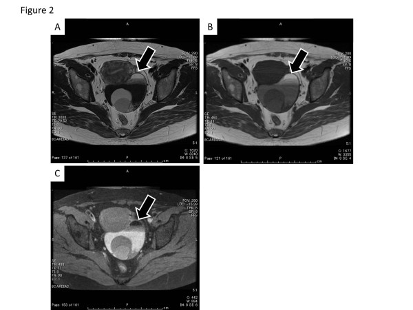
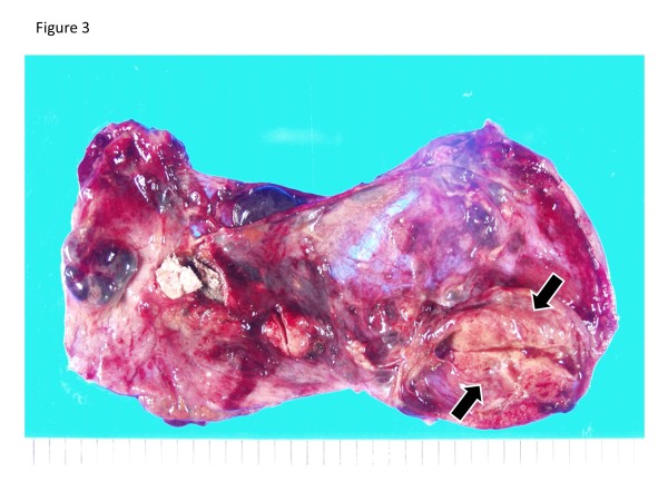
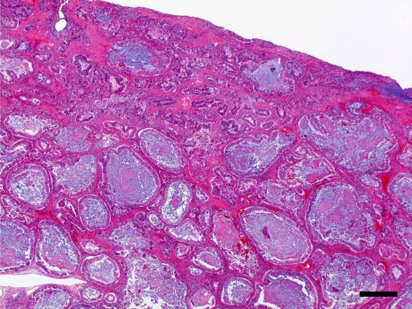
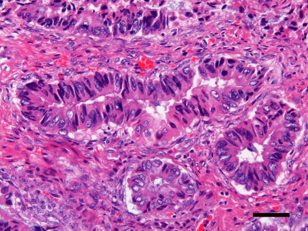
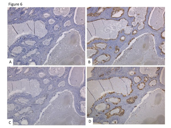
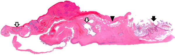
Case: A 49-year-old female underwent surgery for a left ovarian tumor. The histology of the cyst walls revealed a MCT with a few hair shafts and a squamous layer, while another part of the tumor showed adenocarcinoma of the intestinal type. Five years after surgery, she is alive without disease.
Figures
Similar articles
-
An ovarian mature cystic teratoma evolving in squamous cell carcinoma: A case report and review of the literature.Gynecol Oncol Rep. 2016 Dec 18;19:27-30. doi: 10.1016/j.gore.2016.12.005. eCollection 2017 Feb. Gynecol Oncol Rep. 2016. PMID: 28050596 Free PMC article. Review.
-
A rare case report of squamous-cell carcinoma arising from mature cystic teratoma of ovary.G Chir. 2014 Sep-Oct;35(9-10):241-5. G Chir. 2014. PMID: 25419592 Free PMC article.
-
Ovarian mucinous tumors arising from mature cystic teratomas--a molecular genetic approach for understanding the cellular origin.Hum Pathol. 2014 Apr;45(4):717-24. doi: 10.1016/j.humpath.2013.10.031. Epub 2013 Nov 13. Hum Pathol. 2014. PMID: 24485845
-
Intestinal-Type Adenocarcinoma Arising in a Mature Cystic Teratoma of the Ovary.Int J Gynecol Pathol. 2016 Jul;35(4):352-6. doi: 10.1097/PGP.0000000000000258. Int J Gynecol Pathol. 2016. PMID: 26937866 Review.
-
Primary squamous carcinoma of the ovary likely arising from a monodermal cystic mucinous teratoma.Ann Diagn Pathol. 2011 Dec;15(6):446-9. doi: 10.1016/j.anndiagpath.2010.06.005. Epub 2010 Oct 12. Ann Diagn Pathol. 2011. PMID: 20952274
Cited by
-
Adenocarcinoma of intestinal type arising in mature cystic teratoma of ovary: A diagnostic dilemma.Clin Case Rep. 2020 Feb 6;8(4):644-647. doi: 10.1002/ccr3.2718. eCollection 2020 Apr. Clin Case Rep. 2020. PMID: 32274027 Free PMC article.
-
Imaging features of mucinous carcinoma arising from mature teratoma showing cytokeratin 7+ and cytokeratin 20+ expression profile: A case report.Radiol Case Rep. 2024 Jan 13;19(4):1288-1293. doi: 10.1016/j.radcr.2024.01.001. eCollection 2024 Apr. Radiol Case Rep. 2024. PMID: 38292777 Free PMC article.
-
Colorectal Adenocarcinoma Derived From Mature Cystic Teratomas: A Case Report With Review of the Literature.Cureus. 2023 Aug 26;15(8):e44159. doi: 10.7759/cureus.44159. eCollection 2023 Aug. Cureus. 2023. PMID: 37753035 Free PMC article.
-
KRAS mutation in adenocarcinoma of the gastrointestinal type arising from a mature cystic teratoma of the ovary.J Ovarian Res. 2014 Sep 6;7:85. doi: 10.1186/s13048-014-0085-3. J Ovarian Res. 2014. PMID: 25297496 Free PMC article.
-
Experience of applying cytoreductive surgery and hyperthermic intraperitoneal chemotherapy for ovarian teratoma with malignant transformation and peritoneal dissemination.Ther Clin Risk Manag. 2019 Jan 14;15:129-136. doi: 10.2147/TCRM.S190641. eCollection 2019. Ther Clin Risk Manag. 2019. PMID: 30679911 Free PMC article.
References
LinkOut - more resources
Full Text Sources
