Case Reports
. 2012 Dec 5:8:235.
doi: 10.1186/1746-6148-8-235.

Squamous metaplasia of the rete ovarii in a Zebu cow

Affiliations
Case Reports

Squamous metaplasia of the rete ovarii in a Zebu cow

Renato Lima Santos et al. BMC Vet Res. .

Abstract

Background: Stratified keratinizing squamous epithelium in the ovary has been associated with the diagnosis of ovarian teratoma in cows. Recently, the diagnosis of "epidermoid cyst" has been proposed. A case of squamous metaplasia of the rete ovarii in a Zebu cow is described in this report.

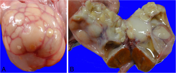
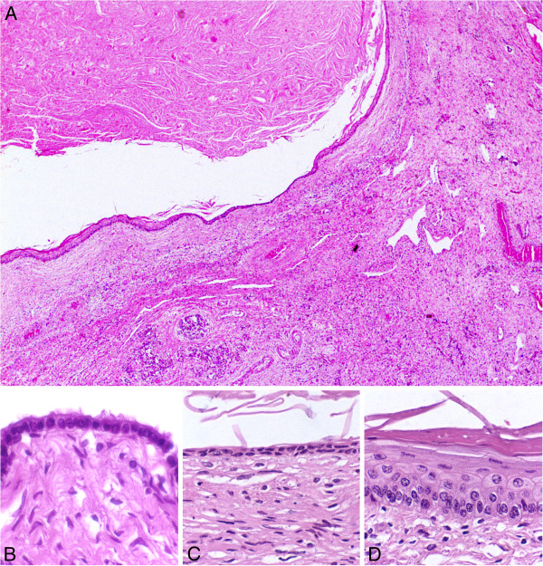
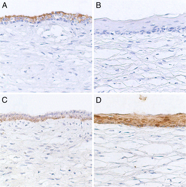
Case presentation: A crossbreed Zebu cow had both ovaries enlarged with multiple cysts. Most cysts were lined by well differentiated keratinizing stratified squamous epithelium and filled with keratinized lamellar material. Some cysts were lined by an epithelial layer that ranged from single cuboidal, double cuboidal epithelium, stratified non keratinized epithelium, and areas of keratinizing stratified squamous epithelium. Single or double layered cuboidal epithelia of the cysts expressed low molecular weight cytokeratin 7, whose expression was absent in the keratinizing stratified squamous epithelia of same cysts. Conversely, high molecular weight cytokeratins 1, 5, 10, and 14 were strongly expressed by the keratinizing stratified epithelium.

Conclusion: Squamous metaplasia of the rete ovarii was diagnosed. Squamous metaplasia of the rete ovarii, may account for some of the previously described squamous lesions in the ovary, which may have been misinterpreted as teratoma or epidermoid cysts.


Figures

Figure 1
Figure 1
Gross appearance of a bovine ovary with squamous metaplasia of the rete ovarii. (A) Enlarged right ovary with an irregular surface containing multiple cysts. (B) Cut surface of the right ovary with multiple cysts and a corpus luteum. Some of the cysts are filled with a translucent or whitish mucous material, while other cysts are filled with a dense keratinized material.
Figure 2
Figure 2
Microscopic features of a bovine ovary with squamous metaplasia of the rete ovarii. (A) Cystic cavity in the ovary filled with keratinized material and lined by single layered cuboidal epithelium (arrowhead) transitioning into a keratinizing stratified squamous epithelium (arrow). (B) Detail of the cystic wall with an area of single layered cuboidal epithelium. (C) Detail of the cystic wall with an area of double layered epithelium. (D) Detail of the cystic wall with an area of keratinizing stratified squamous epithelium. Hematoxylin and eosin.
Figure 3
Figure 3
Immunohistochemical features of a bovine ovary with squamous metaplasia of the rete ovarii. (A) Expression of low molecular weight cytokeratin 7 (clone OV-TL 12/30) in an area of double layered cuboidal non keratinized epithelium. (B) Ovary, cow. Absence of expression of low molecular weight cytokeratin 7 (clone OV-TL 12/30) in an area of keratinizing stratified squamous epithelium. (C) Low levels of expression of high molecular weight cytokeratins 1, 5, 10, and 14 (clone 34BE12) in an area of double layered cuboidal non keratinized epithelium. (D) Strong expression of high molecular weight cytokeratins 1, 5, 10, and 14 (clone 34BE12) in an area of keratinizing stratified squamous epithelium. Streptavidin-biotin-peroxidase, Hematoxylin counterstain.

References

    1. McEntee K. Reproductive pathology of domestic mammals. San Diego: Academic; 1990.
    1. Nascimento EF, Santos RL. Patologia da reprodução dos animais domésticos. 3. Rio de Janeiro: Guanabara Koogan; 2011.
    1. Costa SA. MS thesis. Escola de Veterinária; Brazil: Universidade Federal de Minas Gerais; 1974. Ocorrência de alterações em ovários de vacas azebuadas abatidas em matadouros dos estados de Goiás e Minas Gerais.
    1. Edwards JF. Three cases of ovarian epidermoid cysts in cattle. Vet Pathol. 2002;39:744–746. doi: 10.1354/vp.39-6-744. - DOI - PubMed
    1. Khedmati F, Chirolas C, Seidman JD. Ovarian and paraovarian squamous-lined cysts (epidermoid cysts): a clinicopathologic study of 18 cases with comparison to mature cystic teratomas. Int J Gynecol Pathol. 2009;28:193–196. doi: 10.1097/PGP.0b013e318187e147. - DOI - PubMed

Publication types