. 2012 Dec 5;16(6):801-13.
doi: 10.1016/j.cmet.2012.10.018.

A heme-sensing mechanism in the translational regulation of mitochondrial cytochrome c oxidase biogenesis

Affiliations

A heme-sensing mechanism in the translational regulation of mitochondrial cytochrome c oxidase biogenesis

Iliana C Soto et al. Cell Metab. .

Abstract

Heme plays fundamental roles as cofactor and signaling molecule in multiple pathways devoted to oxygen sensing and utilization in aerobic organisms. For cellular respiration, heme serves as a prosthetic group in electron transfer proteins and redox enzymes. Here we report that in the yeast Saccharomyces cerevisiae, a heme-sensing mechanism translationally controls the biogenesis of cytochrome c oxidase (COX), the terminal mitochondrial respiratory chain enzyme. We show that Mss51, a COX1 mRNA-specific translational activator and Cox1 chaperone, which coordinates Cox1 synthesis in mitoribosomes with its assembly in COX, is a heme-binding protein. Mss51 contains two heme regulatory motifs or Cys-Pro-X domains located in its N terminus. Using a combination of in vitro and in vivo approaches, we have demonstrated that these motifs are important for heme binding and efficient performance of Mss51 functions. We conclude that heme sensing by Mss51 regulates COX biogenesis and aerobic energy production.


Figures

Figure 1
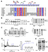
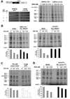
Figure 1. Mss51 contains two conserved CPX motifs and binds heme in vitro
(A) Partial N-terminal sequence alignment of Mss51 from 12 fungal species. Sequences were aligned using the CLUSTALW algorithm (Blosum62 scoring matrix) in Bioedit software. Amino-acids that are conserved in all sequences are shaded red and those conserved in at least 9 of 12 sequences are shaded blue. The conservation of two cysteines in the CPX motifs is marked in yellow. In the linear graph of the protein, all the cysteines (red) and other relevant residues for this study (green or black) are indicated. (B and C) Immunoblot analyses of the binding to hemin-agarose beads of recombinant (B) wild-type Mss51 or (C) CPX mutant Mss51. Mss51 was pre-incubated or not with the indicated concentrations of either hemin or heme A prior to incubation with the beads. To quantify the signals, the images were digitalized and densitometric analyses performed using the histogram function of the Adobe Photoshop program. The graph represents the proportion of bound versus unbound protein for each sample. B, bound; U, unbound; P, protein alone. (D) Difference spectroscopy titration of heme binding to Mss51. Difference absorption spectra and titration curves of wild-type Mss51 (50 nM) with increasing concentrations of Fe3+ hemin (from 0 to 10 µM) as indicated. The curves were generated from fits to an equation describing a single binding site (Y=B max*XKd+X) using GraphPad Prism. (E) Analyses of Mss51 and heme-bound-Mss51 by SDS- and Native-PAGE. The upper panel represents immunoblot analyses with an anti-Mss51 antibody. The lower panel represents in gel heme staining.
Figure 2
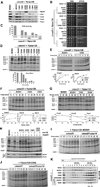
Figure 2. Mutations in Mss51 CPX motifs affect Cox1 synthesis and assembly
(A) Immunoblot analyses of the steady-state levels of Mss51 and COX catalytic core subunits in a wild-type strain (W303) and a Δmss51 strain expressing the indicated wild-type or CPX mutant variants of Mss51. (B) Growth test using serial dilutions of the indicated strains in complete media containing fermentable (glucose, YPD) or non-fermentable (ethanol-glycerol, YPEG) carbon sources. The plates were incubated at 30°C and the pictures taken after 2 days of growth. (C) Cytochrome c oxidase (COX) activity measured spectrophotometrically in the indicated strains. Error bars represent the mean ± SD. (D–J) In vivo mitochondrial protein synthesis in the indicated strains. Pulses were of 30 min (in D and H) or the indicated times. In (G), following a 15 min pulse, labeling was terminated by addition of 12 µg/ml puromycin + 80µM non-radioactive methionine and the products chased during the indicated times. The signals were quantified as in Fig. 1B and used to plot the graphs in the lower panels. (K) Sucrose gradient sedimentation analyses of Mss51 in mitochondrial extracts prepared from the indicated strains. Fractions that contain Mss51-complex peaks are indicated. The gradients were calibrated with lactate dehydrogenase (LDH, 130 kDa) and hemoglobin (Hb, 67 kDa).
Figure 3
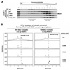
Figure 3. Mitochondrial Mss51 binds heme B
(A) Sucrose gradient sedimentation analyses of Mss51 in mitochondrial extracts prepared from the indicated strains. The indicated fractions containing Mss51-complex peaks were used for GST-pulldown purification of the complexes. (B) Hemes were extracted from 450 kDa and 120 kDa Mss51-containing complexes purified from the indicated strains and analyzed by HPLC on a reverse phase C18 column. The peaks corresponding to heme B (B) are marked. The peak labeled * has not been identified but in some repetitions also appeared in the blank and could be part of the background. The amounts of heme B were calculated from the areas under the peaks.
Figure 4
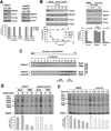
Figure 4. Heme is not required for Mss51 stability but for Mss51 function
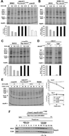
(A) Immunoblot analyses of the steady-state levels of Mss51, Cytochrome b2 and Cox1 in Δhem1 and Δhem1Δcox14 strains grown in the presence of ALA or TEM. The signals were quantified as in Fig. 1B, normalized to porin signal and plotted in the lower panel. (B) Same as in 4A but here a wild-type strain (W303) was treated with the heme biosynthesis inhibitor DHA at the indicated concentrations and periods of time in the presence of TEM. The signals were quantified as in Fig. 1B and plotted in the lower panels. (C) Sucrose gradient sedimentation analyses of Mss51 in mitochondrial extracts prepared from the indicated strains as in Fig. 2I. (D, E) In vivo mitochondrial protein synthesis in the indicated strains and growth conditions performed as in Fig. 2D. The signals were quantified as in Fig. 1B and used to plot the graphs in the lower panels.
Figure 5
Figure 5. Heme is required for efficient COX1 mRNA translation
(A) In vivo mitochondrial protein synthesis in the indicated strains treated or not with DHA. The wild-type (XPM78a) and Δcox14 (LSR39) strains carry an engineered COX1-ARG8 reporter gene in their mitochondrial DNA (upper panel). The synthesized Cox1-Arg8 fusion protein is indicated. The lower panel shows a serial dilution growth test of the strains in synthetic media supplemented or not with DHA and arginine. (B–D) In vivo mitochondrial protein synthesis in the indicated strains treated or not with DHA. The signals were quantified as in Fig. 1B and used to plot the graphs in the lower panels.
Figure 6
Figure 6. Mss51 mutants bypass the requirement of heme for efficient COX1 mRNA translation
(A–E) In vivo mitochondrial protein synthesis in the indicated strains treated or not with DHA in the presence of TEM or in (D) the Δhem1, mss51T167R strain incubated in the presence of either ALA or TEM. In (E), following a 10 minute pulse, the labeled proteins were chased for 30 and 60 min. The signals were quantified as in Fig. 1B and used to plot the graphs in the lower or side panels. (F) Sucrose gradient sedimentation analyses of GST-tagged Mss51F199I in mitochondrial extracts prepared from the indicated strains as in 2I.
Figure 7
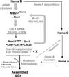
Figure 7. Model of Heme sensing by Mss51 to coordinate Cox1 translation and assembly
See explanation in the Discussion section.

Similar articles

Cited by

References

    1. Barrientos A, Korr D, Tzagoloff A. Shy1p is necessary for full expression of mitochondrial COX1 in the yeast model of Leigh's syndrome. EMBO J. 2002;21:43–52. - PMC - PubMed
    1. Barrientos A, Zambrano A, Tzagoloff A. Mss51p and Cox14p jointly regulate mitochondrial Cox1p expression in Saccharomyces cerevisiae. EMBO J. 2004;23:3472–3482. - PMC - PubMed
    1. Barros MH, Tzagoloff A. Regulation of the heme A biosynthetic pathway in Saccharomyces cerevisiae. FEBS Lett. 2002;516:119–123. - PubMed
    1. Bestwick M, Khalimonchuk O, Pierrel F, Winge DR. The role of Coa2 in hemylation of yeast Cox1 revealed by its genetic interaction with Cox10. Mol Cell Biol. 2010;30:172–185. - PMC - PubMed
    1. Fontanesi F, Clemente P, Barrientos A. Cox25 teams up with Mss51, Ssc1, and Cox14 to regulate mitochondrial cytochrome c oxidase subunit 1 expression and assembly in Saccharomyces cerevisiae. J Biol Chem. 2011;286:555–566. - PMC - PubMed

Publication types

MeSH terms