Case Reports
. 2012 Dec 7;91(6):1095-102.
doi: 10.1016/j.ajhg.2012.10.008.

Cowchock syndrome is associated with a mutation in apoptosis-inducing factor

Affiliations
Case Reports

Cowchock syndrome is associated with a mutation in apoptosis-inducing factor

Carlo Rinaldi et al. Am J Hum Genet. .

Abstract

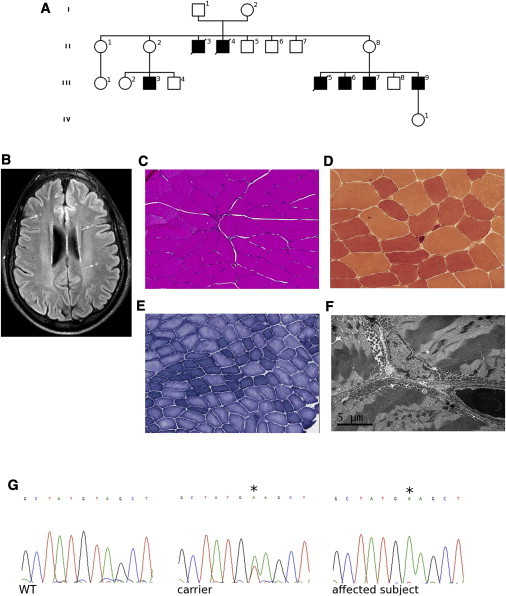
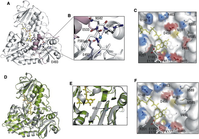
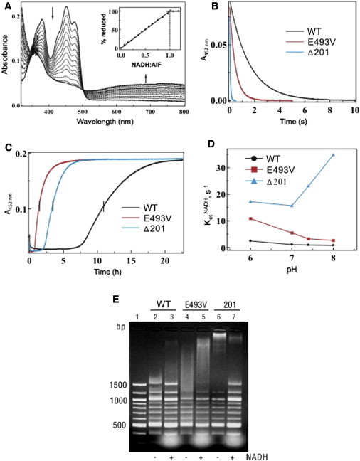
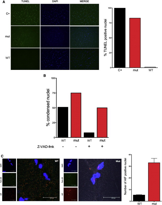
Cowchock syndrome (CMTX4) is a slowly progressive X-linked recessive disorder with axonal neuropathy, deafness, and cognitive impairment. The disease locus was previously mapped to an 11 cM region at chromosome X: q24-q26. Exome sequencing of an affected individual from the originally described family identified a missense change c.1478A>T (p.Glu493Val) in AIFM1, the gene encoding apoptosis-inducing factor (AIF) mitochondrion-associated 1. The change is at a highly conserved residue and cosegregated with the phenotype in the family. AIF is an FAD-dependent NADH oxidase that is imported into mitochondria. With apoptotic insults, a N-terminal transmembrane linker is cleaved off, producing a soluble fragment that is released into the cytosol and then transported into the nucleus, where it triggers caspase-independent apoptosis. Another AIFM1 mutation that predicts p.Arg201del has recently been associated with severe mitochondrial encephalomyopathy in two infants by impairing oxidative phosphorylation. The c.1478A>T (p.Glu493Val) mutation found in the family reported here alters the redox properties of the AIF protein and results in increased cell death via apoptosis, without affecting the activity of the respiratory chain complexes. Our findings expand the spectrum of AIF-related disease and provide insight into the effects of AIFM1 mutations.


Figures

Figure 1
Figure 1
Pedigree, Brain Imaging, and Skeletal Muscle Analysis and Sanger Sequencing (A) Pedigree of the family. White, unaffected; black, affected. (B) Fluid-attenuated inversion recovery (FLAIR) magnetic resonance imaging on a transverse section of supratentorial brain of an affected individual showing multiple punctate hyperintensities in the white matter (arrows; subject III-6). (C–E) Muscle biopsy showing variation in fiber size (hematoxylin and eosin staining, C), esterase-positive angular atrophic myofibers (esterase staining, D), and fiber grouping with some targetoid fibers (nicotinamide adenine dinucleotide-tetrazolium reductase, E) (subject III-9). (F) Electron microscopic image of muscle showing variation in shape and increase in number of mitochondria, mainly in the subsarcolemmal areas (subject III-9). (G) Chromatograms of AIFM1 showing the c.1478A>T (p.Glu493Val) variant (asterisk) in an affected individual (hemizygote) and a carrier (heterozygote).
Figure 2
Figure 2
Structural Comparison of the Wild-Type and p.Glu493Val Mutant of AIF The p.Glu493Val substitution was introduced into the human AIF1 expression plasmid by Stratagene QuikChange kit. Expression, purification, and removal of the C-terminal six-histidine affinity tag were carried out as described previously. (A) Position of Glu493 (indicated by an arrow) in oxidized human AIFwt (PDB code 1M6I). FAD is shown in yellow and a partially disordered regulatory peptide is in pink. (B) Water-bridged contacts involving Glu493 hold the 512–533 helical fragment of the regulatory peptide (pink) close to the active site. (C) Charge distribution near the flavin cofactor. Positively and negatively charged residues are depicted in blue and red, respectively. Glu493 is 10 Å away from FAD and is part of an acidic cluster adjacent to the isoalloxazine ring. (D) Superposition of the structures of the wild-type (gray, PDB code 1M6I) and Glu493Val mutant of human AIF (green, PDB code 4FDC). The structure of AIFE493V was solved at 2.4 Å resolution. Data collection and refinement statistics are given in Table S1. The valine side chain is shown in cyan. (E) A magnified view at the region of AIF near the site of the p.Glu493 residue demonstrating that the substitution does not alter the structure near the active site of AIF. (F) The Glu493Val substitution changes both the surface profile and electrostatic potential, which may affect solvent accessibility and redox properties of FAD.
Figure 3
Figure 3
Comparison of the Redox and Molecular Properties of the Wild-Type and p.Glu493Val and p.Arg201del Mutants of AIF (A) Anaerobic titration of AIFE493V with NADH. Similar to the wild-type protein, the p.Glu493Val variant binds NADH tightly and produces a FADH-NAD charge-transfer complex (CTC) absorbing in the long-wavelength region. Inset shows that an equimolar amount of NADH is required to fully reduce FAD. (B) Kinetics of AIF reduction with NADH. Proteins were mixed with NADH in a stopped flow spectrophotometer, and reduction of FAD was monitored at 452 nm. The derived kinetic parameters are given in Table 1. (C) Kinetics of oxidation of NADH-reduced AIF. Proteins were reduced with a 4-fold excess of NADH before exposure to atmospheric oxygen. Flavin oxidation was monitored at 452 nm. (D) Effect of pH on the kinetics of AIF reduction with NADH. (E) Comparison of DNA binding by wild-type and mutant AIF. Equal amounts of protein (100 μg) were incubated for 15 min with 250 ng of 100 bp DNA ladder (New England Biolabs) in the absence and presence of a 20-fold excess of NADH. After separation on a 2% agarose gel, DNA was visualized with ethidium bromide. Lane 1 is a control (DNA only).
Figure 4
Figure 4
Caspase-Independent Apoptosis and Nuclear Localization of p.Glu493Val AIF (A) TUNEL and DAPI staining of skeletal muscle nuclei from an affected subject (mut) and a control (WT). A normal biopsy treated with DNase was used a positive control (C+). Numerous TUNEL-positive nuclei are present in the affected subject, whereas no TUNEL-positive nuclei are seen in the control. Quantification of TUNEL-positive nuclei (n = 100) on the right. (B) Quantification of altered nuclei in control fibroblasts (WT) and in fibroblasts from an affected individual (mut) after treatment with staurosporine (1 μM for 2 hr) in presence or absence of Z-VAD-fmk (100 μM for 0.5 hr). (C) AIFM1 (1:1,000; Millipore, AB16501) immunostaining of cross section of muscle biopsy showing nuclear localization of mutant AIF in a subject hemizygous for p.Glu493Val (mut) compared to control (WT). Nuclei and mitochondria were counterstained with DAPI and mouse Tom20 antibody (BD Transduction Laboratory), respectively. AIF-positive nuclei were counted and normalized to the total number of DAPI-stained nuclei from three random and nonoverlapping fields (mean; SEM; p < 0.01).

References

    1. Skre H. Genetic and clinical aspects of Charcot-Marie-Tooth’s disease. Clin. Genet. 1974;6:98–118. - PubMed
    1. Pareyson D., Marchesi C. Diagnosis, natural history, and management of Charcot-Marie-Tooth disease. Lancet Neurol. 2009;8:654–667. - PubMed
    1. Boerkoel C.F., Takashima H., Garcia C.A., Olney R.K., Johnson J., Berry K., Russo P., Kennedy S., Teebi A.S., Scavina M. Charcot-Marie-Tooth disease and related neuropathies: mutation distribution and genotype-phenotype correlation. Ann. Neurol. 2002;51:190–201. - PubMed
    1. Bergoffen J., Scherer S.S., Wang S., Scott M.O., Bone L.J., Paul D.L., Chen K., Lensch M.W., Chance P.F., Fischbeck K.H. Connexin mutations in X-linked Charcot-Marie-Tooth disease. Science. 1993;262:2039–2042. - PubMed
    1. Kim H.J., Sohn K.M., Shy M.E., Krajewski K.M., Hwang M., Park J.H., Jang S.Y., Won H.H., Choi B.O., Hong S.H. Mutations in PRPS1, which encodes the phosphoribosyl pyrophosphate synthetase enzyme critical for nucleotide biosynthesis, cause hereditary peripheral neuropathy with hearing loss and optic neuropathy (cmtx5) Am. J. Hum. Genet. 2007;81:552–558. - PMC - PubMed

Publication types

MeSH terms

Substances

Supplementary concepts