Review
. 2012 Nov;45(4):493-521.
doi: 10.1017/S0033583512000145.

The emerging diversity of transpososome architectures

Affiliations
Review

The emerging diversity of transpososome architectures

Fred Dyda et al. Q Rev Biophys. 2012 Nov.

Abstract

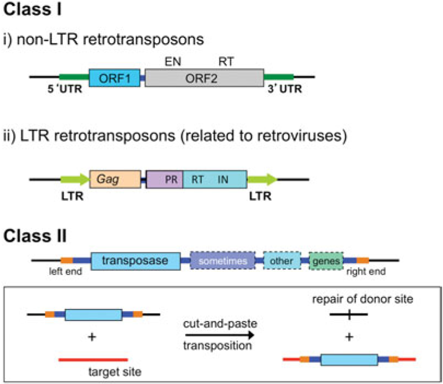
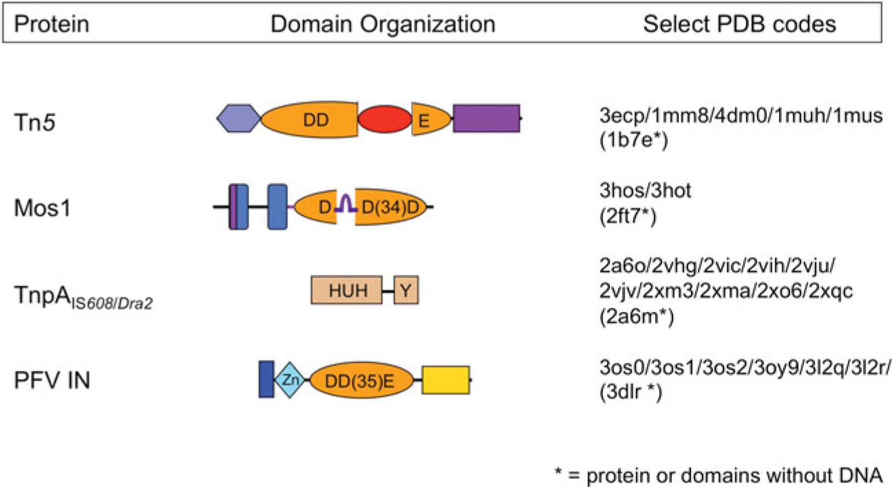
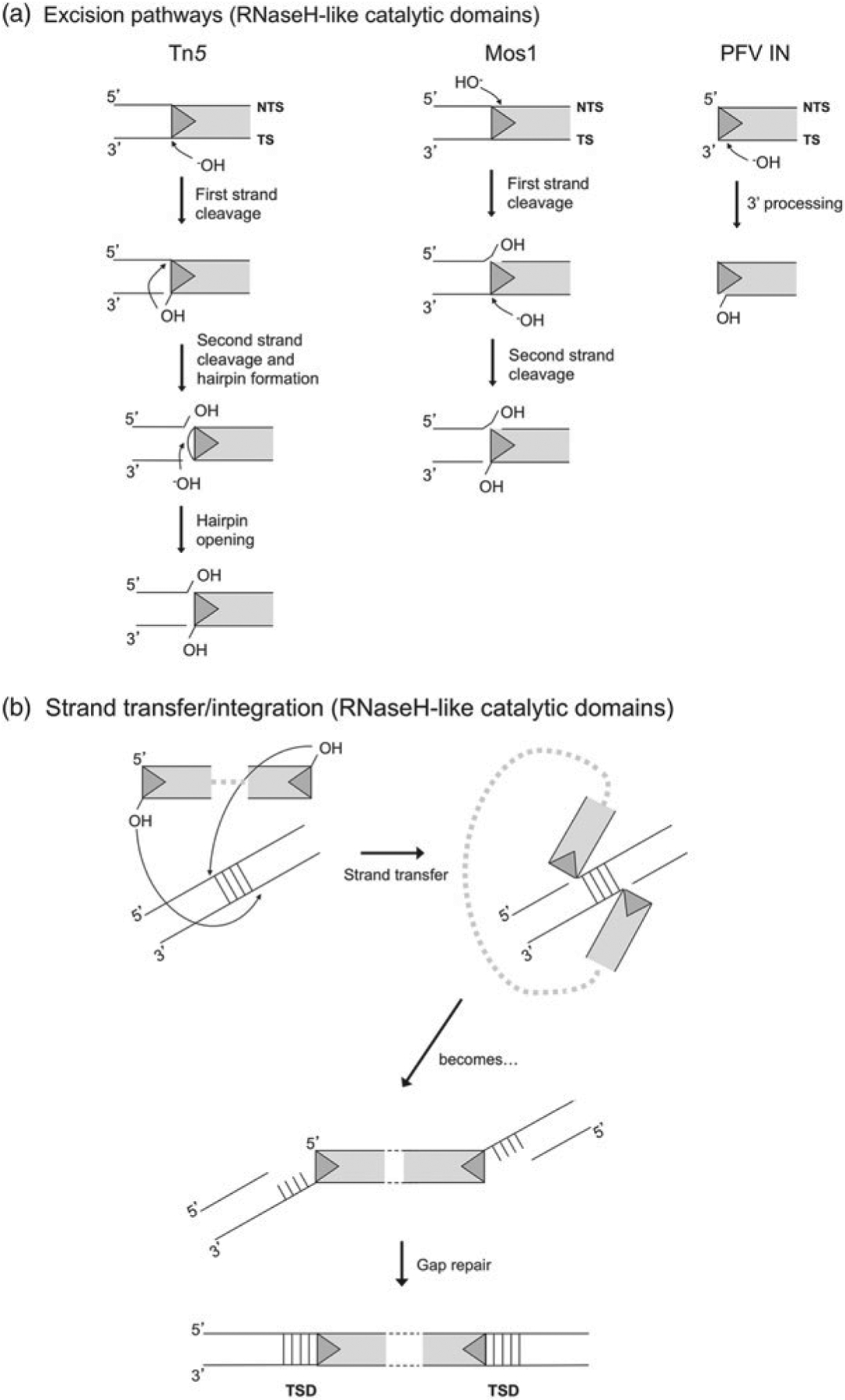
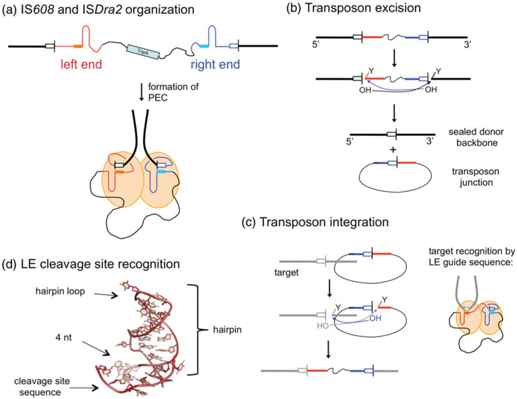
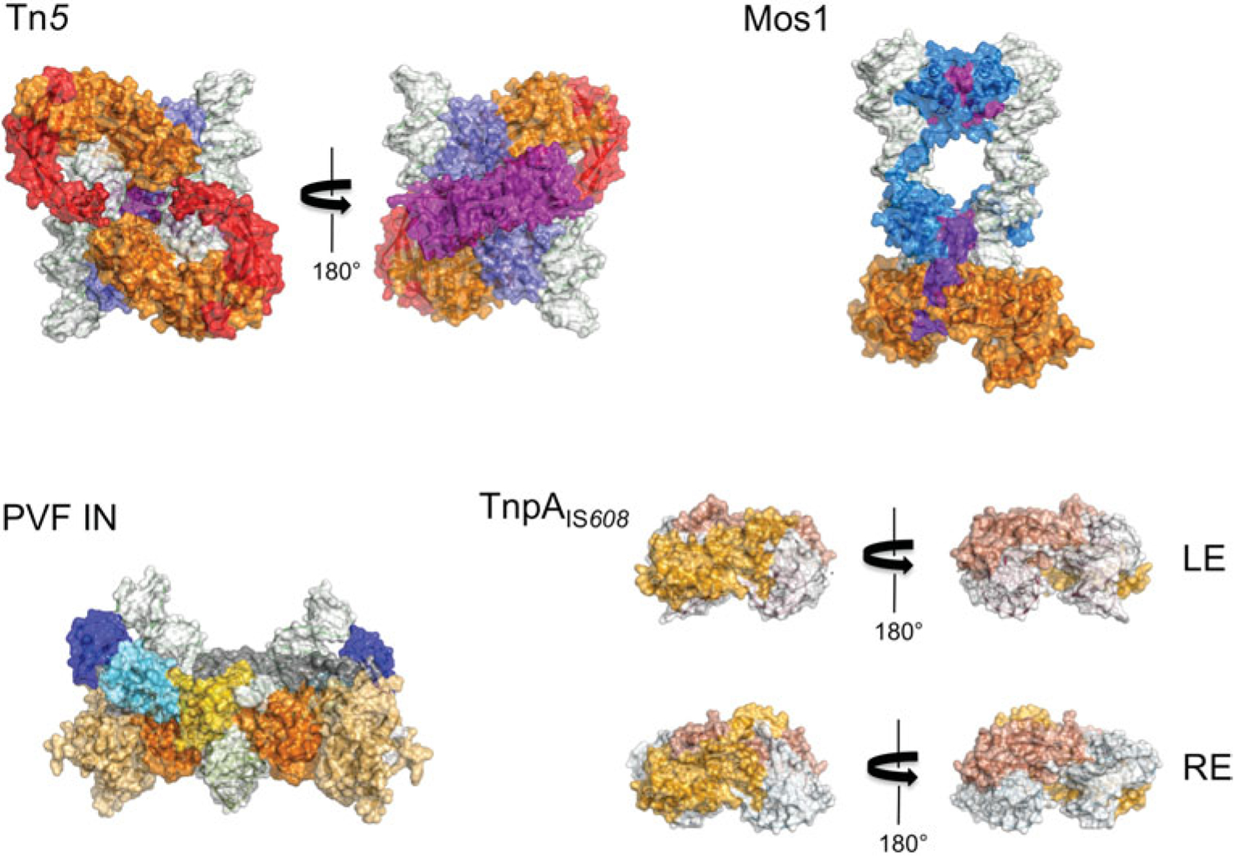
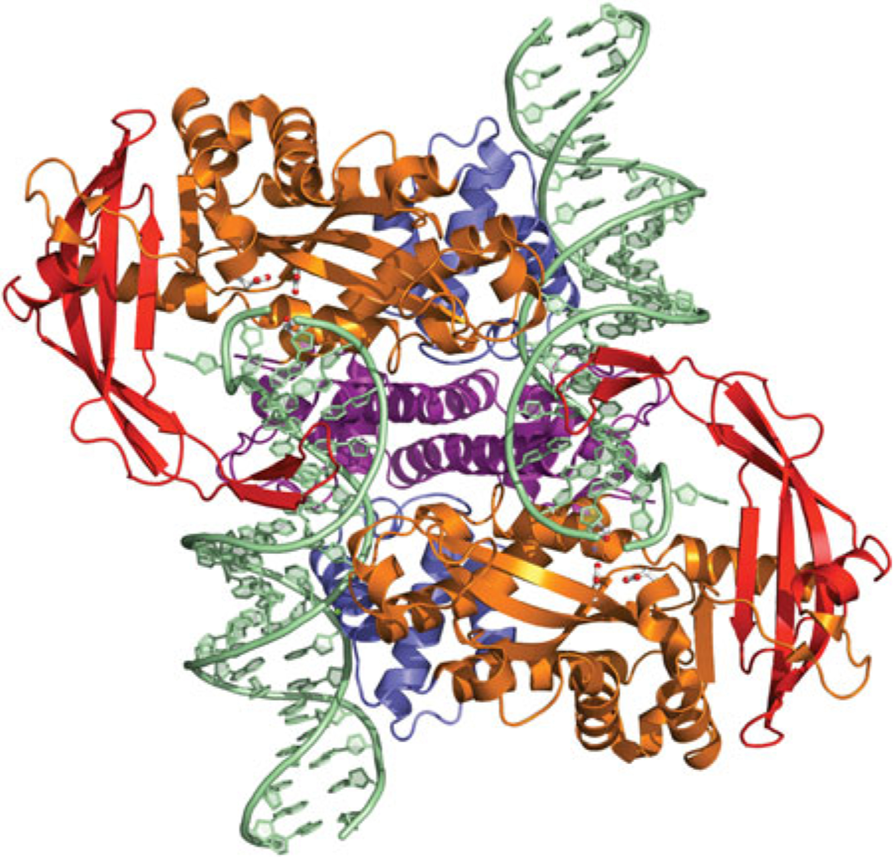
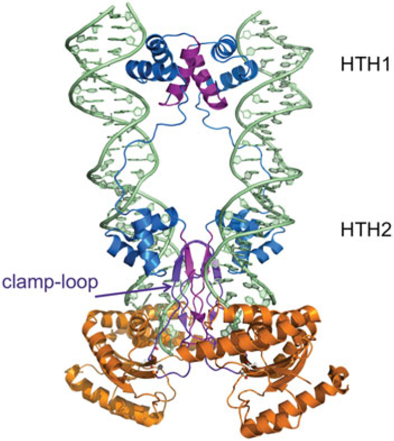
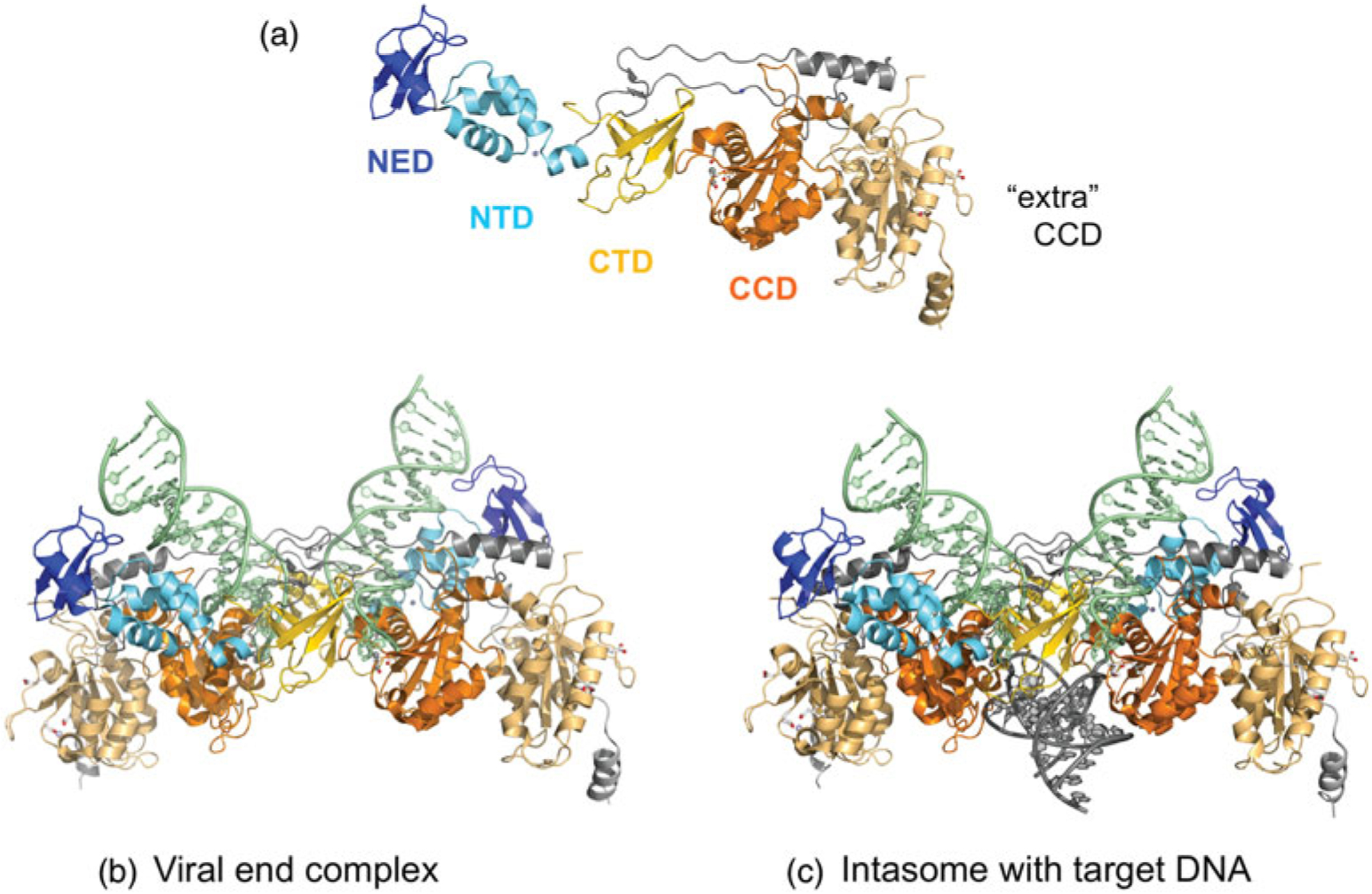
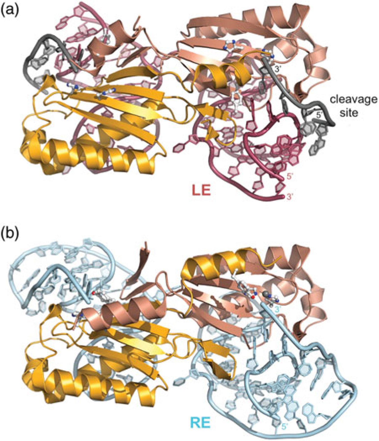
DNA transposases are enzymes that catalyze the movement of discrete pieces of DNA from one location in the genome to another. Transposition occurs through a series of controlled DNA strand cleavage and subsequent integration reactions that are carried out by nucleoprotein complexes known as transpososomes. Transpososomes are dynamic assemblies which must undergo conformational changes that control DNA breaks and ensure that, once started, the transposition reaction goes to completion. They provide a precise architecture within which the chemical reactions involved in transposon movement occur, but adopt different conformational states as transposition progresses. Their components also vary as they must, at some stage, include target DNA and sometimes even host-encoded proteins. A very limited number of transpososome states have been crystallographically captured, and here we provide an overview of the various structures determined to date. These structures include examples of DNA transposases that catalyze transposition by a cut-and-paste mechanism using an RNaseH-like nuclease catalytic domain, those that transpose using only single-stranded DNA substrates and targets, and the retroviral integrases that carry out an integration reaction very similar to DNA transposition. Given that there are a number of common functional requirements for transposition, it is remarkable how these are satisfied by complex assemblies that are so architecturally different.


Figures

Fig. 1.
Fig. 1.
Major transposon types. Among the Class I elements, non-LTR retrotransposons encode two proteins required for retrotransposition: ORF1, a non-specific nucleic acid binding protein, and ORF2 which has endonuclease (EN) and reverse transcriptase (RT) activities. In their simplest form, LTR retrotransposons have a gag gene that encodes structural proteins, and a pol gene that encodes a protease (PR), integrase (IN), and an RT, bounded by LTRs. Class II elements use only DNA intermediates, and although eukaryotic DNA transposons usually encode only the transposase enzyme required for movement (depicted in light blue, and shown in the inset for cut-and-paste DNA transposons), prokaryotic DNA transposons often carry other genes such as those encoding antibiotic resistance proteins (shown in varying shades). Specific binding sites for the transposase (in orange) are located at each end of the transposon.
Fig. 2.
Fig. 2.
Domain organization of select transposases. The four transposases for which there are high resolution transpososome structures are shown schematically, with DNA-binding domains in shades of blue and nuclease catalytic domains in shades of orange. Regions important for multimerization are shown in purple. The Tn5 and Mos1 transposases, and PFV integrase (PFV IN) have RNaseH-like catalytic domains which use three acidic residues (DDD or DDE) to coordinate two divalent metal ions required for catalysis. The TnpA ssDNA transposases use an HUH nuclease domain to coordinate a single metal ion which, in conjunction with a catalytic tyrosine, comprises the enzyme active site. The four beta-strand insertion into the Tn5 catalytic domain is shown in red, and the ‘clamp-loop’ of Mos1, inserted relative to the standard RNaseH topology between beta strands 1 and 2, is shown in purple.
Fig. 3.
Fig. 3.
(a) Excision pathways for Tn5, Mos1, and PFV integrase. Only reactions occurring on the TE LEs are shown. For Tn5, cleavage on both the NTS and the TS occur precisely at the transposon end. For Mos1, NTS cleavage occurs three bases within the transposon end and TS cleavage is precisely at the transposon end. For retroviral integrases such as that of PFV, the product of reverse transcription is a blunt-ended provirus from which integrase removes two 3′-OH nucleotides from the TS in a step known as 3′-processing. (b) Strand transfer pathway for DDE transposases. Shown is the specific pathway for PFV integrase, which catalyzes concerted integration into target DNA with a 4 bp stagger. As a result, after gap repair, the TE is flanked on both sides by a 4 bp TSD. For Tn5, staggered sites for strand transfer are offset by 9 bp and for Mos1 by 2 bp.
Fig. 4.
Fig. 4.
ssDNA transposition. (a) One strand of the transposon is shown, with the LE in red and the RE in blue. Both ends have imperfect palindromic sequences located close to the ends which form hairpins (as shown) that are recognized by the TnpA transposase. In the PEC, dimeric TnpA binds one LE and one RE. The cleavage sites (tetra- or pentanucleotides represented by white boxes) are recognized through non-linear base pairing with DNA (shown as solid boxes) at the base of the hairpins; cleavage occurs at the 3′ ends of the cleavage sites. Flanking DNA is represented as a thick black line, and transposon DNA as a thin black line. (b) Transposon excision. Each active site within the dimer cleaves one end. At the LE, this results in a free 3′-OH on flanking DNA and a covalent 5′-phosphotyrosine intermediate on the transposon end (represented by the ‘Y’). At the RE, the reaction with the same polarity results in a free 3′-OH on the transposon end and a covalent 5′-phosphotyrosine intermediate on flanking DNA. When the 3′-OH of one end attacks the phosphotyrosine intermediate of the other, the resulting products are an excised circular transposon junction and a precisely sealed donor backbone. (c) Transposon integration into a target site proceeds through two cleavage steps of the same polarity as for excision. The subsequent attacks of the 3′-OH groups on the 5′-phosphotyrosine intermediates result in an integrated transposon. The target cleavage site is recognized by non-linear base pairing with the DNA at the base of the LE hairpin, as shown for LE cleavage in (a) and (d).
Fig. 5.
Fig. 5.
Space-filling representation of transpososomes (to scale). The domain colors correspond to those in Fig. 2. DNA is shown with a white surface. In the case of PVF integrase, a target capture complex is shown.
Fig. 6.
Fig. 6.
The dimeric Tn5 transpososome. The domain colors correspond to those shown in Fig. 2, where the NTD is in blue, the catalytic core is in orange, and the C-terminal dimerization domain is in purple. The four-stranded insertion into the RNaseH-like catalytic domain is shown in red. The residues comprising the DDE motif (D97, D188, and E326) are shown in ball-and-stick representation. PDB code used to generate figure: 1MUH.
Fig. 7.
Fig. 7.
The dimeric Mos1 transpososome. The domain colors correspond to those shown in Fig. 2. The N-terminal DNA domain (two HTH domains connected by a long linker) is in blue and the RNaseH-like catalytic core is in orange. Residues involved in multimerization (amino acids 7–21, 112–125, and the clamp–loop residues 162–189) are shown in purple. The residues comprising the DDD motif (D156, D284, and D249) are shown in ball-and-stick representation. PDB code: 3HOT.
Fig. 8.
Fig. 8.
The PFV intasome. (a) The extended string of domains of one monomer of the intasome (domains in color) and its associated unused or ‘extra’ catalytic domain (in light orange). Below are the structures of (b) the intasome with two viral DNA ends and (c) the STC. The domain colors correspond to those shown in Fig. 2, where dark blue is the NED, light blue is the Zn-binding NTD (the Zn2+ ion is shown as a light blue sphere), orange is the CCD (there are two Mn2+ ions bound in the ‘used’ active site), and yellow is the CTD. The two additional CCDs with unused active sites on the periphery of the intasome contributed by outer subunits are shown in light orange in (b) and (c). PDB codes used to generate intasome figure with viral ends (3OY9) and with target DNA (3OS0).
Fig. 9.
Fig. 9.
Dimeric IS608 TnpA transpososomes. (a) TnpA bound to an LE 26-mer (in red) and a 6-mer cleavage or target site (grey). The two monomers are shown in different shades of orange to illustrate the composite active site. Active site residues H64, H66, and Y127 are shown in ball-and-stick representation. PDB code: 2VJV. In this structure, Y127 has been replaced by Phe. (b) TnpA bound to a RE 35-mer (in blue). PDB code: 2VJU.

References

    1. Abdelhakim AH, Sauer RT & Baker TA (2010). The AAA+ ClpX machine unfolds a keystone subunit to remodel the Mu transpososome. Proceedings of the National Academy of Sciences of the United States of America 107, 2437–2442. - PMC - PubMed
    1. Arciszewska LK, Drake D & Craig NL (1989). Transposon Tn7. cis-acting sequences in transposition and transposition immunity. Journal of Molecular Biology 207, 35–52. - PubMed
    1. Arciszewska LK, Mckown RL & Craig NL (1991). Purification of TnsB, a transposition protein that binds to the ends of Tn7. Journal of Biological Chemistry 266, 21736–21744. - PubMed
    1. Augé-Gouillou C, Hamelin MH, Demattei MV, Periquet M & Bigot Y (2001). The wild-type conformation of the Mos-1 inverted terminal repeats is suboptimal for transposition in bacteria. Molecular Genetics and Genomics 265, 51–57. - PubMed
    1. Augé-Gouillou C, Brillet B, Germon S, Hamelin MH & Bigot Y (2005a). Mariner Mos1 transposase dimerizes prior to ITR binding. Journal of Molecular Biology 351, 117–130. - PubMed

Publication types